
Search Count: 75,880
All
Selected
 |
Organism: Homo sapiens
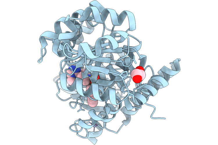
Method: X-RAY DIFFRACTION Resolution:2.42 Å Release Date: 2026-05-13 Classification: HYDROLASE/HYDROLASE INHIBITOR Ligands: ZN, A1C4J, EDO |
 |
Organism: Homo sapiens
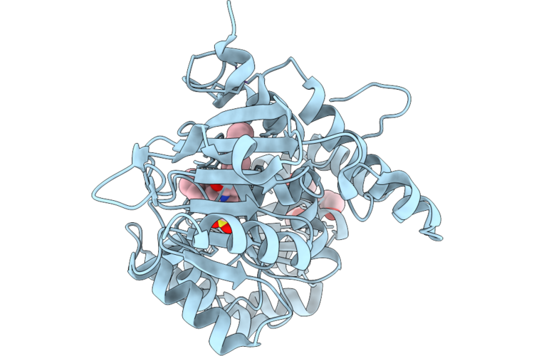
Method: X-RAY DIFFRACTION Resolution:1.37 Å Release Date: 2026-05-13 Classification: HYDROLASE/HYDROLASE INHIBITOR Ligands: ZN, A1C4L, CL, EDO |
 |
Organism: Homo sapiens
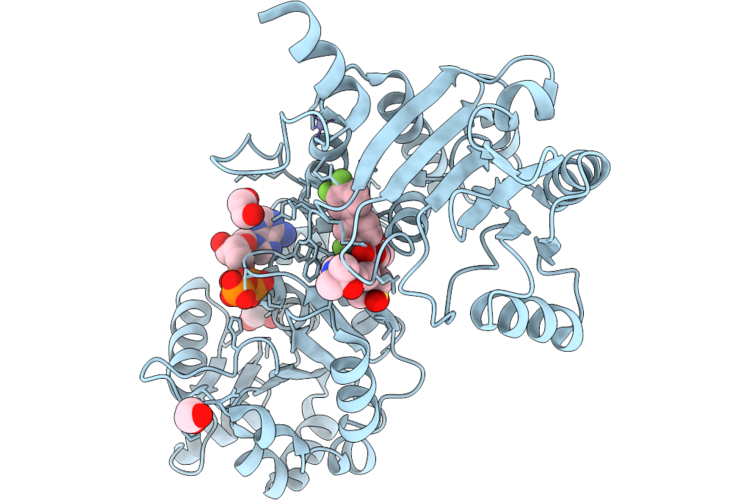
Method: X-RAY DIFFRACTION Resolution:2.58 Å Release Date: 2026-05-13 Classification: HYDROLASE Ligands: A1C4O, ATP, ZN, EDO |
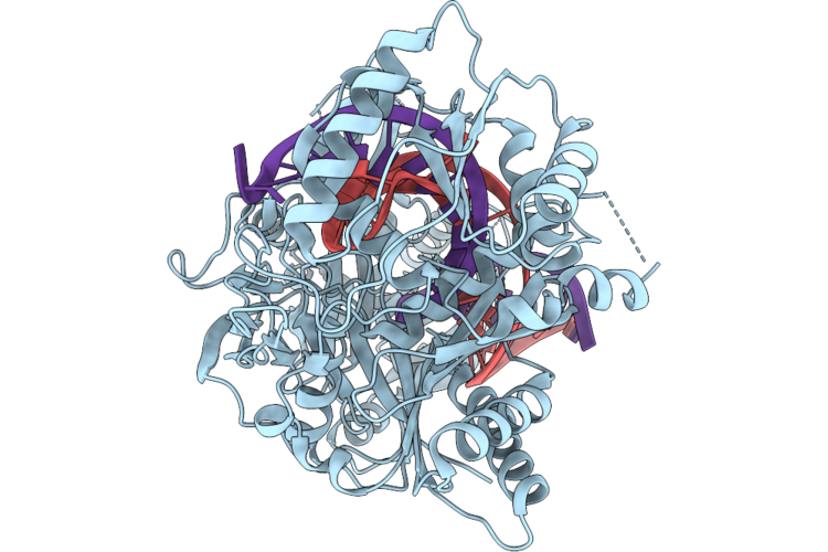 |
Human Argonaute2 Wt - Guide(3 Prime-Amino) Rna In Complex With A Fully Complementary Target
Organism: Homo sapiens, Synthetic construct
Method: ELECTRON MICROSCOPY Release Date: 2026-05-13 Classification: RNA BINDING PROTEIN/RNA Ligands: MG |
 |
Human Argonaute2 R315V/H316A - Guide10U Rna In Complex With A Complementary Target To Position 19
Organism: Homo sapiens, Synthetic construct
Method: ELECTRON MICROSCOPY Release Date: 2026-05-13 Classification: RNA BINDING PROTEIN/RNA Ligands: MG |
 |
Human Argonaute2 R315V/H316A - Guide Rna In Complex With A Fully Complementary Target (Conformation 2)
Organism: Homo sapiens, Synthetic construct
Method: ELECTRON MICROSCOPY Release Date: 2026-05-13 Classification: RNA BINDING PROTEIN/RNA Ligands: MG |
 |
Sars-Cov-2 Spike S2 Trimer Stabilized In The Early Fusion Intermediate Conformation (E-Fics-V3) Bound To The Vn01H1 Fab (Fab Local Refinement)
Organism: Saccharomyces cerevisiae s288c, Severe acute respiratory syndrome coronavirus 2, Homo sapiens
Method: ELECTRON MICROSCOPY Release Date: 2026-05-13 Classification: VIRAL PROTEIN Ligands: NAG |
 |
Sars-Cov-2 Spike S2 Trimer Stabilized In The Early Fusion Intermediate Conformation (E-Fics-V3) Bound To The Vn01H1 Fab (S2 Local Refinement)
Organism: Saccharomyces cerevisiae s288c, Severe acute respiratory syndrome coronavirus 2
Method: ELECTRON MICROSCOPY Release Date: 2026-05-13 Classification: VIRAL PROTEIN Ligands: NAG |
 |
Structure Of The Porcine Deltacoronavirus (Pdcov) Receptor-Binding Domain Bound To The Rbd Minibinder 11, The Pd3 Fab, And The Kappa Light Chain Nanobody (Local Refinement)
Organism: Porcine deltacoronavirus, Synthetic construct
Method: ELECTRON MICROSCOPY Resolution:2.84 Å Release Date: 2026-05-13 Classification: VIRAL PROTEIN |
 |
Structure Of The Porcine Deltacoronavirus (Pdcov) Receptor-Binding Domain Bound To The Rbd Minibinder 11, The Pd3 Fab, And The Kappa Light Chain Nanobody
Organism: Porcine deltacoronavirus, Synthetic construct, Mus musculus, Lama glama
Method: ELECTRON MICROSCOPY Resolution:2.80 Å Release Date: 2026-05-13 Classification: VIRAL PROTEIN |
 |
Organism: Bacillus glycinifermentans
Method: X-RAY DIFFRACTION Resolution:2.20 Å Release Date: 2026-05-13 Classification: HYDROLASE |
 |
Organism: Bacteroides intestinalis dsm 17393
Method: X-RAY DIFFRACTION Resolution:1.80 Å Release Date: 2026-05-13 Classification: HYDROLASE |
 |
Organism: Saccharomyces cerevisiae, Synthetic construct
Method: ELECTRON MICROSCOPY Release Date: 2026-05-13 Classification: REPLICATION Ligands: MG, ZN, ADP, ATP |
 |
Organism: Streptomyces plicatus
Method: X-RAY DIFFRACTION Resolution:1.99 Å Release Date: 2026-05-13 Classification: CARBOHYDRATE Ligands: CL |
 |
Crystal Structure Of Sars-Cov-2 Main Protease (Mpro) In Complex With Covalent Inhibitor A02
Organism: Severe acute respiratory syndrome coronavirus 2
Method: X-RAY DIFFRACTION Resolution:1.76 Å Release Date: 2026-05-13 Classification: HYDROLASE Ligands: A1BGG |
 |
Assembly Intermediate Of Human Mitochondrial Ribosome Small Subunit In Complex With Noa1 And Tfb1M (State N3)
Organism: Homo sapiens
Method: ELECTRON MICROSCOPY Release Date: 2026-05-13 Classification: RIBOSOME Ligands: MG, K, ZN, FES, ATP, GDP |
 |
Organism: Leishmania donovani, Bos taurus
Method: X-RAY DIFFRACTION Resolution:2.00 Å Release Date: 2026-05-13 Classification: HYDROLASE Ligands: CL, EDO, GOL |
 |
Cryo-Electron Microscopic Structure Of A Novel Amidohydrolase Adh3 Triple Mutation
Organism: Stenotrophomonas sp. cw117
Method: ELECTRON MICROSCOPY Release Date: 2026-05-13 Classification: HYDROLASE Ligands: ZN, 97U |
 |
Organism: Lachesis muta
Method: X-RAY DIFFRACTION Resolution:2.36 Å Release Date: 2026-05-13 Classification: TOXIN,HYDROLASE Ligands: MES, CA |
 |
Cryo-Em Structure Of The Pi3K Alpha/Kras/Her3 Phosphopeptide Complex Dimer On Popc/Pops/Pip2 Nanodiscs
Organism: Homo sapiens
Method: ELECTRON MICROSCOPY Release Date: 2026-05-13 Classification: Transferase/Hydrolase Ligands: A1AZD, MG, GNP |