

Search Count: 10
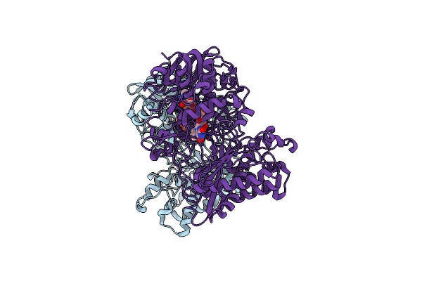 |
Structure Of The Mimivirus Genomic Fibre Asymmetric Unit
Organism: Acanthamoeba polyphaga mimivirus
Method: ELECTRON MICROSCOPY Release Date: 2022-08-10 Classification: VIRAL PROTEIN Ligands: FAD |
Organism: Acanthamoeba polyphaga mimivirus
Method: ELECTRON MICROSCOPY
Release Date: 2022-08-10
Ligands: FAD
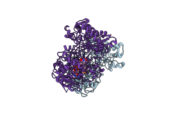 |
Structure Of The Mimivirus Genomic Fibre In Its Compact 6-Start Helix Form
Organism: Acanthamoeba polyphaga mimivirus
Method: ELECTRON MICROSCOPY Release Date: 2022-08-10 Classification: VIRAL PROTEIN Ligands: FAD |
Organism: Acanthamoeba polyphaga mimivirus
Method: ELECTRON MICROSCOPY
Release Date: 2022-08-10
Ligands: FAD
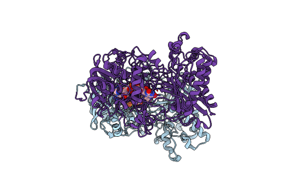 |
Structure Of The Mimivirus Genomic Fibre In Its Compact 5-Start Helix Form
Organism: Acanthamoeba polyphaga mimivirus
Method: ELECTRON MICROSCOPY Release Date: 2022-08-10 Classification: VIRAL PROTEIN Ligands: FAD |
Organism: Acanthamoeba polyphaga mimivirus
Method: ELECTRON MICROSCOPY
Release Date: 2022-08-10
Ligands: FAD
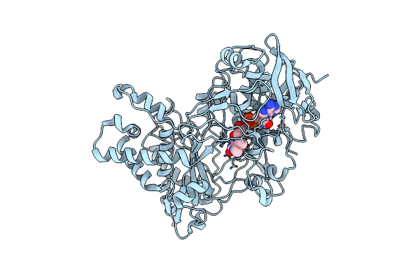 |
Structure Of The Mimivirus Genomic Fibre In Its Relaxed 5-Start Helix Form
Organism: Acanthamoeba polyphaga mimivirus
Method: ELECTRON MICROSCOPY Release Date: 2022-08-10 Classification: VIRAL PROTEIN Ligands: FAD |
Organism: Acanthamoeba polyphaga mimivirus
Method: ELECTRON MICROSCOPY
Release Date: 2022-08-10
Ligands: FAD
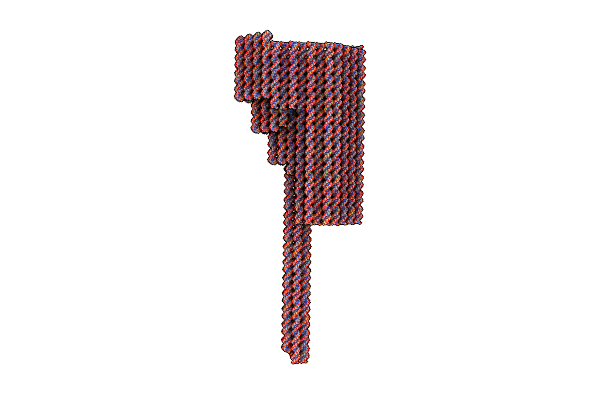 |
Dna Origami Signpost Designed Model
Organism: Escherichia virus m13
Method: ELECTRON MICROSCOPY Release Date: 2021-04-14 Classification: DNA |
Organism: Escherichia virus m13
Method: ELECTRON MICROSCOPY
Release Date: 2021-04-14
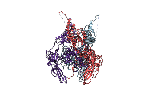 |
Pseudoatomic Model Of The Pre-Fusion Conformation Of Glycoprotein B Of Herpes Simplex Virus 1
Organism: Human herpesvirus 1
Method: ELECTRON MICROSCOPY Release Date: 2020-10-07 Classification: VIRAL PROTEIN |
Organism: Human herpesvirus 1
Method: ELECTRON MICROSCOPY
Release Date: 2020-10-07
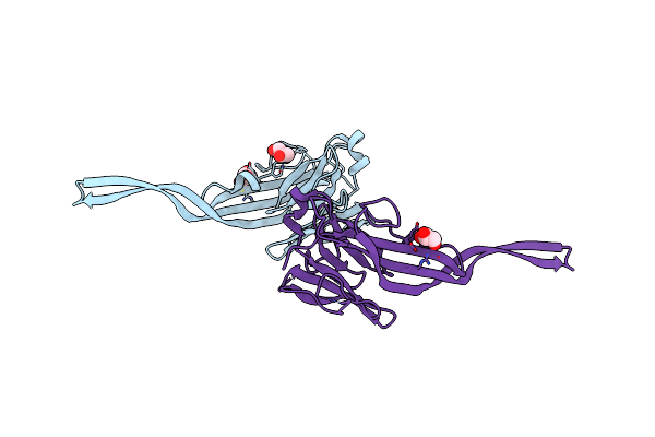 |
Crystal Structure Of Murine Perforin-2 P2 Domain Crystal Form 1
Organism: Mus musculus
Method: X-RAY DIFFRACTION Resolution:2.05 Å Release Date: 2020-02-05 Classification: TOXIN Ligands: GOL, CL |
Organism: Mus musculus
Method: X-RAY DIFFRACTION
Release Date: 2020-02-05
Ligands: GOL, CL
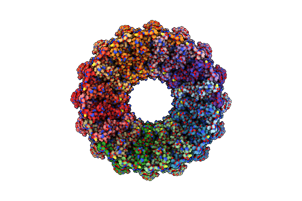 |
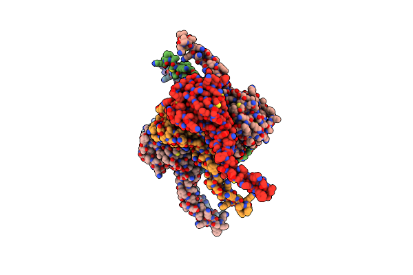
Cryoem Structure Of Murine Perforin-2 Ectodomain In A Pre-Pore Form
Organism: Mus musculus
Method: ELECTRON MICROSCOPY Release Date: 2020-02-05 Classification: TOXIN Ligands: NAG |
Organism: Mus musculus
Method: ELECTRON MICROSCOPY
Release Date: 2020-02-05
Ligands: NAG
 |
Crystal Structure Of Murine Perforin-2 P2 Domain Crystal Form 2
Organism: Mus musculus
Method: X-RAY DIFFRACTION Resolution:3.17 Å Release Date: 2020-02-05 Classification: TOXIN |
Organism: Mus musculus
Method: X-RAY DIFFRACTION
Release Date: 2020-02-05
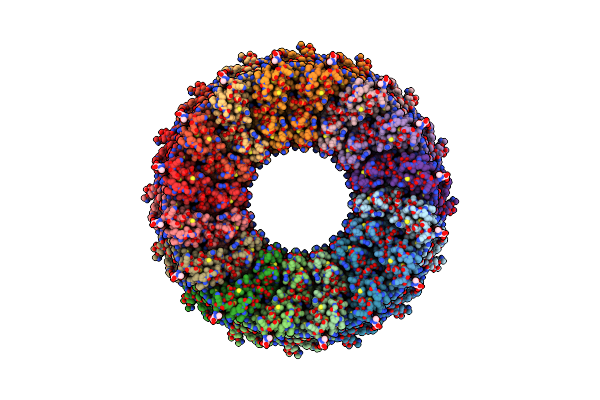 |
Cryoem Structure Of Murine Perforin-2 Ectodomain In A Pore Form
Organism: Mus musculus
Method: ELECTRON MICROSCOPY Release Date: 2020-02-05 Classification: TOXIN Ligands: NAG |
Organism: Mus musculus
Method: ELECTRON MICROSCOPY
Release Date: 2020-02-05
Ligands: NAG