

Search Count: 35
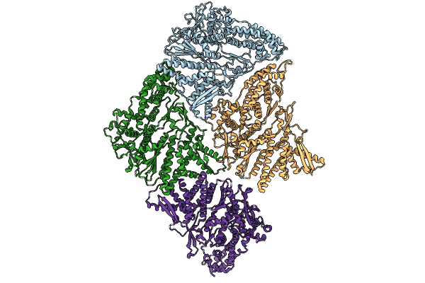 |
Inner Layer Protein P1 Chains In Transcribing Particles Of Bacteriophage Phi6
Organism: Cystovirus phi6
Method: ELECTRON MICROSCOPY Release Date: 2026-01-21 Classification: VIRAL PROTEIN |
Organism: Cystovirus phi6
Method: ELECTRON MICROSCOPY
Release Date: 2026-01-21
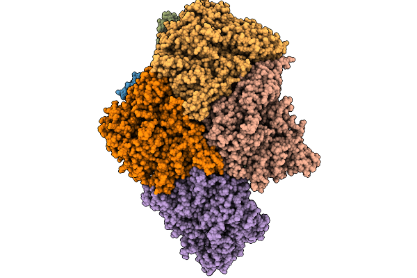 |
Extended And Wrapped Protein P7 Dimers Of Dimers, The P1 Layer And The Rna-Dependent Rna Polymerase P2 In Transcribing Particles Of Bacteriophage Phi6
Organism: Synthetic construct, Cystovirus phi6
Method: ELECTRON MICROSCOPY Release Date: 2026-01-21 Classification: VIRAL PROTEIN |
Organism: Synthetic construct, Cystovirus phi6
Method: ELECTRON MICROSCOPY
Release Date: 2026-01-21
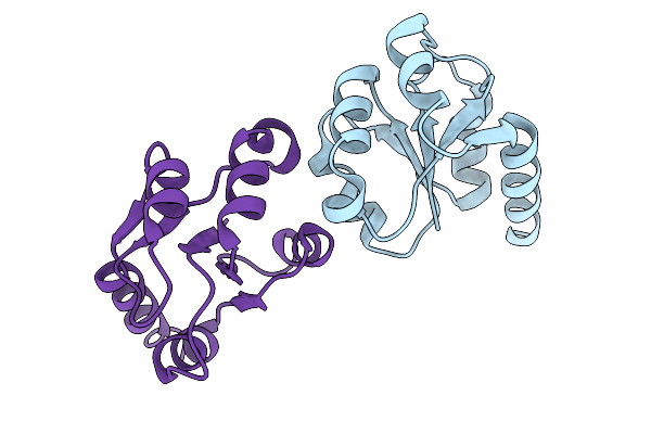 |
Minor Protein P7 Dimer From Transcribing Particles Of Bacteriophage Phi6
Organism: Cystovirus phi6
Method: ELECTRON MICROSCOPY Release Date: 2026-01-14 Classification: VIRUS |
Organism: Cystovirus phi6
Method: ELECTRON MICROSCOPY
Release Date: 2026-01-14
 |
Asymmetric Unit From Transcribing Double-Layered Particle Of Bacteriophage Phi6
Organism: Cystovirus phi6
Method: ELECTRON MICROSCOPY Release Date: 2026-01-14 Classification: VIRUS |
Organism: Cystovirus phi6
Method: ELECTRON MICROSCOPY
Release Date: 2026-01-14
 |
Asymmetric Unit From Transcribing Single-Layered Particle Of Bacteriophage Phi6
Organism: Cystovirus phi6
Method: ELECTRON MICROSCOPY Release Date: 2026-01-14 Classification: VIRUS |
Organism: Cystovirus phi6
Method: ELECTRON MICROSCOPY
Release Date: 2026-01-14
 |
Asymmetric Unit From Transcribing Single-Layered Particle Of Bacteriophage Phi6 In An Over-Expanded State
Organism: Cystovirus phi6
Method: ELECTRON MICROSCOPY Release Date: 2026-01-14 Classification: VIRUS |
Organism: Cystovirus phi6
Method: ELECTRON MICROSCOPY
Release Date: 2026-01-14
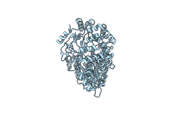 |
Structure Of The Rna-Dependent Rna Polymerase P2 From The Bacteriophage Phi8
Organism: Pseudomonas phage phi8
Method: X-RAY DIFFRACTION Resolution:3.00 Å Release Date: 2024-10-16 Classification: VIRAL PROTEIN Ligands: CA |
Organism: Pseudomonas phage phi8
Method: X-RAY DIFFRACTION
Release Date: 2024-10-16
Ligands: CA
 |
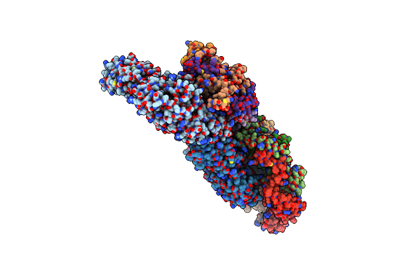
Atomic Models Of P1, P4 C-Terminal Fragment And P8 Fitted In The Bacteriophage Phi6 Nucleocapsid Reconstructed With Icosahedral Symmetry
Organism: Pseudomonas phage phi6
Method: ELECTRON MICROSCOPY Release Date: 2019-06-12 Classification: VIRUS |
Organism: Pseudomonas phage phi6
Method: ELECTRON MICROSCOPY
Release Date: 2019-06-12
 |
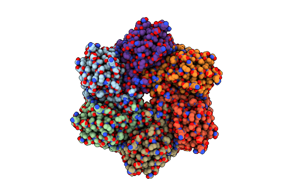
Dsrna Bacteriophage Phi6 Nucleocapsid
Organism: Pseudomonas phage phi6
Method: ELECTRON MICROSCOPY Release Date: 2017-03-22 Classification: VIRUS |
Organism: Pseudomonas phage phi6
Method: ELECTRON MICROSCOPY
Release Date: 2017-03-22
 |
Atomic Structure Fitted Into A Localized Reconstruction Of Bacteriophage Phi6 Packaging Hexamer P4
Organism: Pseudomonas phage phi6
Method: ELECTRON MICROSCOPY Release Date: 2017-03-22 Classification: HYDROLASE Ligands: CA, ADP |
Organism: Pseudomonas phage phi6
Method: ELECTRON MICROSCOPY
Release Date: 2017-03-22
Ligands: CA, ADP
 |
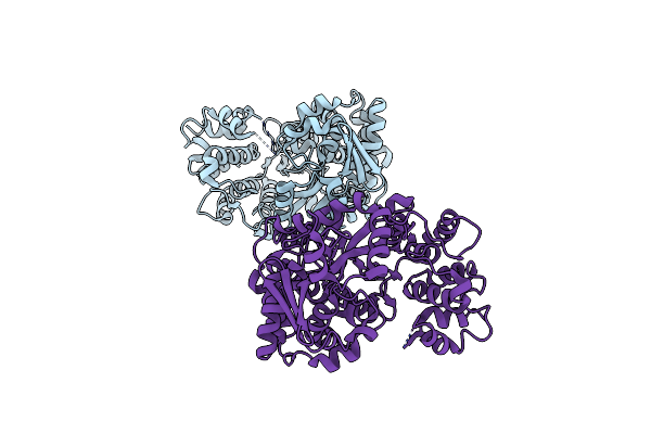
Atomic Structure Of P4 Packaging Enzyme Fitted Into A Localized Reconstruction Of Bacteriophage Phi6 Vertex
Organism: Pseudomonas phage phi6
Method: ELECTRON MICROSCOPY Resolution:9.10 Å Release Date: 2017-03-22 Classification: HYDROLASE Ligands: CA, ADP |
Organism: Pseudomonas phage phi6
Method: ELECTRON MICROSCOPY
Release Date: 2017-03-22
Ligands: CA, ADP
 |
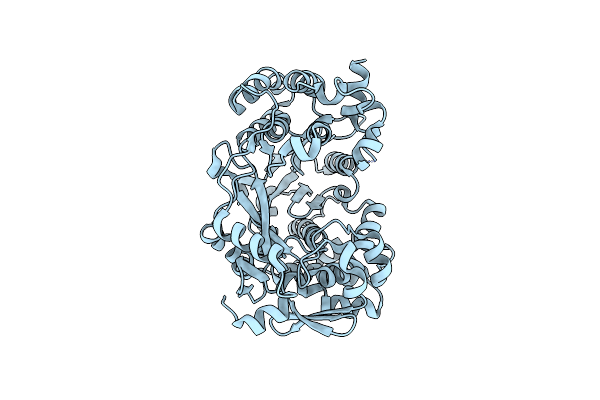
Crystal Structure Of The Rna-Dependent Rna Polymerase Of A Human Picorbirnavirus
Organism: Human picobirnavirus (strain human/thailand/hy005102/-)
Method: X-RAY DIFFRACTION Resolution:2.40 Å Release Date: 2016-03-16 Classification: viral protein, transcription |
Organism: Human picobirnavirus (strain human/thailand/hy005102/-)
Method: X-RAY DIFFRACTION
Release Date: 2016-03-16
 |
Crystal Structure Of The Insertion Loop Deletion Mutant Of The Rna-Dependent Rna Polymerase Of A Human Picorbirnavirus
Organism: Human picobirnavirus (strain human/thailand/hy005102/-)
Method: X-RAY DIFFRACTION Resolution:2.00 Å Release Date: 2016-03-16 Classification: viral protein, replication |
Organism: Human picobirnavirus (strain human/thailand/hy005102/-)
Method: X-RAY DIFFRACTION
Release Date: 2016-03-16
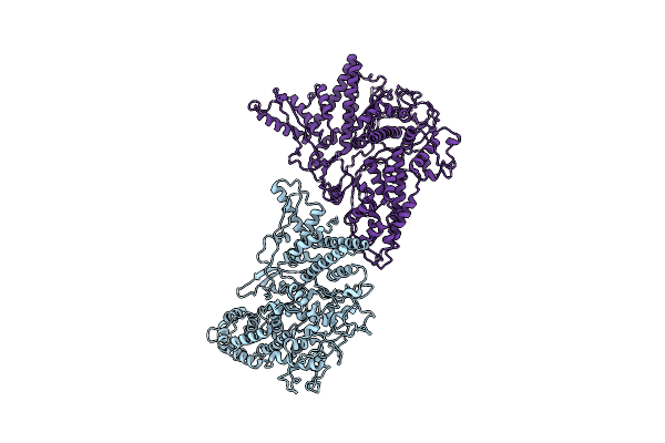 |
Structure Of The In Vitro Assembled Bacteriophage Phi6 Polymerase Complex
Organism: Pseudomonas phage phi6
Method: ELECTRON MICROSCOPY Resolution:4.80 Å Release Date: 2015-11-04 Classification: VIRAL PROTEIN |
Organism: Pseudomonas phage phi6
Method: ELECTRON MICROSCOPY
Release Date: 2015-11-04
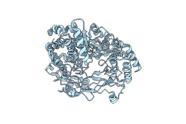 |
Structure Of The P2 Polymerase Inside In Vitro Assembled Bacteriophage Phi6 Polymerase Complex
Organism: Pseudomonas phage phi6
Method: ELECTRON MICROSCOPY Resolution:7.90 Å Release Date: 2015-11-04 Classification: TRANSCRIPTION Ligands: MN |
Organism: Pseudomonas phage phi6
Method: ELECTRON MICROSCOPY
Release Date: 2015-11-04
Ligands: MN
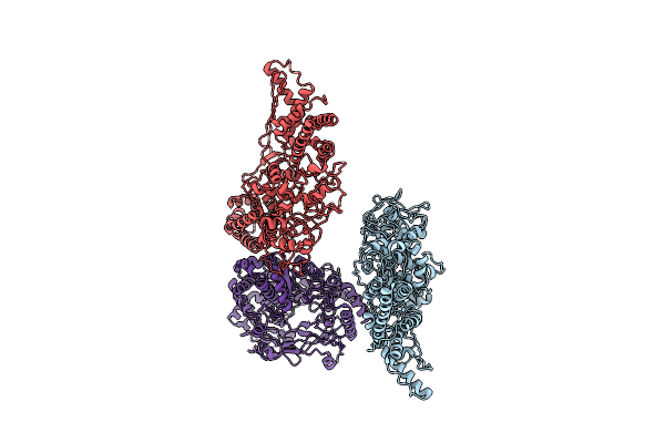 |
Structure Of The P2 Polymerase Inside In Vitro Assembled Bacteriophage Phi6 Polymerase Complex, With P1 Included
Organism: Pseudomonas phage phi6
Method: ELECTRON MICROSCOPY Resolution:7.90 Å Release Date: 2015-11-04 Classification: VIRAL PROTEIN Ligands: MN |
Organism: Pseudomonas phage phi6
Method: ELECTRON MICROSCOPY
Release Date: 2015-11-04
Ligands: MN
 |
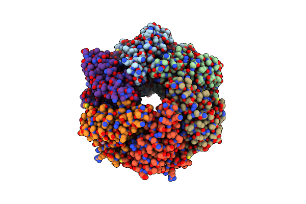
P4 Protein From Bacteriophage Phi6 In Complex With Adp
Organism: Pseudomonas phage phi6
Method: X-RAY DIFFRACTION Resolution:2.80 Å Release Date: 2013-08-21 Classification: HYDROLASE Ligands: CA, ADP |
Organism: Pseudomonas phage phi6
Method: X-RAY DIFFRACTION
Release Date: 2013-08-21
Ligands: CA, ADP
 |
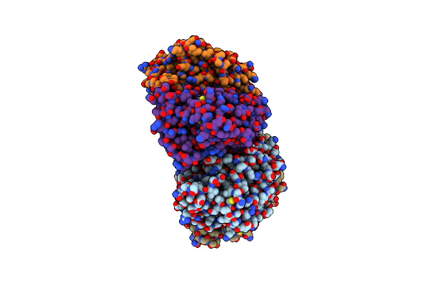
P4 Protein From Bacteriophage Phi13
Organism: Pseudomonas phage phi13
Method: X-RAY DIFFRACTION Resolution:1.70 Å Release Date: 2013-08-21 Classification: HYDROLASE Ligands: FLC, GOL |
Organism: Pseudomonas phage phi13
Method: X-RAY DIFFRACTION
Release Date: 2013-08-21
Ligands: FLC, GOL
 |
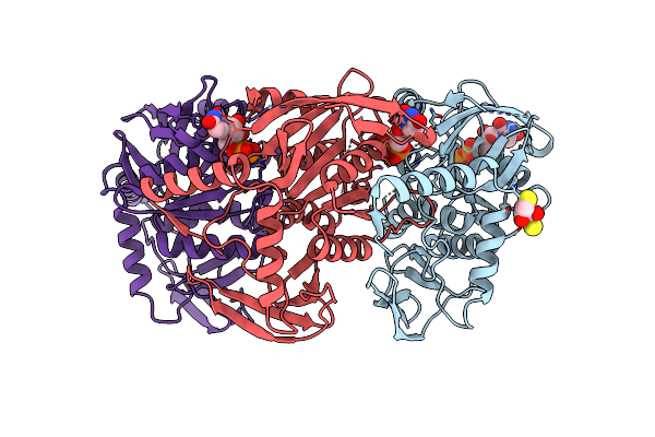
P4 Protein From Bacteriophage Phi8
Organism: Pseudomonas phage phi8
Method: X-RAY DIFFRACTION Resolution:2.79 Å Release Date: 2013-08-21 Classification: HYDROLASE |
Organism: Pseudomonas phage phi8
Method: X-RAY DIFFRACTION
Release Date: 2013-08-21
 |
P4 Protein From Bacteriophage Phi12 In Complex With Utp
Organism: Pseudomonas phage phi12
Method: X-RAY DIFFRACTION Resolution:1.90 Å Release Date: 2013-08-21 Classification: HYDROLASE Ligands: UTP, DTT |
Organism: Pseudomonas phage phi12
Method: X-RAY DIFFRACTION
Release Date: 2013-08-21
Ligands: UTP, DTT