

Search Count: 18
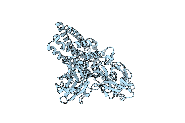 |
Crystal Structure Of Cry1Fa R226Q
Organism: Bacillus thuringiensis
Method: X-RAY DIFFRACTION Resolution:2.02 Å Release Date: 2024-05-29 Classification: TOXIN |
Organism: Bacillus thuringiensis
Method: X-RAY DIFFRACTION
Release Date: 2024-05-29
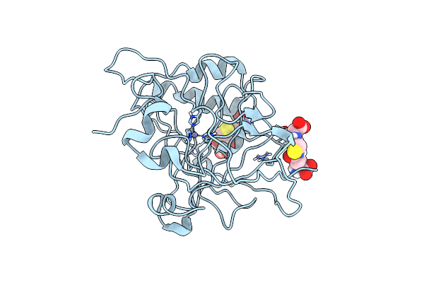 |
Crystal Structure Of S-Glutathiolated Carbonic Anhydrase Iii
Organism: Rattus norvegicus
Method: X-RAY DIFFRACTION Resolution:1.80 Å Release Date: 2000-09-04 Classification: LYASE Ligands: ZN, GSH |
Organism: Rattus norvegicus
Method: X-RAY DIFFRACTION
Release Date: 2000-09-04
Ligands: ZN, GSH
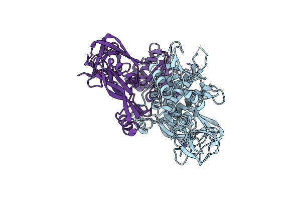 |
Crystal Structure Of Pi-Scei Miniprecursor
Organism: Saccharomyces cerevisiae
Method: X-RAY DIFFRACTION Resolution:2.10 Å Release Date: 2000-06-07 Classification: HYDROLASE Ligands: ZN |
Organism: Saccharomyces cerevisiae
Method: X-RAY DIFFRACTION
Release Date: 2000-06-07
Ligands: ZN
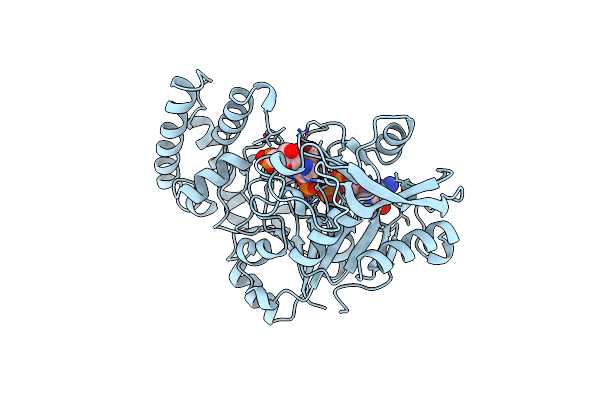 |
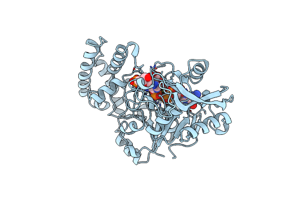
Structure Of Adenylosuccinate Synthetase From E. Coli Complexed With Hadacidin, Gdp, 6-Phosphoryl-Imp, And Mg2+
Organism: Escherichia coli k12
Method: X-RAY DIFFRACTION Resolution:2.50 Å Release Date: 1999-06-17 Classification: LIGASE Ligands: MG, HDA, IMO, GDP |
Organism: Escherichia coli k12
Method: X-RAY DIFFRACTION
Release Date: 1999-06-17
Ligands: MG, HDA, IMO, GDP
 |
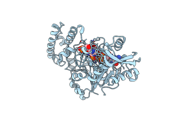
Structure Of The Mutant (K16Q) Of Adenylosuccinate Synthetase From E. Coli Complexed With Hadacidin, Gdp, 6-Phosphoryl-Imp, And Mg2+
Organism: Escherichia coli k12
Method: X-RAY DIFFRACTION Resolution:2.50 Å Release Date: 1999-06-17 Classification: LIGASE Ligands: MG, HDA, IMO, GDP |
Organism: Escherichia coli k12
Method: X-RAY DIFFRACTION
Release Date: 1999-06-17
Ligands: MG, HDA, IMO, GDP
 |
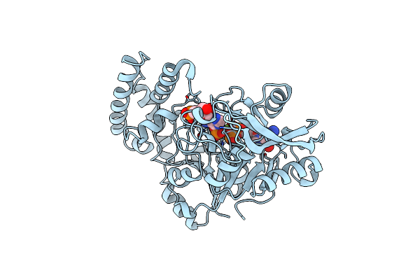
Structure Of The Mutant (R143L) Of Adenylosuccinate Synthetase From E. Coli Complexed With Hadacidin, Gdp, 6-Phosphoryl-Imp, And Mg2+
Organism: Escherichia coli k12
Method: X-RAY DIFFRACTION Resolution:2.50 Å Release Date: 1999-06-17 Classification: LIGASE Ligands: MG, HDA, IMO, GDP |
Organism: Escherichia coli k12
Method: X-RAY DIFFRACTION
Release Date: 1999-06-17
Ligands: MG, HDA, IMO, GDP
 |
Structure Of The Mutant (R303L) Of Adenylosuccinate Synthetase From E. Coli Complexed With, Gdp, 6-Phosphoryl-Imp, And Mg2+
Organism: Escherichia coli k12
Method: X-RAY DIFFRACTION Resolution:2.50 Å Release Date: 1999-06-17 Classification: LIGASE Ligands: MG, IMO, GDP |
Organism: Escherichia coli k12
Method: X-RAY DIFFRACTION
Release Date: 1999-06-17
Ligands: MG, IMO, GDP
 |
Fructose-1,6-Bisphosphatase Complexed With Fructose-6-Phosphate And Zinc Ions
Organism: Sus scrofa
Method: X-RAY DIFFRACTION Resolution:2.27 Å Release Date: 1999-05-28 Classification: HYDROLASE Ligands: F6P, ZN, PO4 |
Organism: Sus scrofa
Method: X-RAY DIFFRACTION
Release Date: 1999-05-28
Ligands: F6P, ZN, PO4
 |
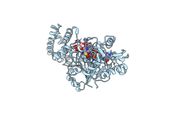
Entrapment Of 6-Thiophosphoryl-Imp In The Active Site Of Crystalline Adenylosuccinate Synthetase From Escherichia Coli, Data Collected At 298K
Organism: Escherichia coli
Method: X-RAY DIFFRACTION Resolution:2.80 Å Release Date: 1997-10-08 Classification: LIGASE Ligands: MG, GDP, HDA, PGS |
Organism: Escherichia coli
Method: X-RAY DIFFRACTION
Release Date: 1997-10-08
Ligands: MG, GDP, HDA, PGS
 |
Entrapment Of 6-Thiophosphoryl-Imp In The Active Site Of Crystalline Adenylosuccinate Synthetase From Escherichia Coli Data Collected At 100K
Organism: Escherichia coli
Method: X-RAY DIFFRACTION Resolution:2.50 Å Release Date: 1997-10-08 Classification: LIGASE Ligands: MG, GDP, HDA, PGS |
Organism: Escherichia coli
Method: X-RAY DIFFRACTION
Release Date: 1997-10-08
Ligands: MG, GDP, HDA, PGS
 |
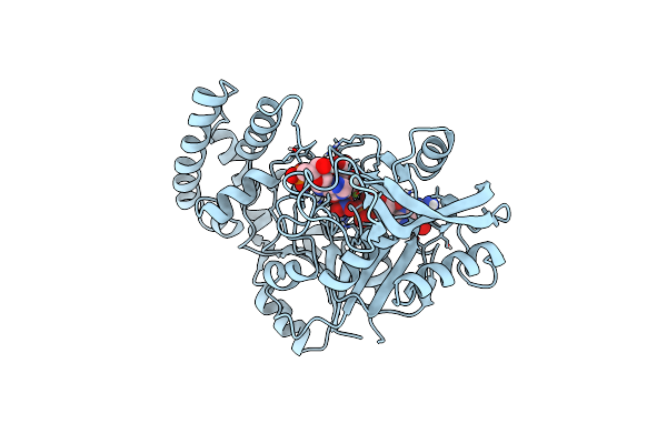
Refined Crystal Structure Of Adenylosuccinate Synthetase From Escherichia Coli Complexed With Hydantocidin 5'-Phosphate Gdp, Hpo4(2-), Mg2+, And Hadacidin
Organism: Escherichia coli
Method: X-RAY DIFFRACTION Resolution:2.50 Å Release Date: 1997-06-24 Classification: LIGASE Ligands: MG, PI, GDP, H5P, HDA |
Organism: Escherichia coli
Method: X-RAY DIFFRACTION
Release Date: 1997-06-24
Ligands: MG, PI, GDP, H5P, HDA
 |
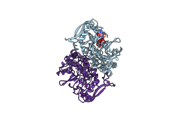
Crystal Structure Of Adenylosuccinate Synthetase From Escherichia Coli Complexed With Gdp, Imp, Hadacidin, No3-, And Mg2+. Data Collected At 298K (Ph 6.5).
Organism: Escherichia coli
Method: X-RAY DIFFRACTION Resolution:2.80 Å Release Date: 1997-02-12 Classification: LIGASE Ligands: MG, NO3, HDA, IMP, GDP |
Organism: Escherichia coli
Method: X-RAY DIFFRACTION
Release Date: 1997-02-12
Ligands: MG, NO3, HDA, IMP, GDP
 |
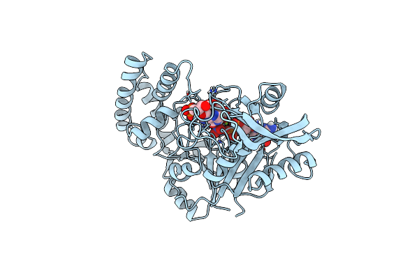
Structure Of Guanine Nucleotide (Gppcp) Complex Of Adenylosuccinate Synthetase From E. Coli At Ph 6.5 And 25 Degrees Celsius
Organism: Escherichia coli
Method: X-RAY DIFFRACTION Resolution:2.30 Å Release Date: 1997-02-12 Classification: LIGASE Ligands: GNH, GNP |
Organism: Escherichia coli
Method: X-RAY DIFFRACTION
Release Date: 1997-02-12
Ligands: GNH, GNP
 |
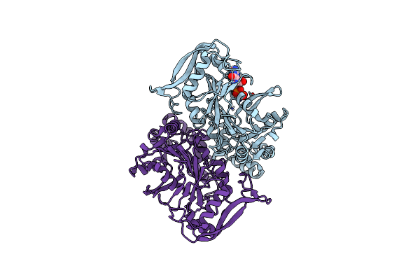
Crystal Structure Of Adenylosuccinate Synthetase From Escherichia Coli Complexed With Gdp, Imp, Hadacidin, No3-, And Mg2+. Data Collected At 100K (Ph 6.5)
Organism: Escherichia coli
Method: X-RAY DIFFRACTION Resolution:2.50 Å Release Date: 1996-12-23 Classification: LIGASE Ligands: MG, NO3, HDA, IMP, GDP |
Organism: Escherichia coli
Method: X-RAY DIFFRACTION
Release Date: 1996-12-23
Ligands: MG, NO3, HDA, IMP, GDP
 |
Structure Of Guanine Nucleotide (Gppcp) Complex Of Adenylosuccinate Synthetase From Escherichia Coli At Ph 6.5 And 25 Degree Celsius
Organism: Escherichia coli
Method: X-RAY DIFFRACTION Resolution:2.30 Å Release Date: 1996-11-08 Classification: LIGASE (SYNTHETASE) Ligands: GNH |
Organism: Escherichia coli
Method: X-RAY DIFFRACTION
Release Date: 1996-11-08
Ligands: GNH
 |
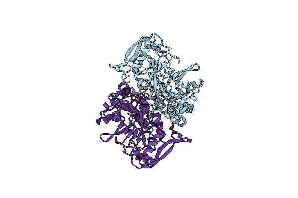
Structure Of Guanine Nucleotide (Gppcp) Complex Of Adenylosuccinate Synthetase From Escherichia Coli At Ph 6.5 And 25 Degrees Celsius
Organism: Escherichia coli
Method: X-RAY DIFFRACTION Resolution:2.30 Å Release Date: 1996-11-08 Classification: LIGASE (SYNTHETASE) Ligands: GCP |
Organism: Escherichia coli
Method: X-RAY DIFFRACTION
Release Date: 1996-11-08
Ligands: GCP
 |
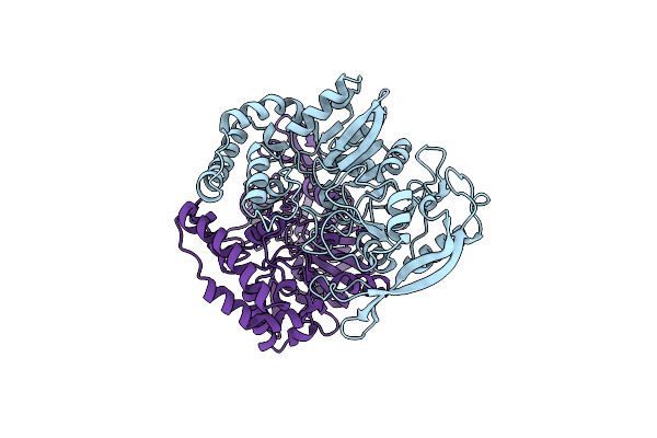
Structure Of Adenylosuccinate Synthetase At Ph 6.5 And 25 Degrees Celsius
Organism: Escherichia coli
Method: X-RAY DIFFRACTION Resolution:2.50 Å Release Date: 1996-06-10 Classification: LIGASE |
Organism: Escherichia coli
Method: X-RAY DIFFRACTION
Release Date: 1996-06-10
 |
Structure Of Adenylosuccinate Synthetase Ph 7 At 25 Degrees Celsius
Organism: Escherichia coli
Method: X-RAY DIFFRACTION Resolution:2.00 Å Release Date: 1996-01-29 Classification: LIGASE (SYNTHETASE) |
Organism: Escherichia coli
Method: X-RAY DIFFRACTION
Release Date: 1996-01-29