
Search Count: 9
 |
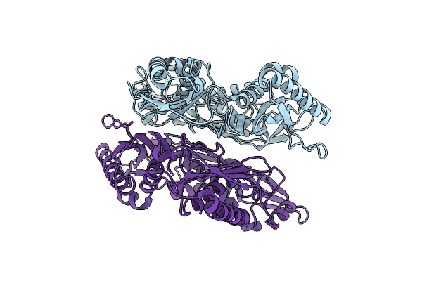
Crystal Structure Of Apo Form L147A/I351A Variant Of S-Adenosylmethionine Synthetase From Methanocaldococcus Jannaschii
Organism: Methanocaldococcus jannaschii (strain atcc 43067 / dsm 2661 / jal-1 / jcm 10045 / nbrc 100440)
Method: X-RAY DIFFRACTION Resolution:2.04 Å Release Date: 2021-11-17 Classification: TRANSFERASE |
 |
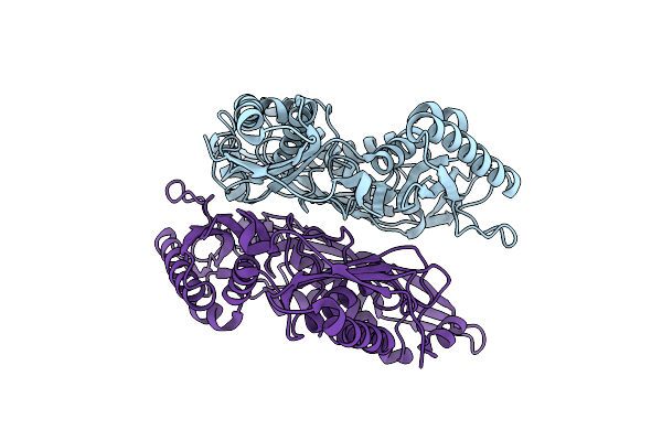
Crystal Structure Of Apo Form Of S-Adenosylmethionine Synthetase From Methanocaldococcus Jannaschii
Organism: Methanocaldococcus jannaschii (strain atcc 43067 / dsm 2661 / jal-1 / jcm 10045 / nbrc 100440)
Method: X-RAY DIFFRACTION Resolution:2.22 Å Release Date: 2021-11-17 Classification: TRANSFERASE |
 |
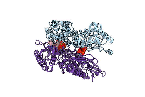
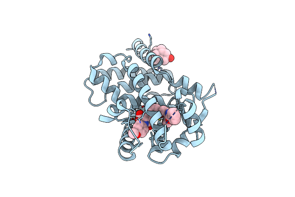
Crystal Structure Of L147A/I351A Variant Of S-Adenosylmethionine Synthetase From Methanocaldococcus Jannaschii In Complex With Onb-Sam (2-Nitro Benzyme S-Adenosyl-Methionine)
Organism: Methanocaldococcus jannaschii (strain atcc 43067 / dsm 2661 / jal-1 / jcm 10045 / nbrc 100440)
Method: X-RAY DIFFRACTION Resolution:2.05 Å Release Date: 2021-11-17 Classification: TRANSFERASE Ligands: 3PO, MG, EU9 |
 |
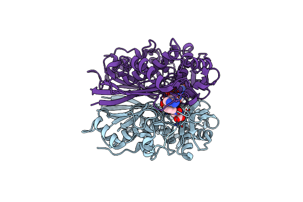
Crystal Structure Of L147A/I351A Variant Of S-Adenosylmethionine Synthetase From Methanocaldococcus Jannaschii In Complex With Dmnb-Sam (4,5-Dimethoxy-2-Nitro Benzyme S-Adenosyl-Methionine)
Organism: Methanocaldococcus jannaschii (strain atcc 43067 / dsm 2661 / jal-1 / jcm 10045 / nbrc 100440)
Method: X-RAY DIFFRACTION Resolution:1.71 Å Release Date: 2021-11-17 Classification: TRANSFERASE Ligands: 3PO, MG, 6IH |
 |
Crystal Structure Of I122A/I330A Variant Of S-Adenosylmethionine Synthetase From Cryptosporidium Hominis In Complex With Onb-Sam (2-Nitro Benzyme S-Adenosyl-Methionine)
Organism: Cryptosporidium hominis
Method: X-RAY DIFFRACTION Resolution:1.87 Å Release Date: 2020-10-21 Classification: TRANSFERASE Ligands: 3PO, MG, EU9 |
 |
Crystal Structure Of Apo Form Of I122A/I330A Variant Of S-Adenosylmethionine Synthetase From Cryptosporidium Hominis
Organism: Cryptosporidium hominis
Method: X-RAY DIFFRACTION Resolution:1.65 Å Release Date: 2020-10-21 Classification: TRANSFERASE Ligands: MG, PO4 |
 |
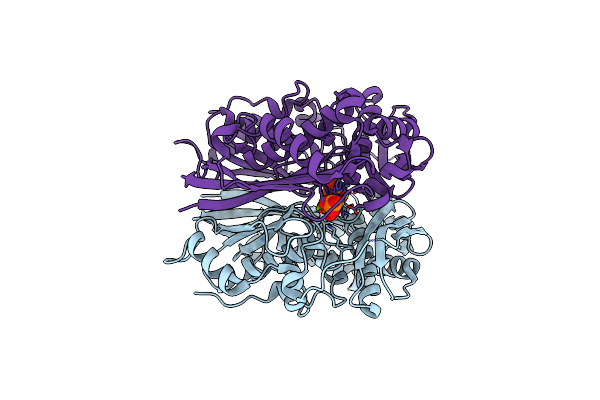
Organism: Mus musculus
Method: X-RAY DIFFRACTION Resolution:3.75 Å Release Date: 2019-07-10 Classification: SIGNALING PROTEIN Ligands: ZN |
 |
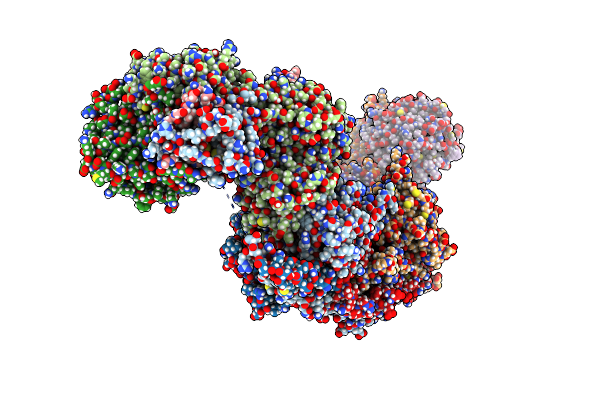
Organism: Homo sapiens
Method: ELECTRON MICROSCOPY Release Date: 2019-07-10 Classification: SIGNALING PROTEIN Ligands: ZN |
 |
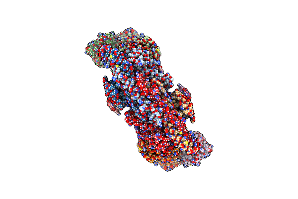
Organism: Homo sapiens
Method: X-RAY DIFFRACTION Resolution:2.35 Å Release Date: 2007-11-27 Classification: TRANSCRIPTION Ligands: CPQ, REW |