

Search Count: 22
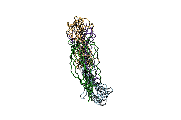 |
Monomer Structure Of Transforming Growth Factor Beta Induced Protein (Tgfbip) G623R Fibril
Organism: Homo sapiens
Method: SOLID-STATE NMR Release Date: 2023-07-26 Classification: PROTEIN FIBRIL |
Organism: Homo sapiens
Method: SOLID-STATE NMR
Release Date: 2023-07-26
 |
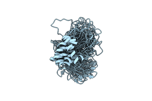
Structure Of Transforming Growth Factor Beta Induced Protein (Tgfbip) G623R Fibril
Organism: Homo sapiens
Method: ELECTRON MICROSCOPY Release Date: 2023-07-26 Classification: PROTEIN FIBRIL |
Organism: Homo sapiens
Method: ELECTRON MICROSCOPY
Release Date: 2023-07-26
 |
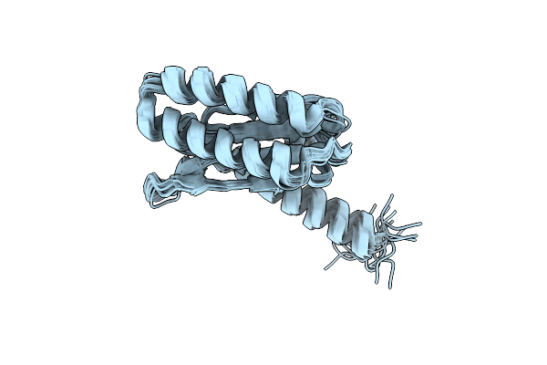
Tertiary Structure Of Barnacle Cement Protein Mrcp20
Organism: Megabalanus rosa
Method: SOLUTION NMR Release Date: 2020-01-15 Classification: STRUCTURAL PROTEIN |
Organism: Megabalanus rosa
Method: SOLUTION NMR
Release Date: 2020-01-15
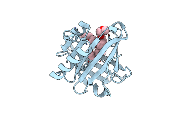 |
Apo Form Crystal Structure Of Human Lipocalin Pgds .
Organism: Homo sapiens
Method: X-RAY DIFFRACTION Resolution:1.45 Å Release Date: 2017-01-25 Classification: CHAPERONE Ligands: PG0, 1PG |
Organism: Homo sapiens
Method: X-RAY DIFFRACTION
Release Date: 2017-01-25
Ligands: PG0, 1PG
 |
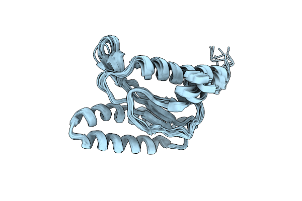
Solution Structure Of E55Q Mutant Of Erf1 N-Domain
|
Organism: Homo sapiens
Method: SOLUTION NMR
Release Date: 2015-07-01
 |
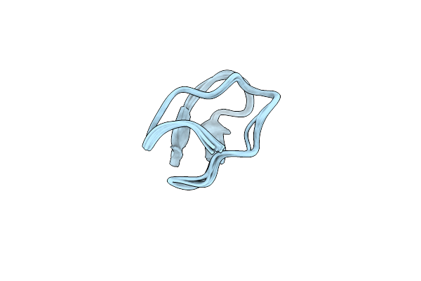
Solution Structure Of Y125F Mutant Of Erf1 N-Domain
|
Organism: Homo sapiens
Method: SOLUTION NMR
Release Date: 2015-06-17
 |
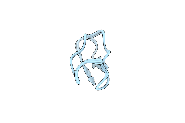
Solution Structure Of Allatide C4, Conformation 1
Organism: Allamanda cathartica
Method: SOLUTION NMR Release Date: 2015-01-14 Classification: HYDROLASE INHIBITOR |
Organism: Allamanda cathartica
Method: SOLUTION NMR
Release Date: 2015-01-14
 |
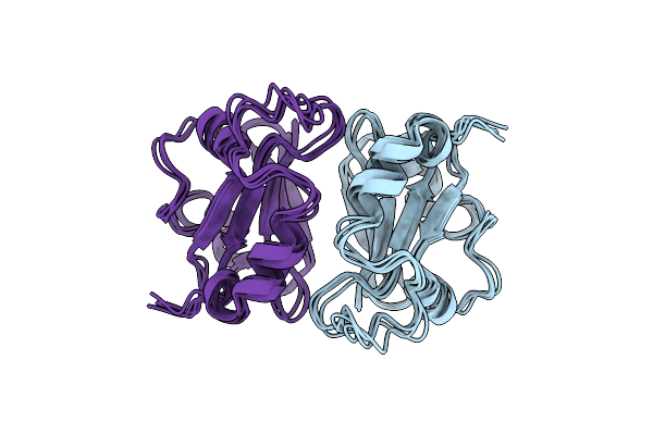
Solution Structure Of Allatide C4, Conformation 2
Organism: Allamanda cathartica
Method: SOLUTION NMR Release Date: 2015-01-14 Classification: HYDROLASE INHIBITOR |
Organism: Allamanda cathartica
Method: SOLUTION NMR
Release Date: 2015-01-14
 |
Haddock Structure Of Gtyybt Pas Homodimer
Organism: Geobacillus thermodenitrificans
Method: SOLUTION NMR Release Date: 2013-03-27 Classification: HYDROLASE |
Organism: Geobacillus thermodenitrificans
Method: SOLUTION NMR
Release Date: 2013-03-27
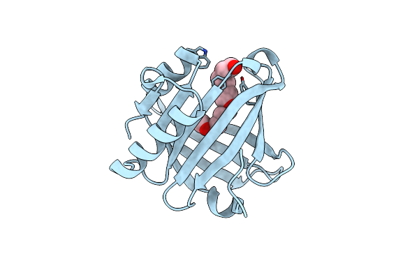 |
Crystal Structure Of Wild Type Human Lipocalin Pgds Bound With Peg Mme 2000
Organism: Homo sapiens
Method: X-RAY DIFFRACTION Resolution:2.09 Å Release Date: 2013-03-20 Classification: ISOMERASE Ligands: 1PG |
Organism: Homo sapiens
Method: X-RAY DIFFRACTION
Release Date: 2013-03-20
Ligands: 1PG
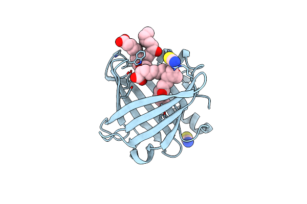 |
Crystal Structure Of Wild Type Human Lipocalin Pgds In Complex With Substrate Analog U44069
Organism: Homo sapiens
Method: X-RAY DIFFRACTION Resolution:1.88 Å Release Date: 2013-03-20 Classification: ISOMERASE Ligands: PWZ, SCN |
Organism: Homo sapiens
Method: X-RAY DIFFRACTION
Release Date: 2013-03-20
Ligands: PWZ, SCN
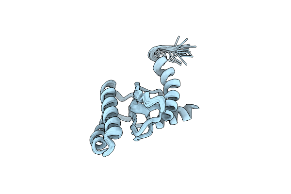 |
Backbone 1H, 13C, And 15N Chemical Shift Assignments For Qfm(Y)F
|
Organism: Homo sapiens
Method: SOLUTION NMR
Release Date: 2012-03-14
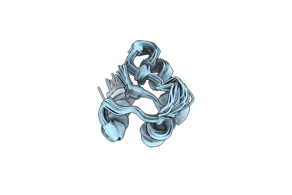 |
Solution Structure Of The Zalpha Domain Mutant Of Adar1 (N43A,Y47A)
|
Organism: Homo sapiens
Method: SOLUTION NMR
Release Date: 2011-01-12
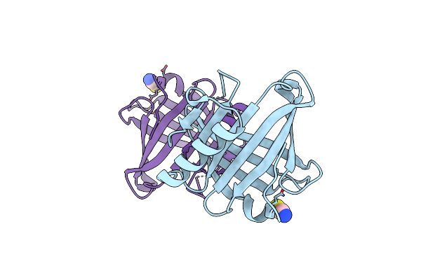 |
Crystal Structure Of The Human Lipocalin-Type Prostaglandin D Synthase
Organism: Homo sapiens
Method: X-RAY DIFFRACTION Resolution:2.00 Å Release Date: 2010-01-12 Classification: ISOMERASE Ligands: CL, SCN |
Organism: Homo sapiens
Method: X-RAY DIFFRACTION
Release Date: 2010-01-12
Ligands: CL, SCN
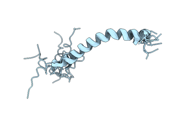 |
The Domain Features Of The Peripheral Stalk Subunit H Of The Methanogenic A1Ao Atp Synthase And The Nmr Solution Structure Of H1-47
Organism: Methanocaldococcus jannaschii
Method: SOLUTION NMR Release Date: 2009-07-21 Classification: STRUCTURAL PROTEIN |
Organism: Methanocaldococcus jannaschii
Method: SOLUTION NMR
Release Date: 2009-07-21
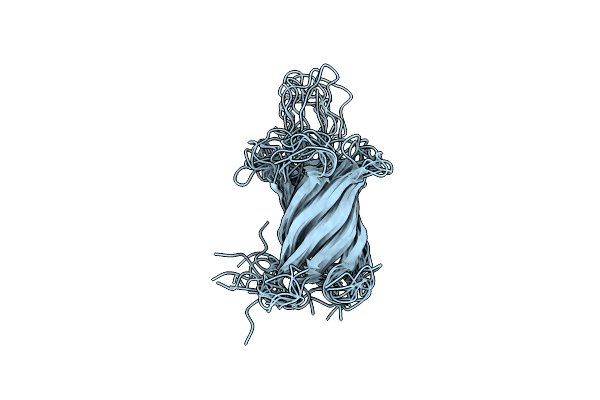 |
Nmr Solution Structure Of A Minimal Transmembrane Beta-Barrel Platform Protein
Organism: Escherichia coli
Method: SOLUTION NMR Release Date: 2007-07-03 Classification: MEMBRANE PROTEIN |
Organism: Escherichia coli
Method: SOLUTION NMR
Release Date: 2007-07-03
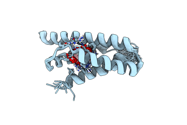 |
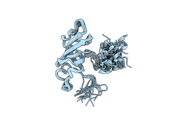
Nmr Structure Of Monomeric Chorismate Mutase From Methanococcus Jannaschii
Organism: Methanocaldococcus jannaschii
Method: SOLUTION NMR Release Date: 2006-10-31 Classification: ISOMERASE Ligands: TSA |
Organism: Methanocaldococcus jannaschii
Method: SOLUTION NMR
Release Date: 2006-10-31
Ligands: TSA
 |
Solution Structure Of The Heme Chaperone Ccme Of Escherichia Coli
|
Organism: Escherichia coli
Method: SOLUTION NMR
Release Date: 2004-04-06
 |
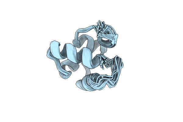
Structural Role Of A Buried Salt Bridge In The 434 Repressor Dna-Binding Domain, Nmr, 20 Structures
Organism: Phage 434
Method: SOLUTION NMR Release Date: 1997-06-16 Classification: GENE REGULATING PROTEIN |
Organism: Phage 434
Method: SOLUTION NMR
Release Date: 1997-06-16
 |
Structural Role Of A Buried Salt Bridge In The 434 Repressor Dna-Binding Domain, Nmr, 20 Structures
Organism: Phage 434
Method: SOLUTION NMR Release Date: 1997-06-16 Classification: GENE REGULATING PROTEIN |
Organism: Phage 434
Method: SOLUTION NMR
Release Date: 1997-06-16