

Search Count: 25
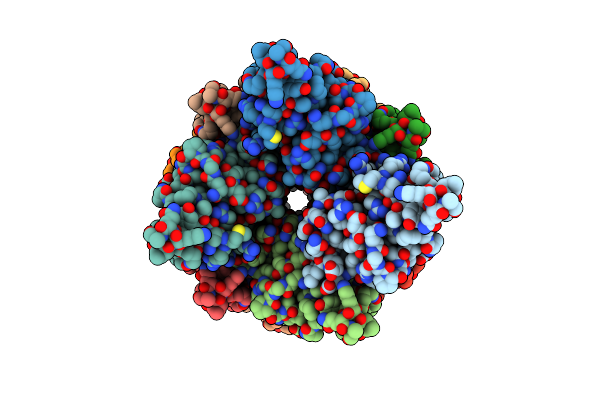 |
Helical Myd88 Death Domain Filament
Organism: Homo sapiens
Method: ELECTRON MICROSCOPY Release Date: 2019-11-20 Classification: SIGNALING PROTEIN |
Organism: Homo sapiens
Method: ELECTRON MICROSCOPY
Release Date: 2019-11-20
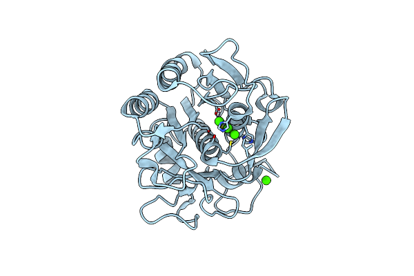 |
Microed Structure Of Proteinase K Recorded On Falcon Iii
Organism: Parengyodontium album
Method: ELECTRON CRYSTALLOGRAPHY Resolution:2.10 Å Release Date: 2019-08-28 Classification: HYDROLASE Ligands: CA |
Organism: Parengyodontium album
Method: ELECTRON CRYSTALLOGRAPHY
Release Date: 2019-08-28
Ligands: CA
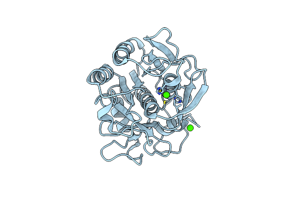 |
Microed Structure Of Proteinase K Recorded On Cetad
Organism: Parengyodontium album
Method: ELECTRON CRYSTALLOGRAPHY Resolution:2.70 Å Release Date: 2019-08-28 Classification: HYDROLASE Ligands: CA |
Organism: Parengyodontium album
Method: ELECTRON CRYSTALLOGRAPHY
Release Date: 2019-08-28
Ligands: CA
 |
Xaxab Pore Complex From Xenorhabdus Nematophila
Organism: Xenorhabdus nematophila atcc 19061
Method: ELECTRON MICROSCOPY Release Date: 2018-07-25 Classification: TOXIN |
Organism: Xenorhabdus nematophila atcc 19061
Method: ELECTRON MICROSCOPY
Release Date: 2018-07-25
 |
Crystal Structure Of Xaxb From Xenorhabdus Nematophil
Organism: Xenorhabdus nematophila (strain atcc 19061 / dsm 3370 / lmg 1036 / ncib 9965 / an6)
Method: X-RAY DIFFRACTION Resolution:3.40 Å Release Date: 2018-07-25 Classification: TOXIN |
 |
Crystal Structure Of Xaxa From Xenorhabdus Nematophila
Organism: Xenorhabdus nematophila (strain atcc 19061 / dsm 3370 / lmg 1036 / ncib 9965 / an6)
Method: X-RAY DIFFRACTION Resolution:2.50 Å Release Date: 2018-07-25 Classification: TOXIN |
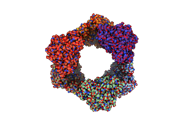 |
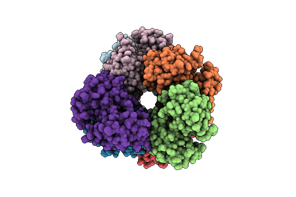
Human Ribonucleotide Reductase Large Subunit (Alpha) With Datp And Cdp
Organism: Homo sapiens
Method: ELECTRON MICROSCOPY Release Date: 2018-04-18 Classification: OXIDOREDUCTASE Ligands: DTP, MG, CDP |
Organism: Homo sapiens
Method: ELECTRON MICROSCOPY
Release Date: 2018-04-18
Ligands: DTP, MG, CDP
 |
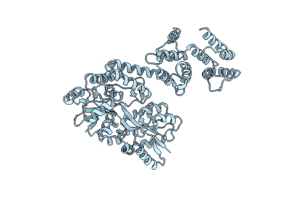
Cryo-Em Structure Of Casp-8 Tded Filament
Organism: Homo sapiens
Method: ELECTRON MICROSCOPY Release Date: 2016-11-02 Classification: APOPTOSIS |
Organism: Homo sapiens
Method: ELECTRON MICROSCOPY
Release Date: 2016-11-02
 |
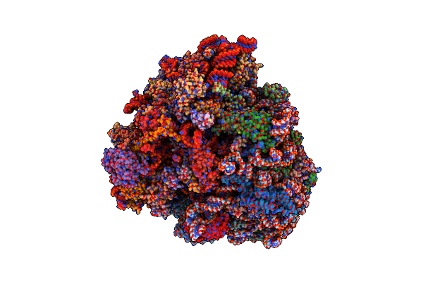
Crystal Structure Of Caspase8 Tded
Organism: Escherichia coli, homo sapiens
Method: X-RAY DIFFRACTION Resolution:3.16 Å Release Date: 2016-10-26 Classification: HYDROLASE |
Organism: Escherichia coli, homo sapiens
Method: X-RAY DIFFRACTION
Release Date: 2016-10-26
 |
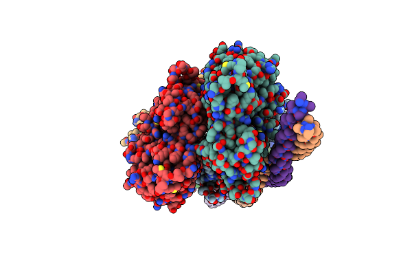
Cryo Electron Microscopy Of Actively Translating Human Polysomes (Post State).
Organism: Homo sapiens, Yersinia pseudotuberculosis
Method: ELECTRON MICROSCOPY Resolution:3.50 Å Release Date: 2015-05-20 Classification: RIBOSOME Ligands: MG, ZN |
Organism: Homo sapiens, Yersinia pseudotuberculosis
Method: ELECTRON MICROSCOPY
Release Date: 2015-05-20
Ligands: MG, ZN
 |
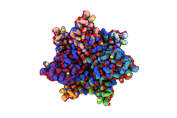
Structure Of The F-Actin-Tropomyosin Complex
Organism: Mus musculus, Oryctolagus cuniculus
Method: ELECTRON MICROSCOPY Resolution:3.70 Å Release Date: 2014-12-10 Classification: STRUCTURAL PROTEIN/HYDROLASE Ligands: MG, ADP |
Organism: Mus musculus, Oryctolagus cuniculus
Method: ELECTRON MICROSCOPY
Release Date: 2014-12-10
Ligands: MG, ADP
 |
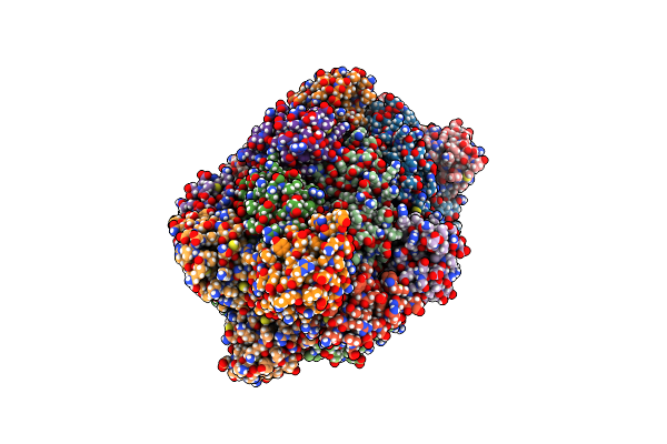
3.6 Angstrom Resolution Mavs Filament Generated From Helical Reconstruction
Organism: Homo sapiens
Method: ELECTRON MICROSCOPY Resolution:3.64 Å Release Date: 2014-07-30 Classification: IMMUNE SYSTEM |
Organism: Homo sapiens
Method: ELECTRON MICROSCOPY
Release Date: 2014-07-30
 |
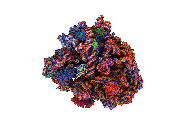
Caught-In-Action Signaling Complex Of Rig-I 2Card Domain And Mavs Card Domain
Organism: Homo sapiens
Method: X-RAY DIFFRACTION Resolution:3.40 Å Release Date: 2014-07-30 Classification: SIGNALING PROTEIN |
Organism: Homo sapiens
Method: X-RAY DIFFRACTION
Release Date: 2014-07-30
 |
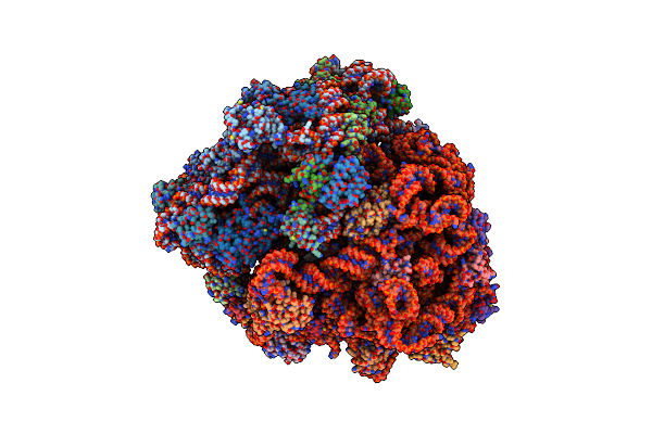
Structure Of The Ribosomal 80S-Eef2-Sordarin Complex From Yeast Obtained By Docking Atomic Models For Rna And Protein Components Into A 11.7 A Cryo-Em Map.
Organism: Saccharomyces cerevisiae
Method: ELECTRON MICROSCOPY Release Date: 2014-07-09 Classification: RIBOSOME |
Organism: Saccharomyces cerevisiae
Method: ELECTRON MICROSCOPY
Release Date: 2014-07-09
 |
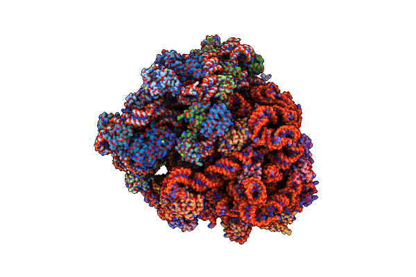
Trna Tranlocation On The 70S Ribosome: The Pre-Translocational Translocation Intermediate Ti(Pre)
Organism: Thermus thermophilus, Escherichia coli
Method: ELECTRON MICROSCOPY Resolution:7.80 Å Release Date: 2014-07-09 Classification: RIBOSOME Ligands: FUA, GDP |
Organism: Thermus thermophilus, Escherichia coli
Method: ELECTRON MICROSCOPY
Release Date: 2014-07-09
Ligands: FUA, GDP
 |
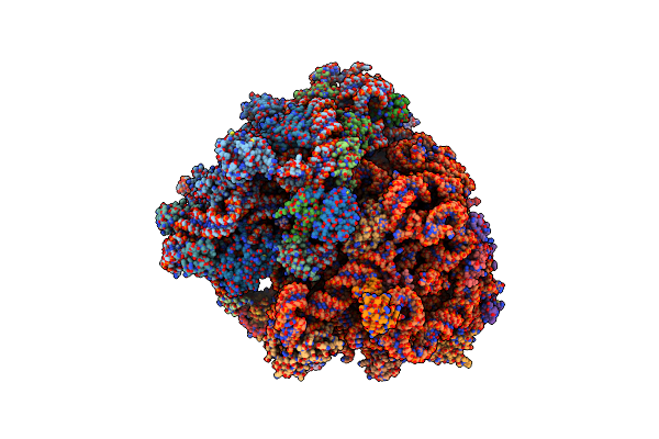
Trna Translocation On The 70S Ribosome: The Post- Translocational Translocation Intermediate Ti(Post)
Organism: Thermus thermophilus, Escherichia coli
Method: ELECTRON MICROSCOPY Resolution:7.60 Å Release Date: 2014-07-09 Classification: RIBOSOME Ligands: FUA, GDP |
Organism: Thermus thermophilus, Escherichia coli
Method: ELECTRON MICROSCOPY
Release Date: 2014-07-09
Ligands: FUA, GDP
 |
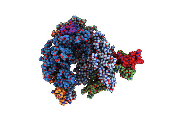
T. Thermophilus 70S Ribosome In Complex With Mrna, Trnas And Ef-Tu.Gdp.Kirromycin Ternary Complex, Fitted To A 6.4 A Cryo-Em Map.
Organism: Thermus thermophilus
Method: ELECTRON MICROSCOPY Release Date: 2014-07-09 Classification: RIBOSOME Ligands: PHA, GDP, MAU, BME |
Organism: Thermus thermophilus
Method: ELECTRON MICROSCOPY
Release Date: 2014-07-09
Ligands: PHA, GDP, MAU, BME
 |
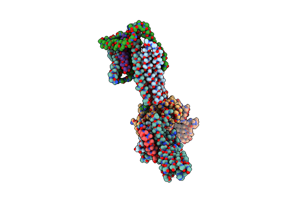
Cryo-Em Map Of A Yeast Minimal Preinitiation Complex Interacting With The Mediator Head Module
Organism: Saccharomyces cerevisiae
Method: ELECTRON MICROSCOPY Release Date: 2013-03-06 Classification: TRANSFERASE |
Organism: Saccharomyces cerevisiae
Method: ELECTRON MICROSCOPY
Release Date: 2013-03-06
 |
Cryo-Em Map Of A Yeast Minimal Preinitiation Complex Interacting With The Mediator Head Module
Organism: Saccharomyces cerevisiae
Method: ELECTRON MICROSCOPY Release Date: 2013-03-06 Classification: TRANSCRIPTION |
Organism: Saccharomyces cerevisiae
Method: ELECTRON MICROSCOPY
Release Date: 2013-03-06
 |
Inward-Facing Conformation Of The Zinc Transporter Yiip Revealed By Cryo-Electron Microscopy
Organism: Shewanella oneidensis
Method: ELECTRON MICROSCOPY Release Date: 2012-10-10 Classification: METAL TRANSPORT |
Organism: Shewanella oneidensis
Method: ELECTRON MICROSCOPY
Release Date: 2012-10-10