

Search Count: 23
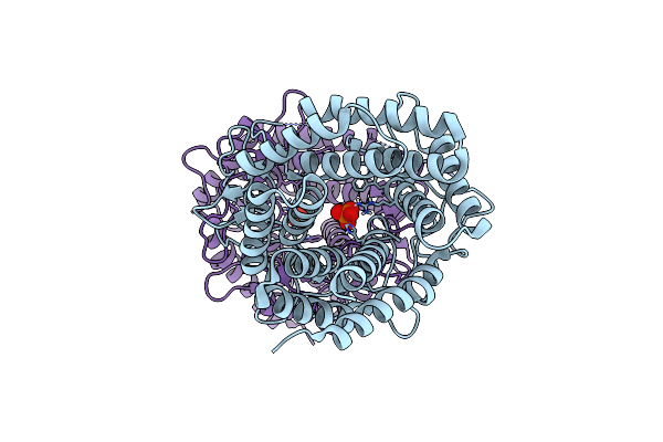 |
Room-Temperature Structure Of Pedobacter Heparinus N-Acetylglucosamine 2-Epimerase At Atmospheric Pressure
Organism: Pedobacter heparinus
Method: X-RAY DIFFRACTION Resolution:1.50 Å Release Date: 2023-12-13 Classification: ISOMERASE Ligands: CL, PO4 |
Organism: Pedobacter heparinus
Method: X-RAY DIFFRACTION
Release Date: 2023-12-13
Ligands: CL, PO4
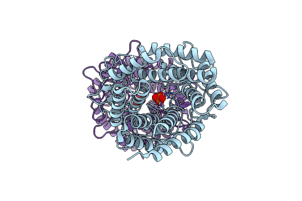 |
Room-Temperature Structure Of Pedobacter Heparinus N-Acetylglucosamine 2-Epimerase At 52 Mpa Helium Gas Pressure In A Sapphire Capillary
Organism: Pedobacter heparinus
Method: X-RAY DIFFRACTION Resolution:1.70 Å Release Date: 2023-12-13 Classification: ISOMERASE Ligands: CL, PO4 |
Organism: Pedobacter heparinus
Method: X-RAY DIFFRACTION
Release Date: 2023-12-13
Ligands: CL, PO4
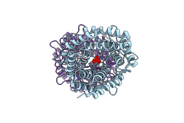 |
Room-Temperature Structure Of Pedobacter Heparinus N-Acetylglucosamine 2-Epimerase At 80 Mpa Helium Gas Pressure In A Sapphire Capillary
Organism: Pedobacter heparinus
Method: X-RAY DIFFRACTION Resolution:1.70 Å Release Date: 2023-12-13 Classification: ISOMERASE Ligands: PO4, CL |
Organism: Pedobacter heparinus
Method: X-RAY DIFFRACTION
Release Date: 2023-12-13
Ligands: PO4, CL
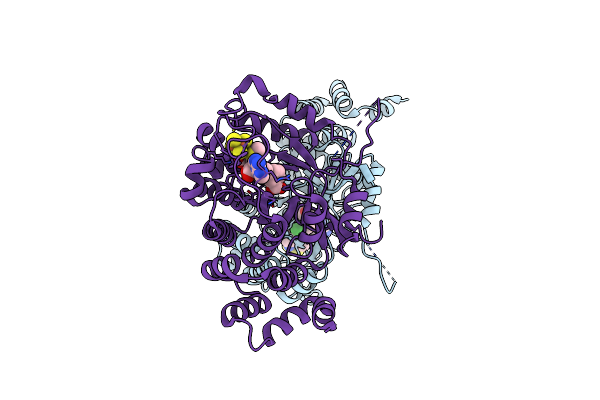 |
X-Ray Crystal Structure Of Mqne From Pedobacter Heparinus In Complex With Aminofutalosine And Methionine
Organism: Pedobacter heparinus (strain atcc 13125 / dsm 2366 / cip 104194 / jcm 7457 / nbrc 12017 / ncimb 9290 / nrrl b-14731 / him 762-3)
Method: X-RAY DIFFRACTION Resolution:2.14 Å Release Date: 2020-07-15 Classification: BIOSYNTHETIC PROTEIN Ligands: SF4, MET, V47, CL |
 |
X-Ray Crystal Structure Of Mqne From Pedobacter Heparinus
Organism: Pedobacter heparinus (strain atcc 13125 / dsm 2366 / cip 104194 / jcm 7457 / nbrc 12017 / ncimb 9290 / nrrl b-14731 / him 762-3)
Method: X-RAY DIFFRACTION Resolution:1.59 Å Release Date: 2020-07-15 Classification: BIOSYNTHETIC PROTEIN Ligands: SF4, TAR |
 |
The Crystal Structure Of Wt Pedobacter Heparinus Smug2
Organism: Pedobacter heparinus dsm 2366
Method: X-RAY DIFFRACTION Resolution:1.80 Å Release Date: 2017-01-18 Classification: LYASE |
Organism: Pedobacter heparinus dsm 2366
Method: X-RAY DIFFRACTION
Release Date: 2017-01-18
 |
The Crystal Structure Of Wt Pedobacter Heparinus Smug2
Organism: Pedobacter heparinus dsm 2366
Method: X-RAY DIFFRACTION Resolution:2.25 Å Release Date: 2017-01-18 Classification: LYASE |
Organism: Pedobacter heparinus dsm 2366
Method: X-RAY DIFFRACTION
Release Date: 2017-01-18
 |
Crystal Structure Of Heparan Sulfate Lyase Hepc From Pedobacter Heparinus
Organism: Pedobacter heparinus
Method: X-RAY DIFFRACTION Resolution:2.20 Å Release Date: 2014-01-29 Classification: LYASE Ligands: CA |
Organism: Pedobacter heparinus
Method: X-RAY DIFFRACTION
Release Date: 2014-01-29
Ligands: CA
 |
Crystal Structure Of Heparan Sulfate Lyase Hepc Mutant From Pedobacter Heparinus
Organism: Pedobacter heparinus
Method: X-RAY DIFFRACTION Resolution:2.40 Å Release Date: 2014-01-29 Classification: LYASE Ligands: CA |
Organism: Pedobacter heparinus
Method: X-RAY DIFFRACTION
Release Date: 2014-01-29
Ligands: CA
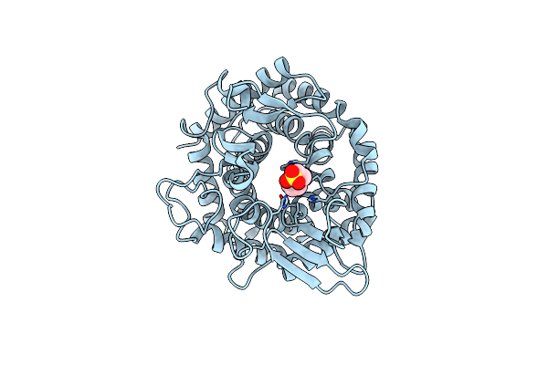 |
Crystal Structure Of Unsaturated Glucuronyl Hydrolase Specific For Heparin
Organism: Pedobacter heparinus
Method: X-RAY DIFFRACTION Resolution:1.35 Å Release Date: 2014-01-08 Classification: HYDROLASE Ligands: EPE |
Organism: Pedobacter heparinus
Method: X-RAY DIFFRACTION
Release Date: 2014-01-08
Ligands: EPE
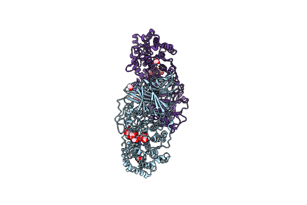 |
Heparinaseii H202A/Y257A Double Mutant Complexed With A Heparan Sulfate Tetrasaccharide Substrate
Organism: Pedobacter heparinus
Method: X-RAY DIFFRACTION Resolution:2.10 Å Release Date: 2008-12-30 Classification: SUGAR BINDING PROTEIN, LYASE Ligands: ZN, ACT |
Organism: Pedobacter heparinus
Method: X-RAY DIFFRACTION
Release Date: 2008-12-30
Ligands: ZN, ACT
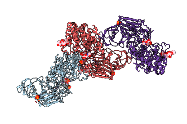 |
Structure Of Heparinase Ii Complexed With Heparan Sulfate Degradation Disaccharide Product
Organism: Pedobacter heparinus
Method: X-RAY DIFFRACTION Resolution:2.35 Å Release Date: 2008-12-30 Classification: SUGAR BINDING PROTEIN, LYASE Ligands: ZN, PO4 |
Organism: Pedobacter heparinus
Method: X-RAY DIFFRACTION
Release Date: 2008-12-30
Ligands: ZN, PO4
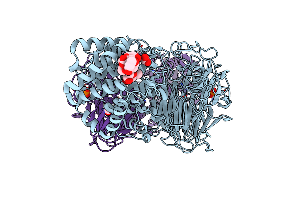 |
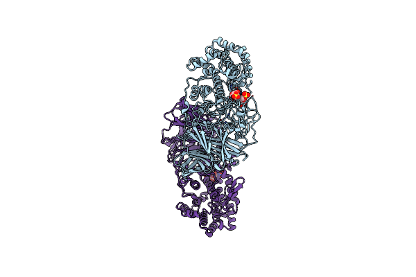
Crystal Structure Of Heparinase Ii
Organism: Pedobacter heparinus
Method: X-RAY DIFFRACTION Resolution:2.15 Å Release Date: 2006-04-18 Classification: SUGAR BINDING PROTEIN Ligands: ZN, PO4, FMT |
Organism: Pedobacter heparinus
Method: X-RAY DIFFRACTION
Release Date: 2006-04-18
Ligands: ZN, PO4, FMT
 |
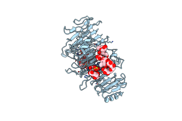
Crystal Structure Of Heparinase Ii Complexed With A Disaccharide Product
Organism: Pedobacter heparinus
Method: X-RAY DIFFRACTION Resolution:2.30 Å Release Date: 2006-04-18 Classification: SUGAR BINDING PROTEIN Ligands: ZN |
Organism: Pedobacter heparinus
Method: X-RAY DIFFRACTION
Release Date: 2006-04-18
Ligands: ZN
 |
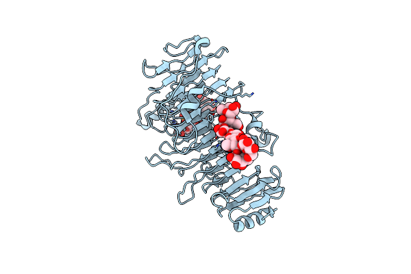
Crystal Structure Of Chondroitinase B Complexed To Dermatan Sulfate Hexasaccharide
Organism: Pedobacter heparinus
Method: X-RAY DIFFRACTION Resolution:1.70 Å Release Date: 2004-06-10 Classification: LYASE Ligands: CA |
Organism: Pedobacter heparinus
Method: X-RAY DIFFRACTION
Release Date: 2004-06-10
Ligands: CA
 |
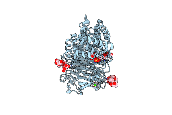
Crystal Structure Of Chondroitinase B Complexed To Chondroitin 4-Sulfate Tetrasaccharide
Organism: Pedobacter heparinus
Method: X-RAY DIFFRACTION Resolution:1.80 Å Release Date: 2004-04-19 Classification: LYASE |
Organism: Pedobacter heparinus
Method: X-RAY DIFFRACTION
Release Date: 2004-04-19
 |
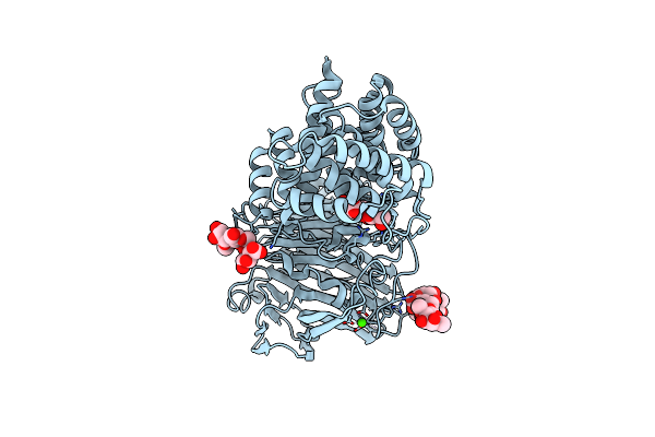
Active Site Of Chondroitinase Ac Lyase Revealed By The Structure Of Enzyme-Oligosaccharide Complexes And Mutagenesis
Organism: Pedobacter heparinus
Method: X-RAY DIFFRACTION Resolution:2.00 Å Release Date: 2001-05-02 Classification: LYASE Ligands: CA |
Organism: Pedobacter heparinus
Method: X-RAY DIFFRACTION
Release Date: 2001-05-02
Ligands: CA
 |
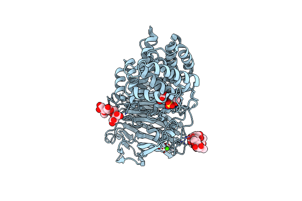
Active Site Of Chondroitinase Ac Lyase Revealed By The Structure Of Enzyme-Oligosaccharide Complexes And Mutagenesis
Organism: Pedobacter heparinus
Method: X-RAY DIFFRACTION Resolution:2.10 Å Release Date: 2001-05-02 Classification: LYASE Ligands: CA |
Organism: Pedobacter heparinus
Method: X-RAY DIFFRACTION
Release Date: 2001-05-02
Ligands: CA
 |
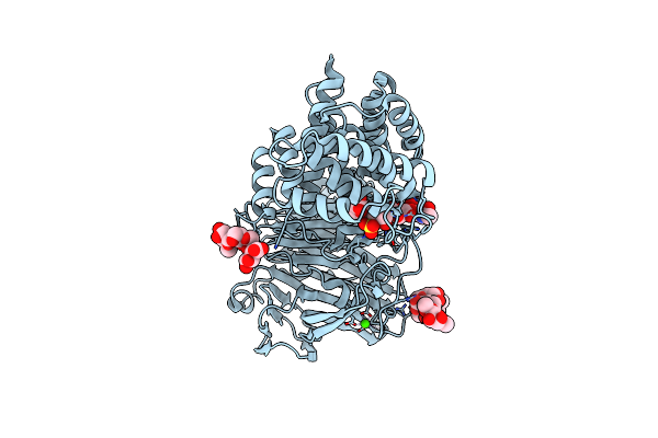
Active Site Of Chondroitinase Ac Lyase Revealed By The Structure Of Enzyme-Oligosaccharide Complexes And Mutagenesis
Organism: Pedobacter heparinus
Method: X-RAY DIFFRACTION Resolution:2.00 Å Release Date: 2001-05-02 Classification: LYASE Ligands: CA |
Organism: Pedobacter heparinus
Method: X-RAY DIFFRACTION
Release Date: 2001-05-02
Ligands: CA
 |
Active Site Of Chondroitinase Ac Lyase Revealed By The Structure Of Enzyme-Oligosaccharide Complexes And Mutagenesis
Organism: Pedobacter heparinus
Method: X-RAY DIFFRACTION Resolution:2.30 Å Release Date: 2001-05-02 Classification: LYASE Ligands: CA |
Organism: Pedobacter heparinus
Method: X-RAY DIFFRACTION
Release Date: 2001-05-02
Ligands: CA