

Search Count: 40
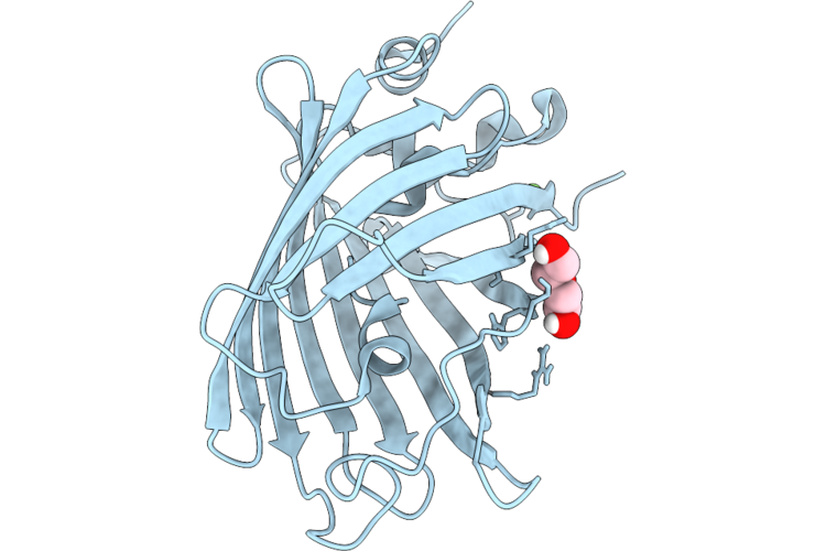 |
Room Temperature Crystal Structure Of Enhanced Green Fluorescent Protein
Organism: Aequorea victoria
Method: X-RAY DIFFRACTION Resolution:1.40 Å Release Date: 2026-07-29 Classification: FLUORESCENT PROTEIN Ligands: CA, PEG |
Organism: Aequorea victoria
Method: X-RAY DIFFRACTION
Release Date: 2026-07-29
Ligands: CA, PEG
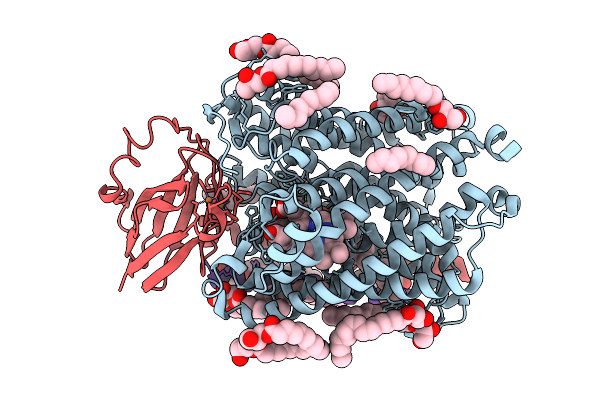 |
Chloride Bound Structure Of Oxidized Ba3-Type Cytochrome C Oxidase Confirmed By Single-Wavelength Anomalous Diffraction
Organism: Thermus thermophilus
Method: X-RAY DIFFRACTION Resolution:2.28 Å Release Date: 2025-08-13 Classification: OXIDOREDUCTASE Ligands: CU, HEM, HAS, OLC, CL, CUA |
Organism: Thermus thermophilus
Method: X-RAY DIFFRACTION
Release Date: 2025-08-13
Ligands: CU, HEM, HAS, OLC, CL, CUA
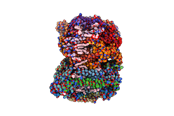 |
Sfx Structure Of Oxidized Cytochrome C Oxidase At 2.38 Angstrom Resolution
Organism: Bos taurus
Method: X-RAY DIFFRACTION Resolution:2.38 Å Release Date: 2023-09-27 Classification: MEMBRANE PROTEIN Ligands: CU, MG, NA, PGV, HEA, OH, CUA, TGL, PSC, CHD, PEK, CDL, DMU, ZN, SAC |
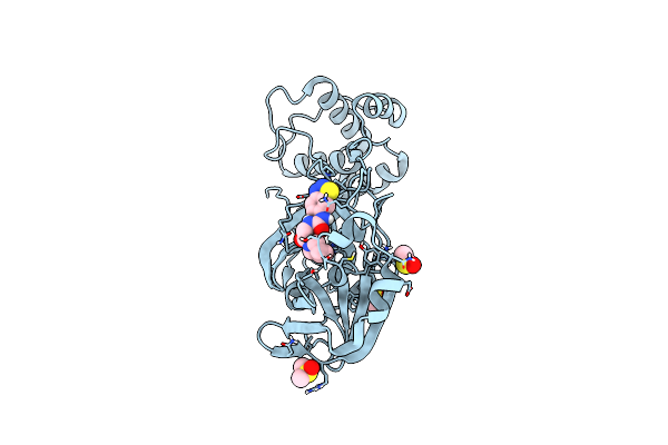 |
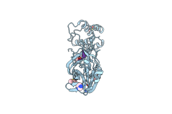
Structure Of Sars-Cov-2 Main Protease Bound To Unc-2327
Organism: Severe acute respiratory syndrome coronavirus 2
Method: X-RAY DIFFRACTION Resolution:1.39 Å Release Date: 2021-03-03 Classification: PEPTIDE BINDING PROTEIN Ligands: CL, DMS, RV5 |
Organism: Severe acute respiratory syndrome coronavirus 2
Method: X-RAY DIFFRACTION
Release Date: 2021-03-03
Ligands: CL, DMS, RV5
 |
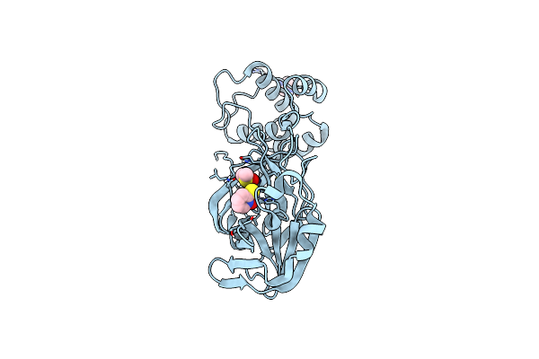
Structure Of The Hemiacetal Complex Between The Sars-Cov-2 Main Protease And Leupeptin
Organism: Severe acute respiratory syndrome coronavirus 2, Streptomyces roseus
Method: X-RAY DIFFRACTION Resolution:1.70 Å Release Date: 2021-03-03 Classification: PEPTIDE BINDING PROTEIN Ligands: DMS, IMD, CL |
Organism: Severe acute respiratory syndrome coronavirus 2, Streptomyces roseus
Method: X-RAY DIFFRACTION
Release Date: 2021-03-03
Ligands: DMS, IMD, CL
 |
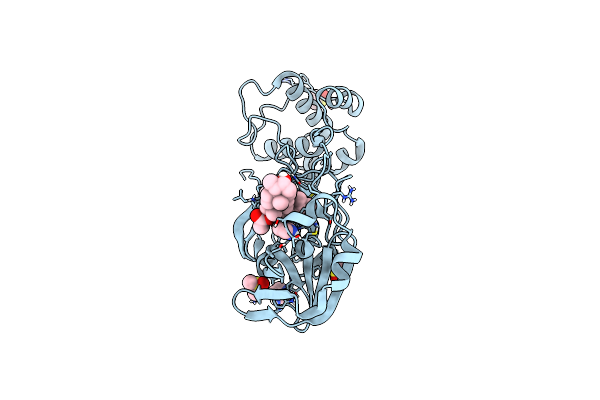
Structure Of Sars-Cov-2 Main Protease Bound To Pyrithione Zinc
Organism: Severe acute respiratory syndrome coronavirus 2
Method: X-RAY DIFFRACTION Resolution:1.80 Å Release Date: 2021-01-13 Classification: PEPTIDE BINDING PROTEIN Ligands: PK8, IMD, DMS, CL |
Organism: Severe acute respiratory syndrome coronavirus 2
Method: X-RAY DIFFRACTION
Release Date: 2021-01-13
Ligands: PK8, IMD, DMS, CL
 |
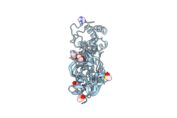
Structure Of Sars-Cov-2 Main Protease Bound To Fusidic Acid.
Organism: Severe acute respiratory syndrome coronavirus 2
Method: X-RAY DIFFRACTION Resolution:1.67 Å Release Date: 2020-12-02 Classification: HYDROLASE Ligands: FUA, DMS, IMD |
Organism: Severe acute respiratory syndrome coronavirus 2
Method: X-RAY DIFFRACTION
Release Date: 2020-12-02
Ligands: FUA, DMS, IMD
 |
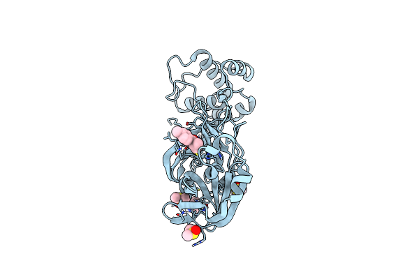
Structure Of Sars-Cov-2 Main Protease Bound To Rs102895
Organism: Severe acute respiratory syndrome coronavirus 2
Method: X-RAY DIFFRACTION Resolution:1.60 Å Release Date: 2020-12-02 Classification: PEPTIDE BINDING PROTEIN Ligands: R6Q, IMD, DMS |
Organism: Severe acute respiratory syndrome coronavirus 2
Method: X-RAY DIFFRACTION
Release Date: 2020-12-02
Ligands: R6Q, IMD, DMS
 |
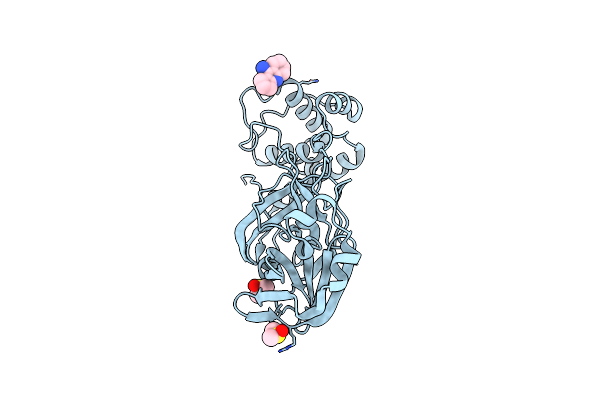
Structure Of Sars-Cov-2 Main Protease Bound To 2,4'-Dimethylpropiophenone.
Organism: Severe acute respiratory syndrome coronavirus 2
Method: X-RAY DIFFRACTION Resolution:1.63 Å Release Date: 2020-12-02 Classification: HYDROLASE Ligands: DMS, R7Q, IMD, CL |
Organism: Severe acute respiratory syndrome coronavirus 2
Method: X-RAY DIFFRACTION
Release Date: 2020-12-02
Ligands: DMS, R7Q, IMD, CL
 |
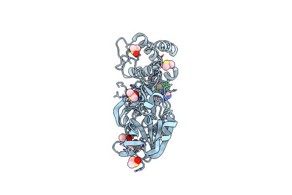
Structure Of Sars-Cov-2 Main Protease Bound To Ipidacrine.
Organism: Severe acute respiratory syndrome coronavirus 2
Method: X-RAY DIFFRACTION Resolution:1.70 Å Release Date: 2020-12-02 Classification: PEPTIDE BINDING PROTEIN Ligands: DMS, CL, R9W |
Organism: Severe acute respiratory syndrome coronavirus 2
Method: X-RAY DIFFRACTION
Release Date: 2020-12-02
Ligands: DMS, CL, R9W
 |
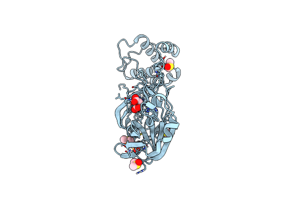
Structure Of Sars-Cov-2 Main Protease Bound To At7519
Organism: Severe acute respiratory syndrome coronavirus 2
Method: X-RAY DIFFRACTION Resolution:1.68 Å Release Date: 2020-12-02 Classification: HYDROLASE Ligands: LZE, CL, DMS |
Organism: Severe acute respiratory syndrome coronavirus 2
Method: X-RAY DIFFRACTION
Release Date: 2020-12-02
Ligands: LZE, CL, DMS
 |
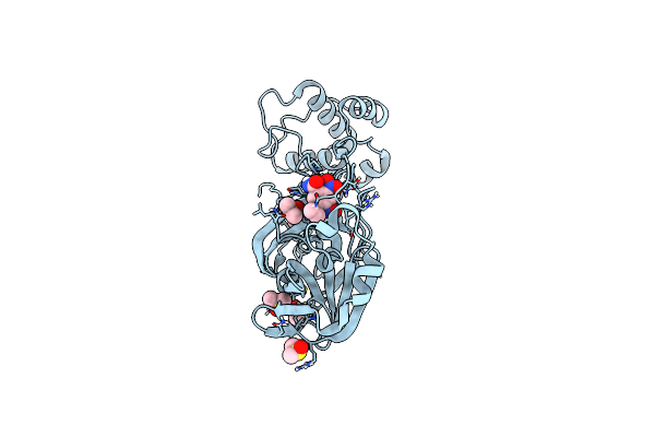
Structure Of Sars-Cov-2 Main Protease Bound To Maleate.
Organism: Severe acute respiratory syndrome coronavirus 2
Method: X-RAY DIFFRACTION Resolution:1.68 Å Release Date: 2020-12-02 Classification: HYDROLASE Ligands: CL, DMS, SIN |
Organism: Severe acute respiratory syndrome coronavirus 2
Method: X-RAY DIFFRACTION
Release Date: 2020-12-02
Ligands: CL, DMS, SIN
 |
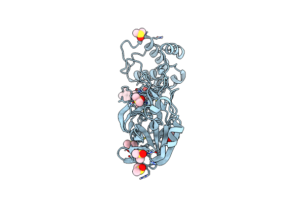
Structure Of Sars-Cov-2 Main Protease Bound To Tretazicar.
Organism: Severe acute respiratory syndrome coronavirus 2
Method: X-RAY DIFFRACTION Resolution:1.63 Å Release Date: 2020-12-02 Classification: PEPTIDE BINDING PROTEIN Ligands: CB1, DMS, CL |
Organism: Severe acute respiratory syndrome coronavirus 2
Method: X-RAY DIFFRACTION
Release Date: 2020-12-02
Ligands: CB1, DMS, CL
 |
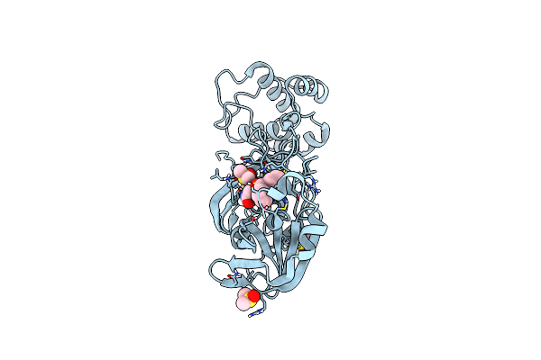
Structure Of Sars-Cov-2 Main Protease Bound To Pd 168568.
Organism: Severe acute respiratory syndrome coronavirus 2
Method: X-RAY DIFFRACTION Resolution:1.59 Å Release Date: 2020-12-02 Classification: HYDROLASE Ligands: CL, DMS, RMZ |
Organism: Severe acute respiratory syndrome coronavirus 2
Method: X-RAY DIFFRACTION
Release Date: 2020-12-02
Ligands: CL, DMS, RMZ
 |
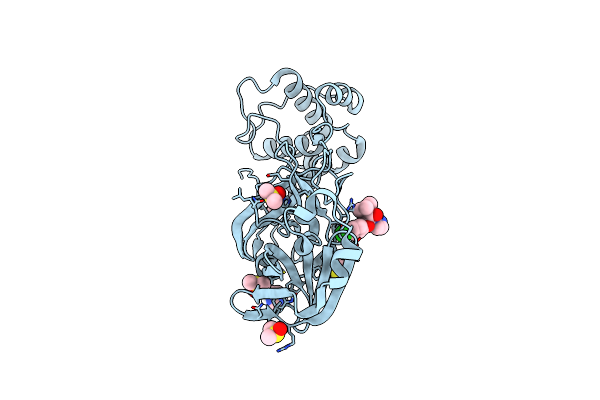
Structure Of Sars-Cov-2 Main Protease Bound To Adrafinil.
Organism: Severe acute respiratory syndrome coronavirus 2
Method: X-RAY DIFFRACTION Resolution:1.70 Å Release Date: 2020-12-02 Classification: PEPTIDE BINDING PROTEIN Ligands: DMS, RNW |
Organism: Severe acute respiratory syndrome coronavirus 2
Method: X-RAY DIFFRACTION
Release Date: 2020-12-02
Ligands: DMS, RNW
 |
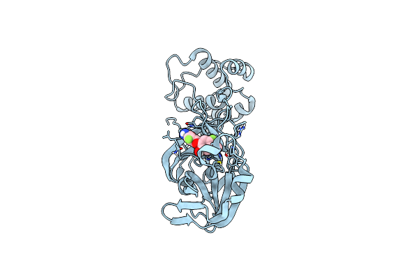
Structure Of Sars-Cov-2 Main Protease Bound To Climbazole
Organism: Severe acute respiratory syndrome coronavirus 2
Method: X-RAY DIFFRACTION Resolution:1.47 Å Release Date: 2020-12-02 Classification: PEPTIDE BINDING PROTEIN Ligands: DMS, IMD, RQH |
Organism: Severe acute respiratory syndrome coronavirus 2
Method: X-RAY DIFFRACTION
Release Date: 2020-12-02
Ligands: DMS, IMD, RQH
 |
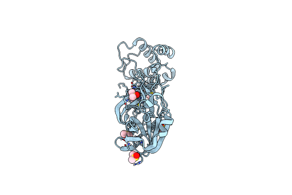
Structure Of Sars-Cov-2 Main Protease Bound To Mut056399.
Organism: Severe acute respiratory syndrome coronavirus 2
Method: X-RAY DIFFRACTION Resolution:1.78 Å Release Date: 2020-12-02 Classification: PEPTIDE BINDING PROTEIN Ligands: RQN |
Organism: Severe acute respiratory syndrome coronavirus 2
Method: X-RAY DIFFRACTION
Release Date: 2020-12-02
Ligands: RQN
 |
Structure Of Sars-Cov-2 Main Protease Bound To Tofogliflozin.
Organism: Severe acute respiratory syndrome coronavirus 2
Method: X-RAY DIFFRACTION Resolution:1.65 Å Release Date: 2020-12-02 Classification: PEPTIDE BINDING PROTEIN Ligands: RT2, DMS, CL |
Organism: Severe acute respiratory syndrome coronavirus 2
Method: X-RAY DIFFRACTION
Release Date: 2020-12-02
Ligands: RT2, DMS, CL
 |
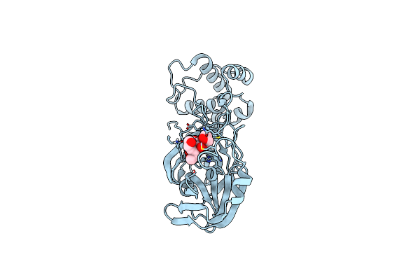
Structure Of Sars-Cov-2 Main Protease Bound To Ifenprodil
Organism: Severe acute respiratory syndrome coronavirus 2
Method: X-RAY DIFFRACTION Resolution:1.70 Å Release Date: 2020-12-02 Classification: HYDROLASE Ligands: QEL, DMS, IMD |
Organism: Severe acute respiratory syndrome coronavirus 2
Method: X-RAY DIFFRACTION
Release Date: 2020-12-02
Ligands: QEL, DMS, IMD
 |
Structure Of Sars-Cov-2 Main Protease Bound To Triglycidyl Isocyanurate.
Organism: Severe acute respiratory syndrome coronavirus 2
Method: X-RAY DIFFRACTION Resolution:2.59 Å Release Date: 2020-12-02 Classification: PEPTIDE BINDING PROTEIN Ligands: S7H, RV8 |
Organism: Severe acute respiratory syndrome coronavirus 2
Method: X-RAY DIFFRACTION
Release Date: 2020-12-02
Ligands: S7H, RV8