

Search Count: 10
 |
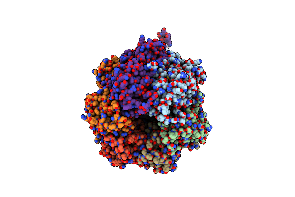
Crystal Structure Of The Phosphorylation-Site Mutant T432A Of The Kaic Circadian Clock Protein
Organism: Synechococcus elongatus pcc 7942
Method: X-RAY DIFFRACTION Resolution:2.90 Å Release Date: 2010-03-31 Classification: CIRCADIAN CLOCK PROTEIN, TRANSFERASE Ligands: ATP, MG |
Organism: Synechococcus elongatus pcc 7942
Method: X-RAY DIFFRACTION
Release Date: 2010-03-31
Ligands: ATP, MG
 |
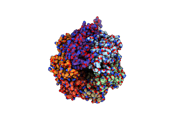
Crystal Structure Of The Phosphorylation-Site Mutant S431D Of The Kaic Circadian Clock Protein
Organism: Synechococcus elongatus pcc 7942
Method: X-RAY DIFFRACTION Resolution:3.20 Å Release Date: 2010-03-31 Classification: CIRCADIAN CLOCK PROTEIN, TRANSFERASE Ligands: MG, ATP |
Organism: Synechococcus elongatus pcc 7942
Method: X-RAY DIFFRACTION
Release Date: 2010-03-31
Ligands: MG, ATP
 |
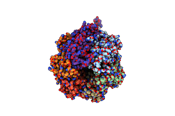
Crystal Structure Of The Phosphorylation-Site Mutant S431A Of The Kaic Circadian Clock Protein
Organism: Synechococcus elongatus pcc 7942
Method: X-RAY DIFFRACTION Resolution:3.00 Å Release Date: 2010-03-31 Classification: CIRCADIAN CLOCK PROTEIN, TRANSFERASE Ligands: ATP, MG |
Organism: Synechococcus elongatus pcc 7942
Method: X-RAY DIFFRACTION
Release Date: 2010-03-31
Ligands: ATP, MG
 |
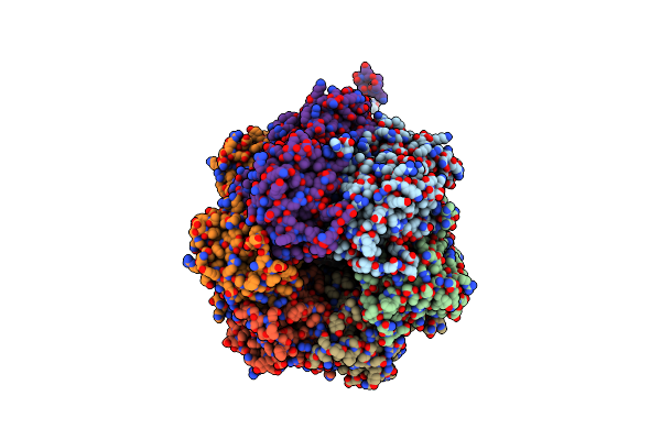
Crystal Structure Of The Phosphorylation-Site Double Mutant S431A/T432E Of The Kaic Circadian Clock Protein
Organism: Synechococcus elongatus pcc 7942
Method: X-RAY DIFFRACTION Resolution:3.30 Å Release Date: 2010-03-31 Classification: CIRCADIAN CLOCK PROTEIN, TRANSFERASE Ligands: ATP, MG |
Organism: Synechococcus elongatus pcc 7942
Method: X-RAY DIFFRACTION
Release Date: 2010-03-31
Ligands: ATP, MG
 |
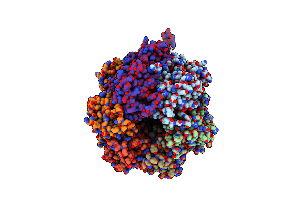
Crystal Structure Of The Phosphorylation-Site Mutant T426N Of The Kaic Circadian Clock Protein
Organism: Synechococcus elongatus pcc 7942
Method: X-RAY DIFFRACTION Resolution:3.20 Å Release Date: 2010-03-31 Classification: CIRCADIAN CLOCK PROTEIN, TRANSFERASE Ligands: MG, ATP |
Organism: Synechococcus elongatus pcc 7942
Method: X-RAY DIFFRACTION
Release Date: 2010-03-31
Ligands: MG, ATP
 |
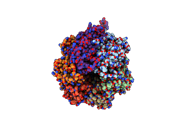
Crystal Structure Of The Phosphorylation-Site Double Mutant T426A/T432A Of The Kaic Circadian Clock Protein
Organism: Synechococcus elongatus pcc 7942
Method: X-RAY DIFFRACTION Resolution:3.00 Å Release Date: 2010-03-31 Classification: CIRCADIAN CLOCK PROTEIN, TRANSFERASE Ligands: MG, ATP |
Organism: Synechococcus elongatus pcc 7942
Method: X-RAY DIFFRACTION
Release Date: 2010-03-31
Ligands: MG, ATP
 |
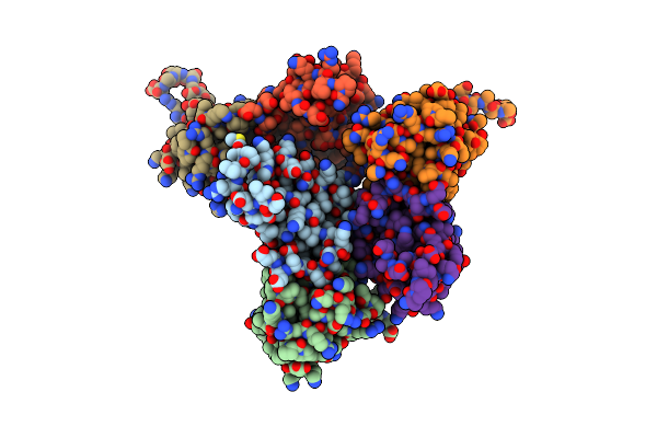
Crystal Structure Of Full Length Circadian Clock Protein Kaic With Correct Geometry At Phosphorylation Sites
Organism: Synechococcus elongatus
Method: X-RAY DIFFRACTION Resolution:2.80 Å Release Date: 2009-07-28 Classification: CIRCADIAN CLOCK PROTEIN, Transferase Ligands: MG, ATP |
Organism: Synechococcus elongatus
Method: X-RAY DIFFRACTION
Release Date: 2009-07-28
Ligands: MG, ATP
 |
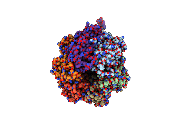
Wild Type Crystal Structure Of Full Length Circadian Clock Protein Kaib From Thermosynechococcus Elongatus Bp-1
Organism: Synechococcus elongatus
Method: X-RAY DIFFRACTION Resolution:2.70 Å Release Date: 2008-06-17 Classification: CIRCADIAN CLOCK PROTEIN |
Organism: Synechococcus elongatus
Method: X-RAY DIFFRACTION
Release Date: 2008-06-17
 |
Crystal Structure Of Full Length Circadian Clock Protein Kaic With Phosphorylation Sites
Organism: Synechococcus elongatus
Method: X-RAY DIFFRACTION Resolution:2.80 Å Release Date: 2007-01-23 Classification: CIRCADIAN CLOCK PROTEIN, TRANSFERASE Ligands: MG, ATP |
Organism: Synechococcus elongatus
Method: X-RAY DIFFRACTION
Release Date: 2007-01-23
Ligands: MG, ATP
 |
Crystal Structure Of Circadian Clock Protein Kaic With Phosphorylation Sites
Organism: Synechococcus elongatus
Method: X-RAY DIFFRACTION Resolution:2.80 Å Release Date: 2005-04-19 Classification: CIRCADIAN CLOCK PROTEIN Ligands: MG, ATP |
Organism: Synechococcus elongatus
Method: X-RAY DIFFRACTION
Release Date: 2005-04-19
Ligands: MG, ATP