

Search Count: 26
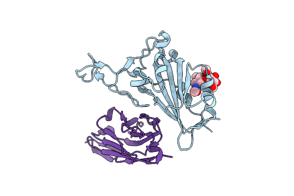 |
Crystal Structure Of The Sars-Cov-2 Neutralizing Vhh 7A9 Bound To The Spike Receptor Binding Domain
Organism: Severe acute respiratory syndrome coronavirus 2, Lama glama
Method: X-RAY DIFFRACTION Resolution:2.01 Å Release Date: 2023-08-16 Classification: ANTIVIRAL PROTEIN |
Organism: Severe acute respiratory syndrome coronavirus 2, Lama glama
Method: X-RAY DIFFRACTION
Release Date: 2023-08-16
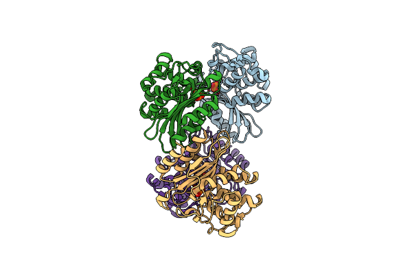 |
Beta-Lactamase, Unmixed Shards Crystal Form
Organism: Mycobacterium tuberculosis (strain atcc 25618 / h37rv)
Method: X-RAY DIFFRACTION Resolution:2.45 Å Release Date: 2018-06-27 Classification: HYDROLASE Ligands: PO4 |
Organism: Mycobacterium tuberculosis (strain atcc 25618 / h37rv)
Method: X-RAY DIFFRACTION
Release Date: 2018-06-27
Ligands: PO4
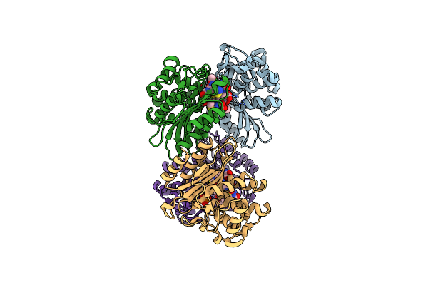 |
Beta-Lactamase, Mixed With Ceftriaxone, 30Ms Time Point, Shards Crystal Form
Organism: Mycobacterium tuberculosis (strain atcc 25618 / h37rv)
Method: X-RAY DIFFRACTION Resolution:2.75 Å Release Date: 2018-06-27 Classification: HYDROLASE Ligands: PO4, 9F2 |
Organism: Mycobacterium tuberculosis (strain atcc 25618 / h37rv)
Method: X-RAY DIFFRACTION
Release Date: 2018-06-27
Ligands: PO4, 9F2
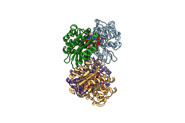 |
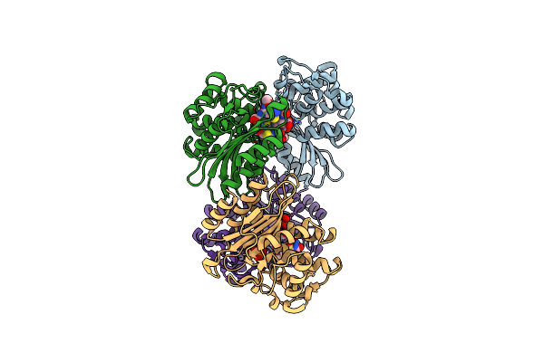
Beta-Lactamase, 100Ms Timepoint, Mixed, Shards Crystal Form
Organism: Mycobacterium tuberculosis (strain atcc 25618 / h37rv)
Method: X-RAY DIFFRACTION Resolution:2.15 Å Release Date: 2018-06-27 Classification: Hydrolase/Antibiotic Ligands: PO4, 9F2, FZS |
Organism: Mycobacterium tuberculosis (strain atcc 25618 / h37rv)
Method: X-RAY DIFFRACTION
Release Date: 2018-06-27
Ligands: PO4, 9F2, FZS
 |
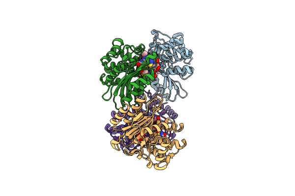
Beta-Lactamase, 500Ms Timepoint, Mixed, Shards Crystal Form
Organism: Mycobacterium tuberculosis (strain atcc 25618 / h37rv)
Method: X-RAY DIFFRACTION Resolution:2.20 Å Release Date: 2018-06-27 Classification: HYDROLASE/ANTIBIOTIC Ligands: PO4, 9F2, CUG, FZS |
Organism: Mycobacterium tuberculosis (strain atcc 25618 / h37rv)
Method: X-RAY DIFFRACTION
Release Date: 2018-06-27
Ligands: PO4, 9F2, CUG, FZS
 |
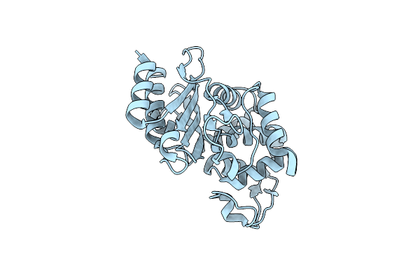
Beta-Lactamase, 2Secs Timepoint, Mixed, Shards Crystal Form
Organism: Mycobacterium tuberculosis (strain atcc 25618 / h37rv)
Method: X-RAY DIFFRACTION Resolution:2.30 Å Release Date: 2018-06-27 Classification: HYDROLASE/ANTIBIOTIC Ligands: PO4, 9F2, FZS |
Organism: Mycobacterium tuberculosis (strain atcc 25618 / h37rv)
Method: X-RAY DIFFRACTION
Release Date: 2018-06-27
Ligands: PO4, 9F2, FZS
 |
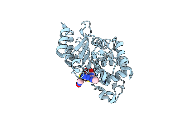
Beta-Lactamase, Unmixed Needles Crystal Form
Organism: Mycobacterium tuberculosis (strain atcc 25618 / h37rv)
Method: X-RAY DIFFRACTION Resolution:1.80 Å Release Date: 2018-06-27 Classification: HYDROLASE |
Organism: Mycobacterium tuberculosis (strain atcc 25618 / h37rv)
Method: X-RAY DIFFRACTION
Release Date: 2018-06-27
 |
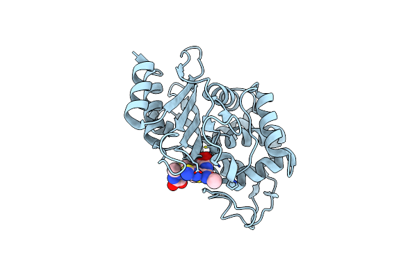
Beta-Lactamase, Mixed With Ceftriaxone, Needles Crystal Form, 30Ms
Organism: Mycobacterium tuberculosis (strain atcc 25618 / h37rv)
Method: X-RAY DIFFRACTION Resolution:1.90 Å Release Date: 2018-06-27 Classification: HYDROLASE/Antibiotic Ligands: 9F2 |
Organism: Mycobacterium tuberculosis (strain atcc 25618 / h37rv)
Method: X-RAY DIFFRACTION
Release Date: 2018-06-27
Ligands: 9F2
 |
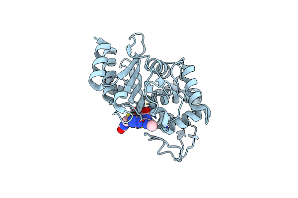
Beta-Lactamase, Mixed With Ceftriaxone, Needles Crystal Form, 100Ms
Organism: Mycobacterium tuberculosis (strain atcc 25618 / h37rv)
Method: X-RAY DIFFRACTION Resolution:1.80 Å Release Date: 2018-06-27 Classification: HYDROLASE/ANTIBIOTIC Ligands: FZS, 9F2 |
Organism: Mycobacterium tuberculosis (strain atcc 25618 / h37rv)
Method: X-RAY DIFFRACTION
Release Date: 2018-06-27
Ligands: FZS, 9F2
 |
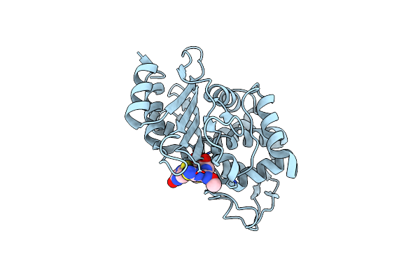
Beta-Lactamase, Mixed With Ceftriaxone, Needles Crystal Form, 500Ms
Organism: Mycobacterium tuberculosis (strain atcc 25618 / h37rv)
Method: X-RAY DIFFRACTION Resolution:1.90 Å Release Date: 2018-06-27 Classification: HYDROLASE/Antibiotic Ligands: FZS, 9F2 |
Organism: Mycobacterium tuberculosis (strain atcc 25618 / h37rv)
Method: X-RAY DIFFRACTION
Release Date: 2018-06-27
Ligands: FZS, 9F2
 |
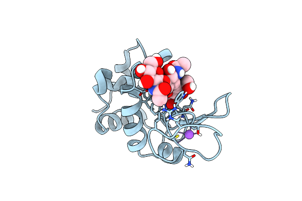
Beta-Lactamase, Mixed With Ceftriaxone, Needles Crystal Form, 2Sec
Organism: Mycobacterium tuberculosis (strain atcc 25618 / h37rv)
Method: X-RAY DIFFRACTION Resolution:2.05 Å Release Date: 2018-06-27 Classification: Hydrolase/ANTIBIOTIC Ligands: 9F2 |
Organism: Mycobacterium tuberculosis (strain atcc 25618 / h37rv)
Method: X-RAY DIFFRACTION
Release Date: 2018-06-27
Ligands: 9F2
 |
Mix-And-Diffuse Serial Synchrotron Crystallography: Structure Of N,N',N''-Triacetylchitotriose Bound To Lysozyme With 1S Time-Delay, Phased With 1Hew
Organism: Gallus gallus
Method: X-RAY DIFFRACTION Resolution:1.70 Å Release Date: 2017-10-18 Classification: HYDROLASE Ligands: CL, NA |
Organism: Gallus gallus
Method: X-RAY DIFFRACTION
Release Date: 2017-10-18
Ligands: CL, NA
 |
Mix-And-Diffuse Serial Synchrotron Crystallography: Structure Of N,N',N''-Triacetylchitotriose Bound To Lysozyme With 1S Time-Delay, Phased With 4Et8
Organism: Gallus gallus
Method: X-RAY DIFFRACTION Resolution:1.70 Å Release Date: 2017-10-18 Classification: HYDROLASE Ligands: CL, NA |
Organism: Gallus gallus
Method: X-RAY DIFFRACTION
Release Date: 2017-10-18
Ligands: CL, NA
 |
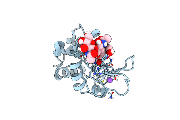
Mix-And-Diffuse Serial Synchrotron Crystallography: Structure Of N,N',N''-Triacetylchitotriose Bound To Lysozyme With 50S Time-Delay, Phased With 4Et8
Organism: Gallus gallus
Method: X-RAY DIFFRACTION Resolution:1.70 Å Release Date: 2017-10-18 Classification: HYDROLASE Ligands: CL, NA |
Organism: Gallus gallus
Method: X-RAY DIFFRACTION
Release Date: 2017-10-18
Ligands: CL, NA
 |
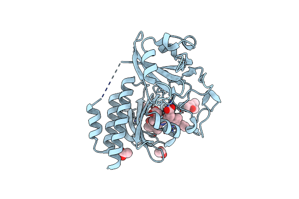
Mix-And-Diffuse Serial Synchrotron Crystallography: Structure Of N,N',N''-Triacetylchitotriose Bound To Lysozyme With 50S Time-Delay, Phased With 1Hew
Organism: Gallus gallus
Method: X-RAY DIFFRACTION Resolution:1.70 Å Release Date: 2017-10-18 Classification: HYDROLASE Ligands: CL, NA |
Organism: Gallus gallus
Method: X-RAY DIFFRACTION
Release Date: 2017-10-18
Ligands: CL, NA
 |
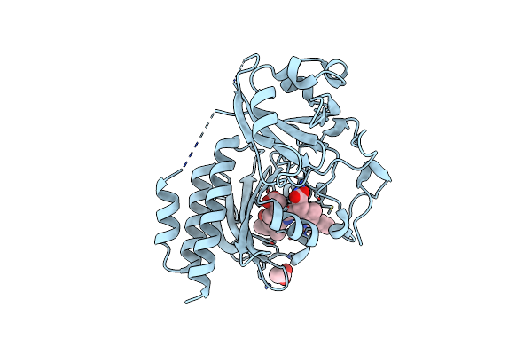
Wild-Type Pas-Gaf Fragment From Deinococcus Radiodurans Bphp
Organism: Deinococcus radiodurans
Method: X-RAY DIFFRACTION Resolution:1.35 Å Release Date: 2016-10-26 Classification: TRANSFERASE Ligands: ACT, MPD, LBV |
Organism: Deinococcus radiodurans
Method: X-RAY DIFFRACTION
Release Date: 2016-10-26
Ligands: ACT, MPD, LBV
 |
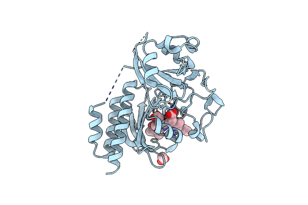
Wild-Type Pas-Gaf Fragment From Deinococcus Radiodurans Bphp Collected At Lcls
Organism: Deinococcus radiodurans
Method: X-RAY DIFFRACTION Resolution:2.10 Å Release Date: 2016-10-26 Classification: TRANSFERASE Ligands: LBV, ACT |
Organism: Deinococcus radiodurans
Method: X-RAY DIFFRACTION
Release Date: 2016-10-26
Ligands: LBV, ACT
 |
Wild-Type Pas-Gaf Fragment From Deinococcus Radiodurans Bphp Collected At Sacla
Organism: Deinococcus radiodurans
Method: X-RAY DIFFRACTION Resolution:2.20 Å Release Date: 2016-10-26 Classification: TRANSFERASE Ligands: LBV, ACT |
Organism: Deinococcus radiodurans
Method: X-RAY DIFFRACTION
Release Date: 2016-10-26
Ligands: LBV, ACT
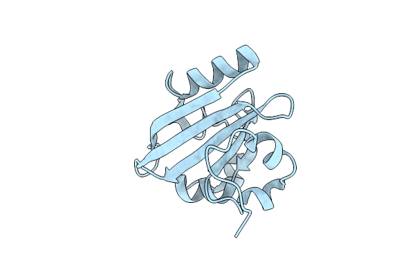 |
Femtosecond Structural Dynamics Drives The Trans/Cis Isomerization In Photoactive Yellow Protein: 200 Ns Time Delay Photo-Activated (Light) Structure
Organism: Halorhodospira halophila
Method: X-RAY DIFFRACTION Resolution:1.60 Å Release Date: 2016-05-25 Classification: SIGNALING PROTEIN |
Organism: Halorhodospira halophila
Method: X-RAY DIFFRACTION
Release Date: 2016-05-25
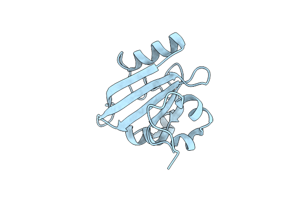 |
Femtosecond Structural Dynamics Drives The Trans/Cis Isomerization In Photoactive Yellow Protein: 100 Fs To 400 Fs Structure
Organism: Halorhodospira halophila
Method: X-RAY DIFFRACTION Resolution:1.60 Å Release Date: 2016-05-25 Classification: SIGNALING PROTEIN |
Organism: Halorhodospira halophila
Method: X-RAY DIFFRACTION
Release Date: 2016-05-25