

Search Count: 13,852
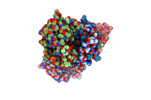 |
Crystal Structure Of Zika Ns2B-Ns3 Protease With Compound 5
Organism: Zika virus, Synthetic construct
Method: X-RAY DIFFRACTION Resolution:1.90 Å Release Date: 2021-04-14 Classification: VIRAL PROTEIN |
Organism: Zika virus, Synthetic construct
Method: X-RAY DIFFRACTION
Release Date: 2021-04-14
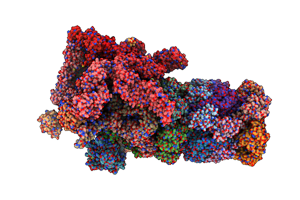 |
26S Proteasome In Presence Of Befx (S4)
Organism: Saccharomyces cerevisiae (strain atcc 204508 / s288c)
Method: ELECTRON MICROSCOPY Release Date: 2017-03-08 Classification: HYDROLASE Ligands: ATP, MG, ADP |
Organism: Saccharomyces cerevisiae (strain atcc 204508 / s288c)
Method: ELECTRON MICROSCOPY
Release Date: 2017-03-08
Ligands: ATP, MG, ADP
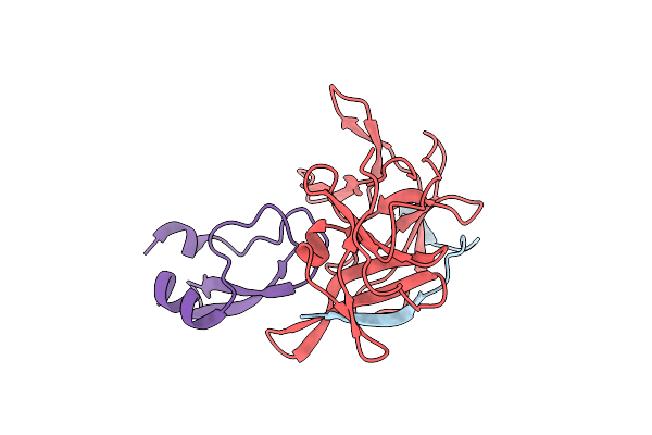 |
Aprotinin Bound To Dengue Virus Protease
Organism: Dengue virus 3, Bos taurus
Method: X-RAY DIFFRACTION Resolution:1.80 Å Release Date: 2011-11-09 Classification: HYDROLASE/HYDROLASE INHIBITOR |
Organism: Dengue virus 3, Bos taurus
Method: X-RAY DIFFRACTION
Release Date: 2011-11-09
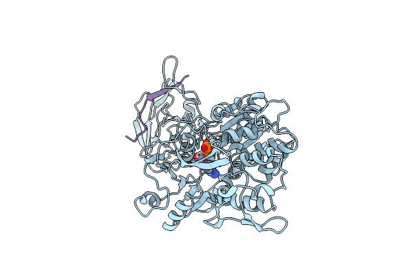 |
A Second Conformation Of The Ns3 Protease-Helicase From Dengue Virus
Organism: Dengue virus 4
Method: X-RAY DIFFRACTION Resolution:2.20 Å Release Date: 2010-04-07 Classification: HYDROLASE Ligands: ADP, MN, CL |
Organism: Dengue virus 4
Method: X-RAY DIFFRACTION
Release Date: 2010-04-07
Ligands: ADP, MN, CL
 |
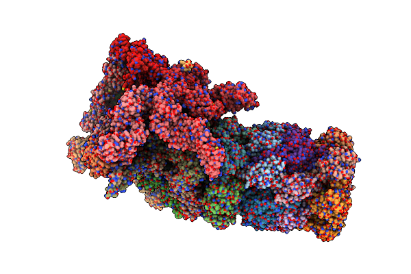
26S Proteasome In Presence Of Atp (S1)
Organism: Saccharomyces cerevisiae (strain atcc 204508 / s288c)
Method: ELECTRON MICROSCOPY Release Date: 2017-03-08 Classification: HYDROLASE Ligands: ATP, MG, ADP |
Organism: Saccharomyces cerevisiae (strain atcc 204508 / s288c)
Method: ELECTRON MICROSCOPY
Release Date: 2017-03-08
Ligands: ATP, MG, ADP
 |
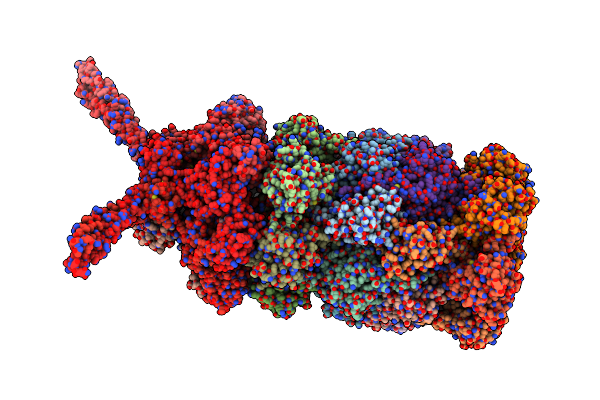
26S Proteasome In Presence Of Amp-Pnp (S3)
Organism: Saccharomyces cerevisiae (strain atcc 204508 / s288c)
Method: ELECTRON MICROSCOPY Release Date: 2017-03-08 Classification: HYDROLASE Ligands: ANP, MG, ADP |
Organism: Saccharomyces cerevisiae (strain atcc 204508 / s288c)
Method: ELECTRON MICROSCOPY
Release Date: 2017-03-08
Ligands: ANP, MG, ADP
 |
26S Proteasome In Presence Of Atp (S2)
Organism: Saccharomyces cerevisiae (strain atcc 204508 / s288c)
Method: ELECTRON MICROSCOPY Release Date: 2017-03-08 Classification: HYDROLASE Ligands: MG, ATP, ADP |
Organism: Saccharomyces cerevisiae (strain atcc 204508 / s288c)
Method: ELECTRON MICROSCOPY
Release Date: 2017-03-08
Ligands: MG, ATP, ADP
 |
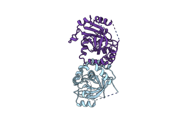
Catalytic Fragment Of Masp-1 In Complex With Its Specific Inhibitor Developed By Directed Evolution On Sgci Scaffold
Organism: Homo sapiens, Schistocerca gregaria
Method: X-RAY DIFFRACTION Resolution:3.20 Å Release Date: 2012-04-25 Classification: HYDROLASE/HYDROLASE INHIBITOR |
Organism: Homo sapiens, Schistocerca gregaria
Method: X-RAY DIFFRACTION
Release Date: 2012-04-25
 |
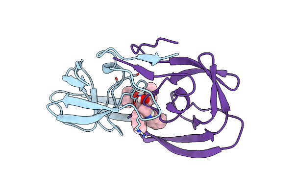
Human Cytomegalovirus Protease
Organism: Human herpesvirus 5
Method: X-RAY DIFFRACTION Resolution:2.27 Å Release Date: 1997-09-04 Classification: SERINE PROTEASE |
Organism: Human herpesvirus 5
Method: X-RAY DIFFRACTION
Release Date: 1997-09-04
 |
A New Class Of Hiv-1 Protease Inhibitor: The Crystallographic Structure, Inhibition And Chemical Synthesis Of An Aminimide Peptide Isostere
Organism: Human immunodeficiency virus 1
Method: X-RAY DIFFRACTION Resolution:2.50 Å Release Date: 1997-09-17 Classification: ASPARTYL PROTEASE Ligands: ARQ |
Organism: Human immunodeficiency virus 1
Method: X-RAY DIFFRACTION
Release Date: 1997-09-17
Ligands: ARQ
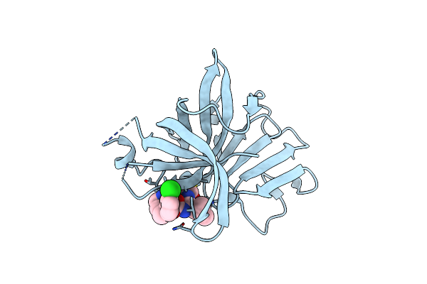 |
1.40 A Resolution Structure Of Norovirus 3Cl Protease In Complex With The A M-Chlorophenyl Substituted Macrocyclic Inhibitor (17-Mer)
Organism: Norwalk virus
Method: X-RAY DIFFRACTION Resolution:1.40 Å Release Date: 2017-01-11 Classification: PROTEASE/PROTEASE INHIBITOR Ligands: CL, V56 |
Organism: Norwalk virus
Method: X-RAY DIFFRACTION
Release Date: 2017-01-11
Ligands: CL, V56
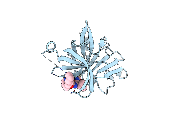 |
1.75 A Resolution Structure Of Norovirus 3Cl Protease In Complex With The A N-Pentyl Substituted Macrocyclic Inhibitor (17-Mer)
Organism: Norwalk virus
Method: X-RAY DIFFRACTION Resolution:1.75 Å Release Date: 2017-01-11 Classification: PROTEASE/PROTEASE INHIBITOR Ligands: V69 |
Organism: Norwalk virus
Method: X-RAY DIFFRACTION
Release Date: 2017-01-11
Ligands: V69
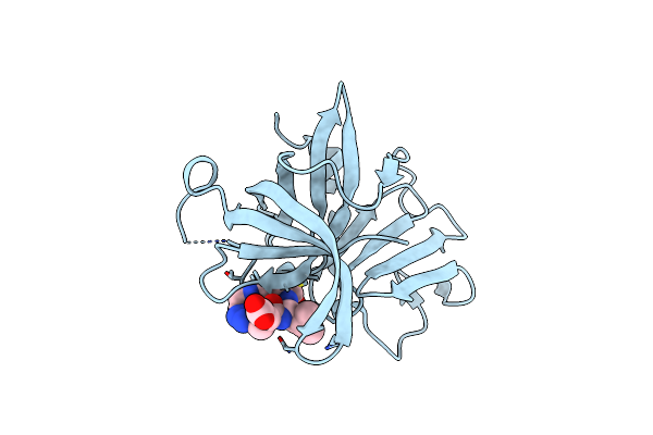 |
1.20 A Resolution Structure Of Norovirus 3Cl Protease In Complex With A Triazole-Based Macrocyclic (17-Mer) Inhibitor
Organism: Norwalk virus
Method: X-RAY DIFFRACTION Resolution:1.20 Å Release Date: 2016-05-04 Classification: PROTEASE/PROTEASE INHIBITOR Ligands: CL, 5LG |
Organism: Norwalk virus
Method: X-RAY DIFFRACTION
Release Date: 2016-05-04
Ligands: CL, 5LG
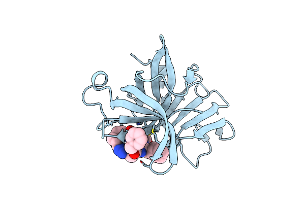 |
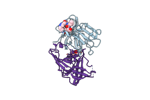
1.20 A Resolution Structure Of Norovirus 3Cl Protease In Complex With A Triazole-Based Macrocyclic (21-Mer) Inhibitor
Organism: Norwalk virus
Method: X-RAY DIFFRACTION Resolution:1.20 Å Release Date: 2016-05-04 Classification: PROTEASE/PROTEASE INHIBITOR Ligands: CL, 5LJ |
Organism: Norwalk virus
Method: X-RAY DIFFRACTION
Release Date: 2016-05-04
Ligands: CL, 5LJ
 |
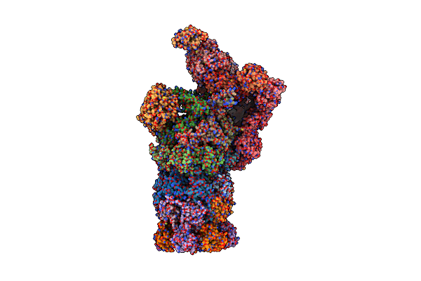
1.95 A Resolution Structure Of Norovirus 3Cl Protease In Complex With A Triazole-Based Macrocyclic (18-Mer) Inhibitor
Organism: Norwalk virus
Method: X-RAY DIFFRACTION Resolution:1.95 Å Release Date: 2016-05-04 Classification: PROTEASE/PROTEASE INHIBITOR Ligands: 5LH, GOL |
Organism: Norwalk virus
Method: X-RAY DIFFRACTION
Release Date: 2016-05-04
Ligands: 5LH, GOL
 |
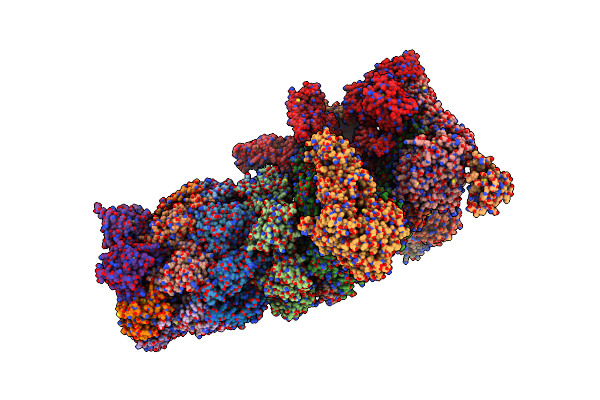
The Resting State Of Yeast Proteasome
Organism: Saccharomyces cerevisiae (strain atcc 204508 / s288c)
Method: ELECTRON MICROSCOPY Release Date: 2017-03-22 Classification: HYDROLASE |
Organism: Saccharomyces cerevisiae (strain atcc 204508 / s288c)
Method: ELECTRON MICROSCOPY
Release Date: 2017-03-22
 |
Yeast Proteasome In Translocation Competent State (C3-A)
Organism: Saccharomyces cerevisiae s288c
Method: ELECTRON MICROSCOPY Release Date: 2019-03-13 Classification: HYDROLASE |
Organism: Saccharomyces cerevisiae s288c
Method: ELECTRON MICROSCOPY
Release Date: 2019-03-13
 |
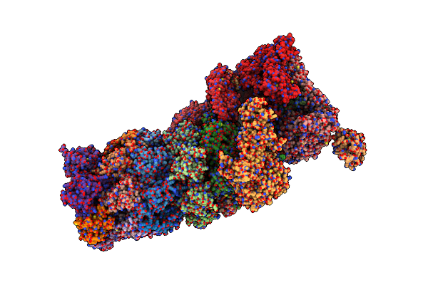
26S Proteasome Trimer
Organism: Saccharomyces cerevisiae s288c
Method: ELECTRON MICROSCOPY Release Date: 2026-01-28 Classification: HYDROLASE |
Organism: Saccharomyces cerevisiae s288c
Method: ELECTRON MICROSCOPY
Release Date: 2026-01-28
 |
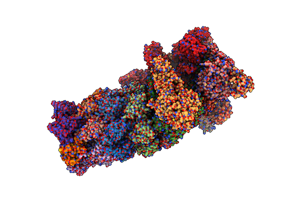
Yeast Proteasome In Ub-Accepted State (C1-B)
Organism: Saccharomyces cerevisiae s288c
Method: ELECTRON MICROSCOPY Release Date: 2019-03-13 Classification: HYDROLASE |
Organism: Saccharomyces cerevisiae s288c
Method: ELECTRON MICROSCOPY
Release Date: 2019-03-13
 |
Yeast Proteasome In Substrate-Processing State (C3-B)
Organism: Saccharomyces cerevisiae s288c
Method: ELECTRON MICROSCOPY Release Date: 2019-03-20 Classification: HYDROLASE |
Organism: Saccharomyces cerevisiae s288c
Method: ELECTRON MICROSCOPY
Release Date: 2019-03-20