

Search Count: 48
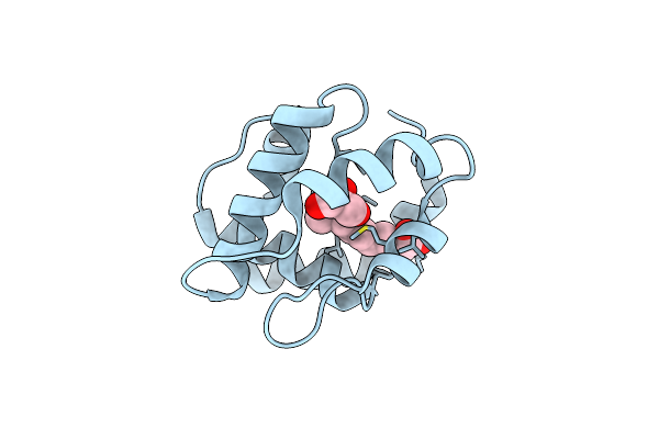 |
Crystal Structure Of A Pheromone Binding Protein From Apis Mellifera In Complex With The Queen Mandibular Pheromone
Organism: Apis mellifera
Method: X-RAY DIFFRACTION Resolution:2.25 Å Release Date: 2008-06-10 Classification: PHEROMONE BINDING PROTEIN Ligands: 9OD, GOL |
Organism: Apis mellifera
Method: X-RAY DIFFRACTION
Release Date: 2008-06-10
Ligands: 9OD, GOL
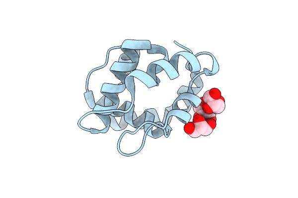 |
Crystal Structure Of A Pheromone Binding Protein From Apis Mellifera Soaked At Ph 7.0
Organism: Apis mellifera
Method: X-RAY DIFFRACTION Resolution:1.95 Å Release Date: 2008-06-10 Classification: PHEROMONE-BINDING PROTEIN Ligands: GOL |
Organism: Apis mellifera
Method: X-RAY DIFFRACTION
Release Date: 2008-06-10
Ligands: GOL
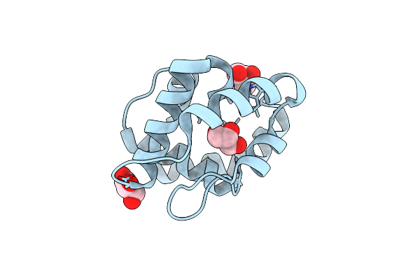 |
Crystal Structure Of A Pheromone Binding Protein From Apis Mellifera Soaked At Ph 4.0
Organism: Apis mellifera
Method: X-RAY DIFFRACTION Resolution:2.00 Å Release Date: 2008-06-10 Classification: PHEROMONE-BINDING PROTEIN Ligands: CL, GOL |
Organism: Apis mellifera
Method: X-RAY DIFFRACTION
Release Date: 2008-06-10
Ligands: CL, GOL
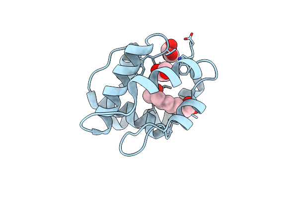 |
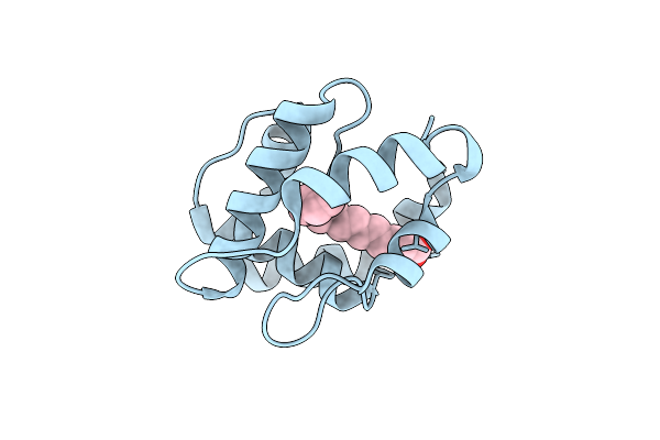
Crystal Structure Of A Pheromone Binding Protein From Apis Mellifera In Complex With The 9-Keto-2(E)-Decenoic Acid
Organism: Apis mellifera
Method: X-RAY DIFFRACTION Resolution:2.15 Å Release Date: 2008-06-10 Classification: PHEROMONE BINDING PROTEIN Ligands: CL, 9OD, GOL |
Organism: Apis mellifera
Method: X-RAY DIFFRACTION
Release Date: 2008-06-10
Ligands: CL, 9OD, GOL
 |
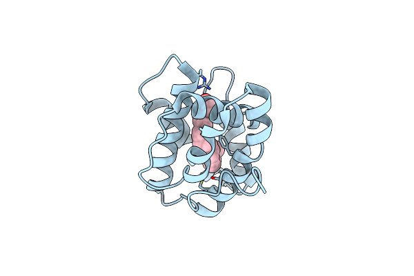
Crystal Structure Of A Pheromone Binding Protein From Apis Mellifera In Complex With Hexadecanoic Acid
Organism: Apis mellifera
Method: X-RAY DIFFRACTION Resolution:2.00 Å Release Date: 2008-06-10 Classification: PHEROMONE BINDING PROTEIN Ligands: CL, PLM |
Organism: Apis mellifera
Method: X-RAY DIFFRACTION
Release Date: 2008-06-10
Ligands: CL, PLM
 |
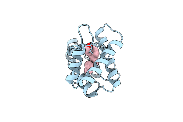
Structure Of Pheromone-Binding Protein 1 In Complex With (11Z,13Z)-Hexadecadienal
Organism: Amyelois transitella
Method: X-RAY DIFFRACTION Resolution:1.14 Å Release Date: 2013-03-06 Classification: pheromone-binding protein Ligands: 1EY |
Organism: Amyelois transitella
Method: X-RAY DIFFRACTION
Release Date: 2013-03-06
Ligands: 1EY
 |
Structure Of Pheromone-Binding Protein 1 In Complex With (Z,Z)-11,13- Hexadecadienol
Organism: Amyelois transitella
Method: X-RAY DIFFRACTION Resolution:1.85 Å Release Date: 2013-03-06 Classification: pheromone-binding protein Ligands: 1EX |
Organism: Amyelois transitella
Method: X-RAY DIFFRACTION
Release Date: 2013-03-06
Ligands: 1EX
 |
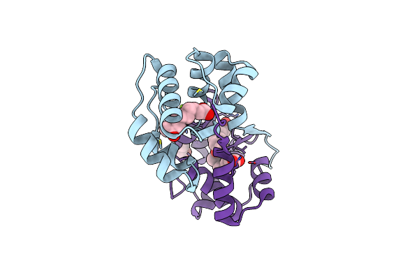
Dimeric Crystal Structure Of A Pheromone Binding Protein From Apis Mellifera In Complex With The N-Butyl Benzene Sulfonamide At Ph 7.0
Organism: Apis mellifera
Method: X-RAY DIFFRACTION Resolution:1.50 Å Release Date: 2009-04-28 Classification: Pheromone Binding Protein Ligands: NBB, GOL, MG, CL |
Organism: Apis mellifera
Method: X-RAY DIFFRACTION
Release Date: 2009-04-28
Ligands: NBB, GOL, MG, CL
 |
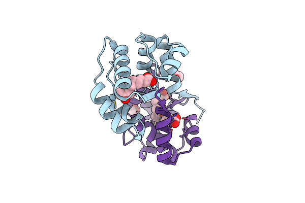
Dimeric Crystal Structure Of A Pheromone Binding Protein From Apis Mellifera In Complex With The N-Butyl Benzene Sulfonamide At Ph 7.0
Organism: Apis mellifera
Method: X-RAY DIFFRACTION Resolution:1.70 Å Release Date: 2009-04-28 Classification: Pheromone Binding Protein Ligands: 9OD, CL, MG, GOL |
Organism: Apis mellifera
Method: X-RAY DIFFRACTION
Release Date: 2009-04-28
Ligands: 9OD, CL, MG, GOL
 |
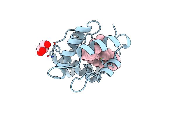
Dimeric Crystal Structure Of A Pheromone Binding Protein From Apis Mellifera In Complex With 9-Keto-2(E)-Decenoic Acid At Ph 7.0
Organism: Apis mellifera
Method: X-RAY DIFFRACTION Resolution:1.80 Å Release Date: 2009-04-28 Classification: Pheromone Binding Protein Ligands: 9OD, CL, MG, GOL |
Organism: Apis mellifera
Method: X-RAY DIFFRACTION
Release Date: 2009-04-28
Ligands: 9OD, CL, MG, GOL
 |
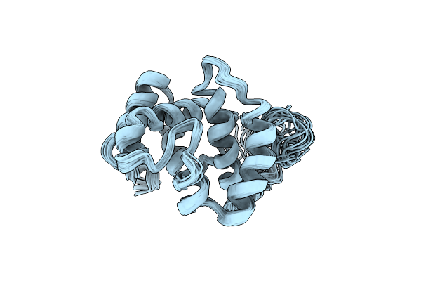
Crystal Structure Of A Pheromone Binding Protein From Apis Mellifera With A Serendipitous Ligand At Ph 5.5
Organism: Apis mellifera
Method: X-RAY DIFFRACTION Resolution:1.80 Å Release Date: 2009-12-01 Classification: Pheromone binding protein Ligands: CMJ, GOL, CL |
Organism: Apis mellifera
Method: X-RAY DIFFRACTION
Release Date: 2009-12-01
Ligands: CMJ, GOL, CL
 |
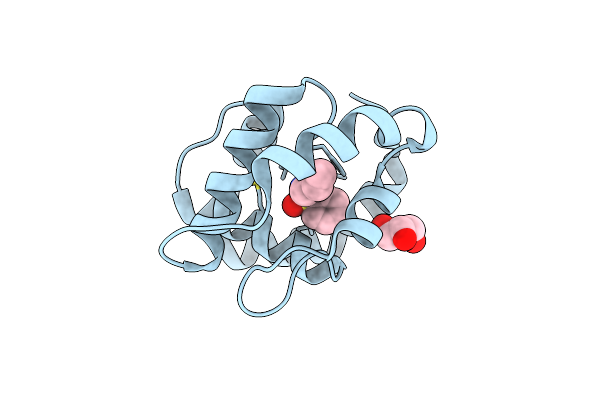
Nmr Structure Of The Pheromone Binding Protein From Antheraea Polyphemus At Acidic Ph
Organism: Antheraea polyphemus
Method: SOLUTION NMR Release Date: 2005-10-25 Classification: pheromone binding protein |
Organism: Antheraea polyphemus
Method: SOLUTION NMR
Release Date: 2005-10-25
 |
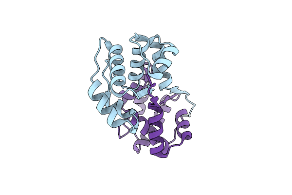
Soft-Sad Crystal Structure Of A Pheromone Binding Protein From The Honeybee Apis Mellifera L.
Organism: Apis mellifera
Method: X-RAY DIFFRACTION Resolution:1.60 Å Release Date: 2007-12-18 Classification: PHEROMONE BINDING PROTEIN Ligands: NBB, GOL |
Organism: Apis mellifera
Method: X-RAY DIFFRACTION
Release Date: 2007-12-18
Ligands: NBB, GOL
 |
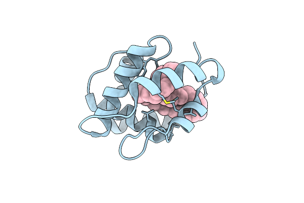
Dimeric Crystal Structure Of A Pheromone Binding Protein From Apis Mellifera At Ph 7.0
Organism: Apis mellifera
Method: X-RAY DIFFRACTION Resolution:2.50 Å Release Date: 2009-04-28 Classification: Pheromone Binding Protein Ligands: CL |
Organism: Apis mellifera
Method: X-RAY DIFFRACTION
Release Date: 2009-04-28
Ligands: CL
 |
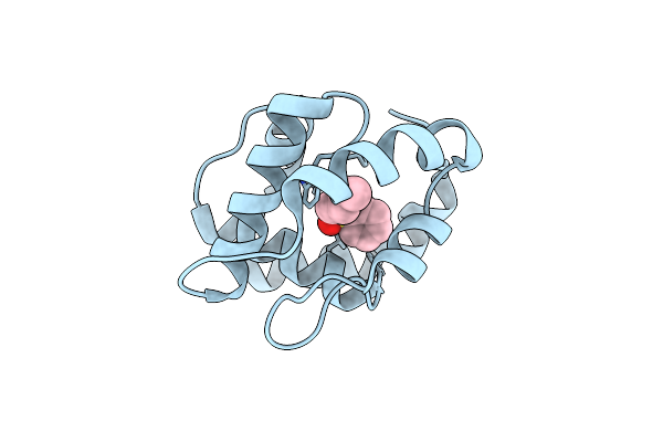
Crystal Structure Of A Pheromone Binding Protein From Apis Mellifera With A Serendipitous Ligand Soaked At Ph 7.0
Organism: Apis mellifera
Method: X-RAY DIFFRACTION Resolution:1.75 Å Release Date: 2009-12-01 Classification: Pheromone binding protein Ligands: CMJ, CL |
Organism: Apis mellifera
Method: X-RAY DIFFRACTION
Release Date: 2009-12-01
Ligands: CMJ, CL
 |
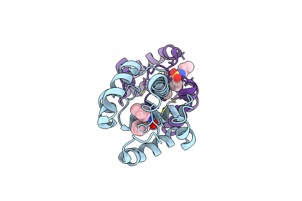
Crystal Structure Of A Pheromone Binding Protein Mutant D35N, From Apis Mellifera, At Ph 5.5
Organism: Apis mellifera
Method: X-RAY DIFFRACTION Resolution:2.30 Å Release Date: 2009-05-26 Classification: Pheromone Binding Protein Ligands: NBB |
Organism: Apis mellifera
Method: X-RAY DIFFRACTION
Release Date: 2009-05-26
Ligands: NBB
 |
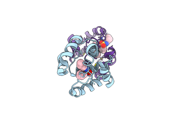
Crystal Structure Of A Pheromone Binding Protein Mutant D35A, From Apis Mellifera, At Ph 7.0
Organism: Apis mellifera
Method: X-RAY DIFFRACTION Resolution:2.03 Å Release Date: 2009-05-26 Classification: Pheromone Binding Protein Ligands: NBB |
Organism: Apis mellifera
Method: X-RAY DIFFRACTION
Release Date: 2009-05-26
Ligands: NBB
 |
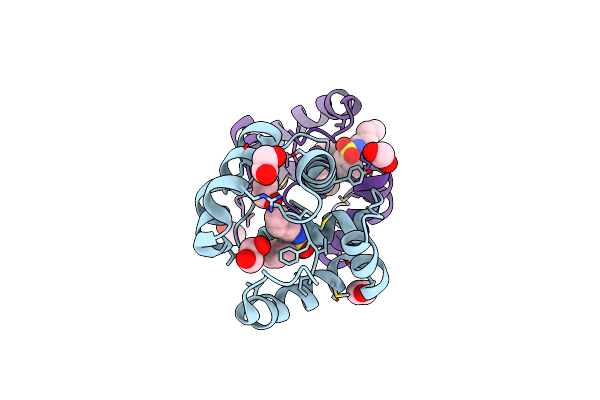
Crystal Structure Of A Pheromone Binding Protein Mutant D35A, From Apis Mellifera, Soaked At Ph 5.5
Organism: Apis mellifera
Method: X-RAY DIFFRACTION Resolution:2.10 Å Release Date: 2009-05-26 Classification: Pheromone Binding Protein Ligands: NBB |
Organism: Apis mellifera
Method: X-RAY DIFFRACTION
Release Date: 2009-05-26
Ligands: NBB
 |
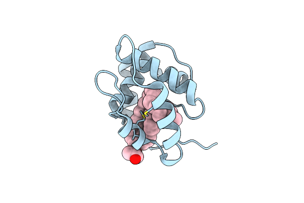
Dimeric Crystal Structure Of A Pheromone Binding Protein Mutant D35N, From Apis Mellifera, At Ph 7.0
Organism: Apis mellifera
Method: X-RAY DIFFRACTION Resolution:1.60 Å Release Date: 2009-05-26 Classification: Pheromone binding protein Ligands: NBB, EDO |
Organism: Apis mellifera
Method: X-RAY DIFFRACTION
Release Date: 2009-05-26
Ligands: NBB, EDO
 |
Crystal Structure Of A Pheromone Binding Protein From Apis Mellifera With A Serendipitous Ligand Soaked At Ph 4.0
Organism: Apis mellifera
Method: X-RAY DIFFRACTION Resolution:1.90 Å Release Date: 2009-12-01 Classification: Pheromone binding protein Ligands: CMJ, GOL, CL |
Organism: Apis mellifera
Method: X-RAY DIFFRACTION
Release Date: 2009-12-01
Ligands: CMJ, GOL, CL