
Search Count: 27
All
Selected
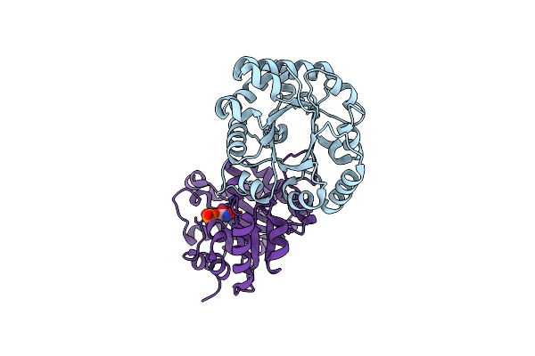 |
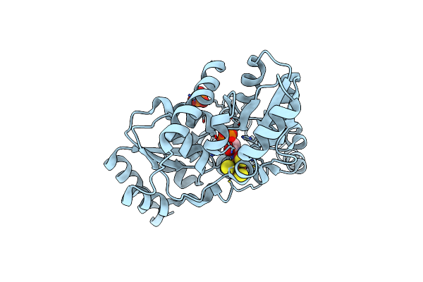
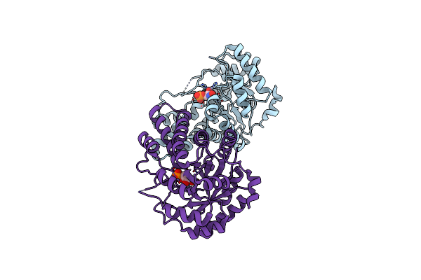
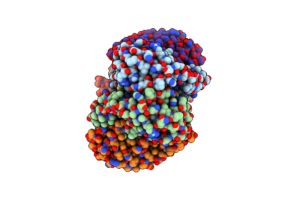
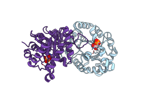
Crystal Structure Of The Worst Case Of The Reconstruction Of The Ancestral Triosephosphate Isomerase Of The Last Opisthokont Common Ancestor Obtained By Maximum Likelihood With Pgh
Organism: Synthetic construct
Method: X-RAY DIFFRACTION Resolution:2.05 Å Release Date: 2024-09-04 Classification: ISOMERASE Ligands: PGH |
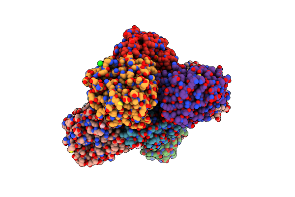 |
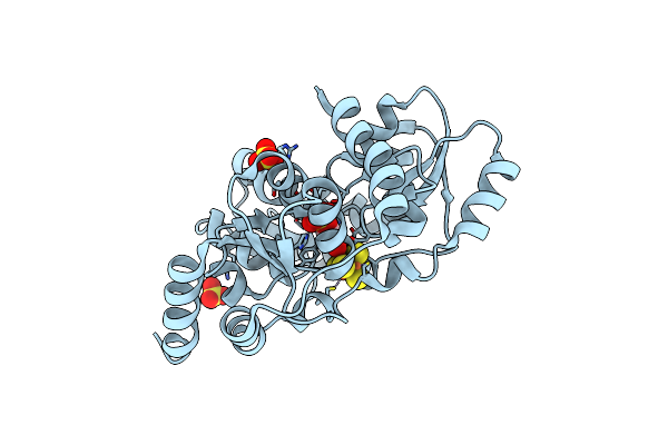
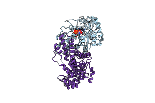
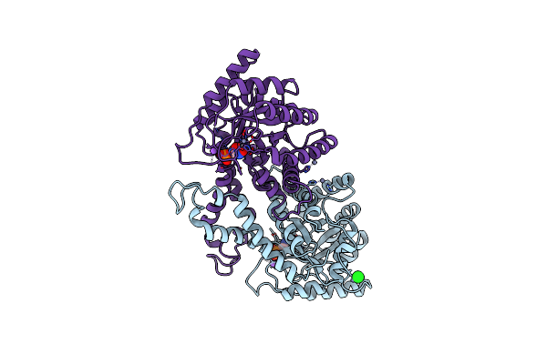
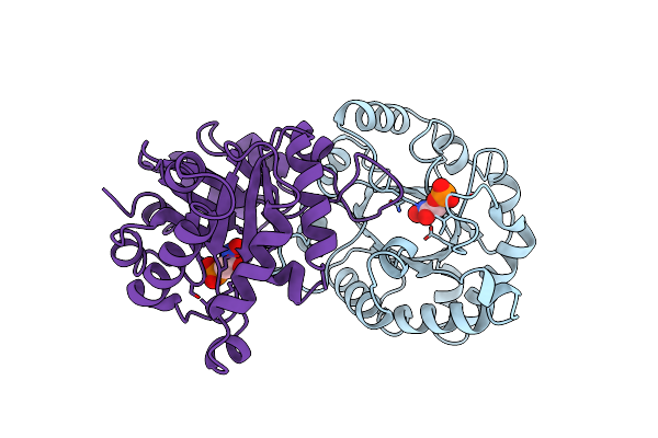
Crystal Structure Of The Reconstruction Of The Ancestral Triosephosphate Isomerase Of The Last Opisthokont Common Ancestor Obtained By Maximum Likelihood With Pgh
Organism: Synthetic construct
Method: X-RAY DIFFRACTION Resolution:2.06 Å Release Date: 2024-09-04 Classification: ISOMERASE Ligands: PGH, ACY, PGE, ACT, NA, EDO, PEG, CL |
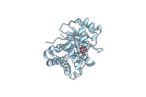 |
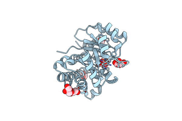
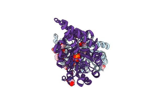
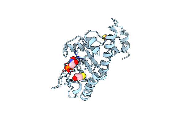
Organism: Leishmania mexicana
Method: X-RAY DIFFRACTION, NEUTRON DIFFRACTION Resolution:1.10 Å, 1.8000 Å Release Date: 2021-07-28 Classification: ISOMERASE Ligands: PGH |
 |
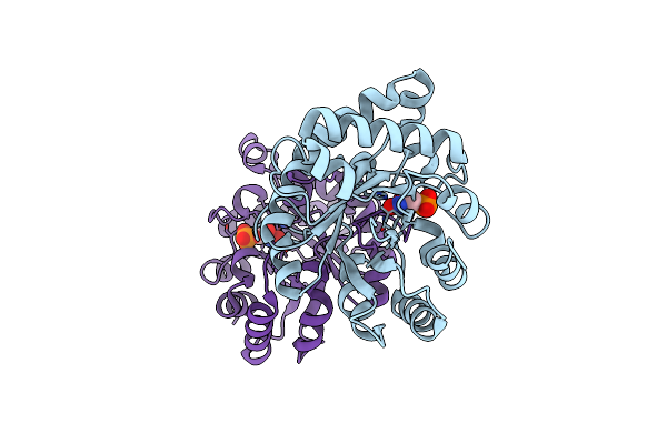
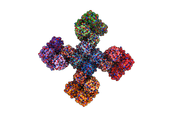
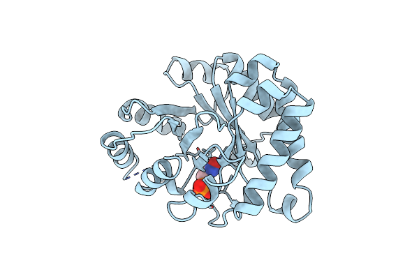
Organism: Leishmania mexicana
Method: X-RAY DIFFRACTION, NEUTRON DIFFRACTION Resolution:1.10 Å, 1.8000 Å Release Date: 2021-07-28 Classification: ISOMERASE Ligands: PGH |
 |
Crystal Structure Of A Reconstructed Ancestor Of Triosephosphate Isomerase From Eukaryotes
Organism: Synthetic construct
Method: X-RAY DIFFRACTION Resolution:1.90 Å Release Date: 2019-01-09 Classification: ISOMERASE Ligands: PGH |
 |
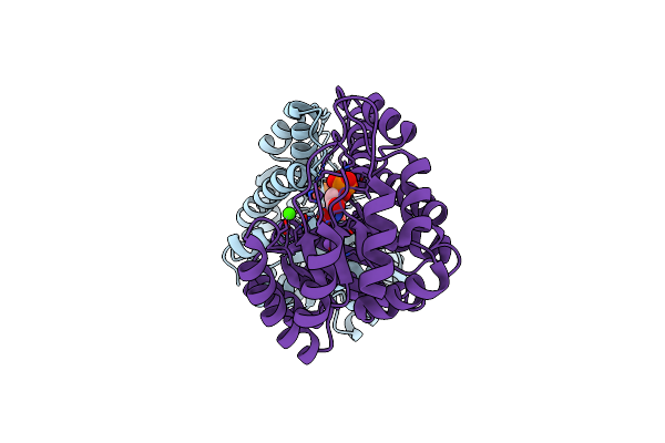
Organism: Thermotoga maritima
Method: X-RAY DIFFRACTION Resolution:2.00 Å Release Date: 2018-04-25 Classification: TRANSFERASE Ligands: SF4, PGH, PO4, CL |
 |
Structure Of Quinolinate Synthase In Complex With Phosphoglycolohydroxamate
Organism: Thermotoga maritima msb8
Method: X-RAY DIFFRACTION Resolution:1.45 Å Release Date: 2016-09-07 Classification: TRANSFERASE Ligands: SF4, PGH, SO4 |
 |
Structure Of Mycobacterium Tuberculosis Triosephosphate Isomerase Bound To Phosphoglycolohydroxamate
Organism: Mycobacterium tuberculosis
Method: X-RAY DIFFRACTION Resolution:1.45 Å Release Date: 2011-11-30 Classification: ISOMERASE Ligands: PGH |
 |
Organism: Leishmania mexicana
Method: X-RAY DIFFRACTION Resolution:0.82 Å Release Date: 2009-07-14 Classification: ISOMERASE Ligands: PGH, PGA, GOL, ACT |
 |
Class Ii Fructose-1,6-Bisphosphate Aldolase From Helicobacter Pylori In Complex With Phosphoglycolohydroxamic Acid, A Competitive Inhibitor
Organism: Helicobacter pylori
Method: X-RAY DIFFRACTION Resolution:2.30 Å Release Date: 2008-08-26 Classification: LYASE Ligands: ZN, CA, NA, PGH |
 |
Structure Of Giardia Fructose-1,6-Biphosphate Aldolase In Complex With Phosphoglycolohydroxamate
Organism: Giardia intestinalis
Method: X-RAY DIFFRACTION Resolution:2.30 Å Release Date: 2006-12-12 Classification: LYASE Ligands: ZN, PGH |
 |
Structure Of Giardia Fructose-1,6-Biphosphate Aldolase In Complex With Phosphoglycolohydroxamate
Organism: Giardia intestinalis
Method: X-RAY DIFFRACTION Resolution:1.75 Å Release Date: 2006-12-12 Classification: LYASE Ligands: ZN, PGH |
 |
Organism: Escherichia coli
Method: X-RAY DIFFRACTION Resolution:1.45 Å Release Date: 2002-06-18 Classification: LYASE Ligands: PGH, ZN, NA, EDO |
 |
Organism: Escherichia coli
Method: X-RAY DIFFRACTION Resolution:2.70 Å Release Date: 2002-05-03 Classification: LYASE Ligands: ZN, PGH |
 |
X-Ray Structure Of Methylglyoxal Synthase From E. Coli Complexed With Phosphoglycolohydroxamic Acid
Organism: Escherichia coli
Method: X-RAY DIFFRACTION Resolution:2.00 Å Release Date: 2001-09-26 Classification: LYASE Ligands: PGH |
 |
Class Ii Fructose-1,6-Bisphosphate Aldolase In Complex With Phosphoglycolohydroxamate
Organism: Escherichia coli
Method: X-RAY DIFFRACTION Resolution:2.00 Å Release Date: 2000-01-07 Classification: LYASE Ligands: ZN, NA, CL, PGH |
 |
Organism: Escherichia coli
Method: X-RAY DIFFRACTION Resolution:2.43 Å Release Date: 1996-10-14 Classification: LYASE (ALDEHYDE) Ligands: ZN, SO4, BME, PGH |
 |
Three New Crystal Structures Of Point Mutation Variants Of Monotim: Conformational Flexibility Of Loop-1,Loop-4 And Loop-8
Organism: Trypanosoma brucei brucei
Method: X-RAY DIFFRACTION Resolution:2.40 Å Release Date: 1995-09-15 Classification: ISOMERASE(INTRAMOLECULAR OXIDOREDUCTASE) Ligands: PGH |
 |
S96P Change Is A Second-Site Suppressor For H95N Sluggish Mutant Triosephosphate Isomerase
Organism: Gallus gallus
Method: X-RAY DIFFRACTION Resolution:1.90 Å Release Date: 1995-04-20 Classification: ISOMERASE(INTRAMOLECULAR OXIDOREDUCTASE) Ligands: PGH |
 |
S96P Change Is A Second-Site Suppressor For H95N Sluggish Mutant Triosephosphate Isomerase
Organism: Gallus gallus
Method: X-RAY DIFFRACTION Resolution:1.90 Å Release Date: 1995-04-20 Classification: ISOMERASE(INTRAMOLECULAR OXIDOREDUCTASE) Ligands: PGH |