

Search Count: 102
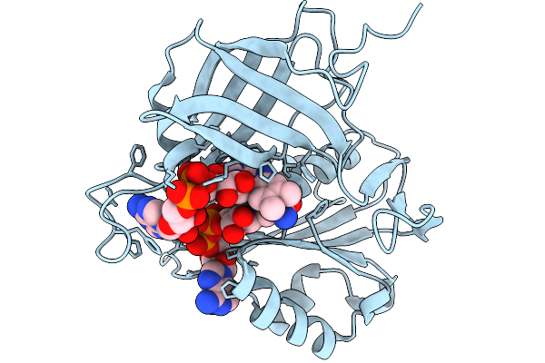 |
X-Ray Crystal Structure Of The Two-Electron Reduced Form Of Wild Type B5R
Organism: Sus scrofa
Method: X-RAY DIFFRACTION Resolution:0.96 Å Release Date: 2025-11-19 Classification: OXIDOREDUCTASE Ligands: FDA, NAD |
Organism: Sus scrofa
Method: X-RAY DIFFRACTION
Release Date: 2025-11-19
Ligands: FDA, NAD
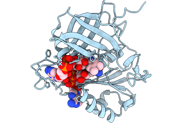 |
X-Ray Crystal Structure Of The T66V Mutant B5R Co-Crystallized With Nadh
Organism: Sus scrofa
Method: X-RAY DIFFRACTION Resolution:0.94 Å Release Date: 2025-11-19 Classification: OXIDOREDUCTASE Ligands: FAD, NAI |
Organism: Sus scrofa
Method: X-RAY DIFFRACTION
Release Date: 2025-11-19
Ligands: FAD, NAI
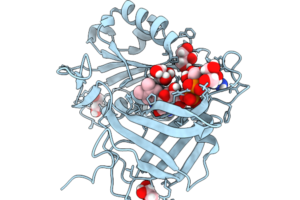 |
Neutron Crystal Structure Of The Oxidized Form Of B5R At Pd 6.5
Organism: Sus scrofa
Method: NEUTRON DIFFRACTION Resolution:1.60 Å Release Date: 2025-11-19 Classification: OXIDOREDUCTASE Ligands: FAD, GOL |
Organism: Sus scrofa
Method: NEUTRON DIFFRACTION
Release Date: 2025-11-19
Ligands: FAD, GOL
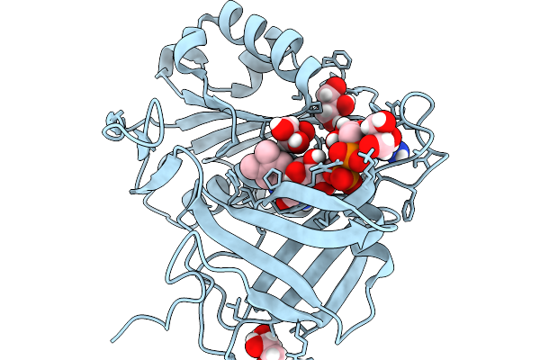 |
Neutron Crystal Structure Of The Oxidized Form Of B5R At Pd 7.5
Organism: Sus scrofa
Method: NEUTRON DIFFRACTION Resolution:1.45 Å Release Date: 2025-11-19 Classification: OXIDOREDUCTASE Ligands: FAD, GOL |
Organism: Sus scrofa
Method: NEUTRON DIFFRACTION
Release Date: 2025-11-19
Ligands: FAD, GOL
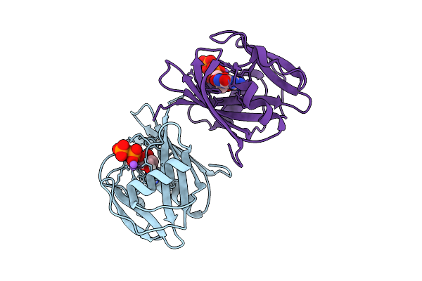 |
Neutron Crystal Structure Of Human Mth1(G2K/C87A/C104S Mutant) In Complex With 8-Oxo-Dgtp
Organism: Homo sapiens
Method: X-RAY DIFFRACTION, NEUTRON DIFFRACTION Resolution:1.40 Å, 1.7 Å Release Date: 2025-07-30 Classification: HYDROLASE Ligands: 8DG, NA |
Organism: Homo sapiens
Method: X-RAY DIFFRACTION, NEUTRON DIFFRACTION
Release Date: 2025-07-30
Ligands: 8DG, NA
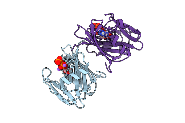 |
Neutron Crystal Structure Of Human Mth1(G2K/C87A/C104S Mutant) In Complex With 2-Oxo-Datp
Organism: Homo sapiens
Method: X-RAY DIFFRACTION, NEUTRON DIFFRACTION Resolution:1.50 Å, 2.1 Å Release Date: 2025-07-30 Classification: HYDROLASE Ligands: 6U4, NA |
Organism: Homo sapiens
Method: X-RAY DIFFRACTION, NEUTRON DIFFRACTION
Release Date: 2025-07-30
Ligands: 6U4, NA
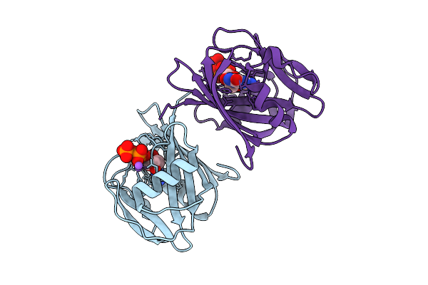 |
8-Oxo-Dgtp Hydrolysis In Human Mth1(G2K Mutant) Crystal Using Mn2+: The Es Complex
Organism: Homo sapiens
Method: X-RAY DIFFRACTION Resolution:1.20 Å Release Date: 2025-07-30 Classification: HYDROLASE Ligands: 8DG, NA |
Organism: Homo sapiens
Method: X-RAY DIFFRACTION
Release Date: 2025-07-30
Ligands: 8DG, NA
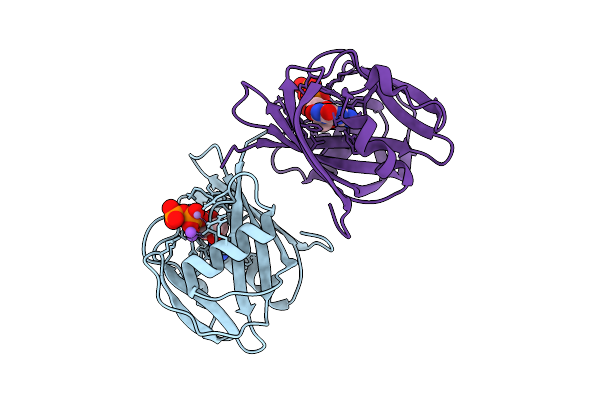 |
8-Oxo-Dgtp Hydrolysis In Human Mth1(G2K Mutant) Crystal Using Mn2+: The Es-3M Complex
Organism: Homo sapiens
Method: X-RAY DIFFRACTION Resolution:1.21 Å Release Date: 2025-07-30 Classification: HYDROLASE Ligands: 8DG, NA, MN |
Organism: Homo sapiens
Method: X-RAY DIFFRACTION
Release Date: 2025-07-30
Ligands: 8DG, NA, MN
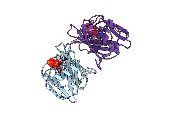 |
8-Oxo-Dgtp Hydrolysis In Human Mth1(G2K Mutant) Crystal Using Mn2+: The Es/Ep-3M Complex
Organism: Homo sapiens
Method: X-RAY DIFFRACTION Resolution:1.09 Å Release Date: 2025-07-30 Classification: HYDROLASE Ligands: MN, 8DG, 8OG |
Organism: Homo sapiens
Method: X-RAY DIFFRACTION
Release Date: 2025-07-30
Ligands: MN, 8DG, 8OG
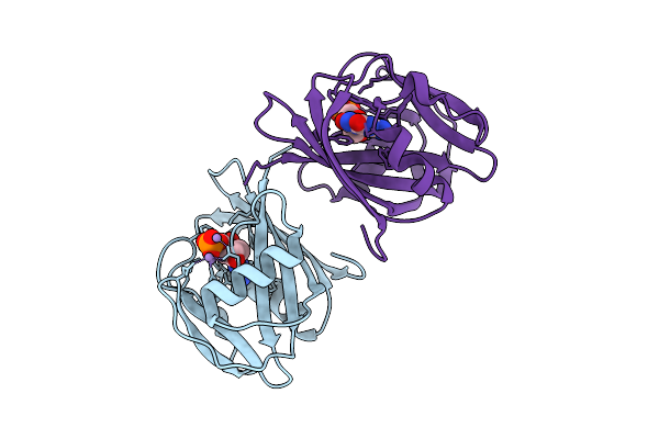 |
8-Oxo-Dgtp Hydrolysis In Human Mth1(G2K Mutant) Crystal Using Mn2+: The Ep-M Complex
Organism: Homo sapiens
Method: X-RAY DIFFRACTION Resolution:1.57 Å Release Date: 2025-07-30 Classification: HYDROLASE Ligands: 8OG, MN |
Organism: Homo sapiens
Method: X-RAY DIFFRACTION
Release Date: 2025-07-30
Ligands: 8OG, MN
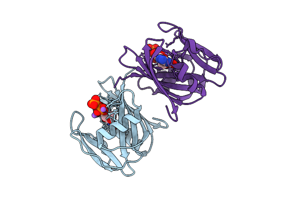 |
2-Oxo-Datp Hydrolysis In Human Mth1(G2K Mutant) Crystal Using Mn2+: The Es Complex
Organism: Homo sapiens
Method: X-RAY DIFFRACTION Resolution:1.18 Å Release Date: 2025-07-30 Classification: HYDROLASE Ligands: 6U4, NA |
Organism: Homo sapiens
Method: X-RAY DIFFRACTION
Release Date: 2025-07-30
Ligands: 6U4, NA
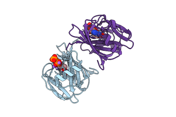 |
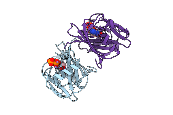
2-Oxo-Datp Hydrolysis In Human Mth1(G2K Mutant) Crystal Using Mn2+: The Es-2M Complex
Organism: Homo sapiens
Method: X-RAY DIFFRACTION Resolution:1.37 Å Release Date: 2025-07-30 Classification: HYDROLASE Ligands: 6U4, MN, NA |
Organism: Homo sapiens
Method: X-RAY DIFFRACTION
Release Date: 2025-07-30
Ligands: 6U4, MN, NA
 |
2-Oxo-Datp Hydrolysis In Human Mth1(G2K Mutant) Crystal Using Mn2+: The Es/Ep-3M Complex
Organism: Homo sapiens
Method: X-RAY DIFFRACTION Resolution:1.42 Å Release Date: 2025-07-30 Classification: HYDROLASE Ligands: 6U4, IGU, MN |
Organism: Homo sapiens
Method: X-RAY DIFFRACTION
Release Date: 2025-07-30
Ligands: 6U4, IGU, MN
 |
2-Oxo-Datp Hydrolysis In Human Mth1(G2K Mutant) Crystal Using Mn2+: The Ep-M Complex
Organism: Homo sapiens
Method: X-RAY DIFFRACTION Resolution:1.55 Å Release Date: 2025-07-30 Classification: HYDROLASE Ligands: IGU, MN |
Organism: Homo sapiens
Method: X-RAY DIFFRACTION
Release Date: 2025-07-30
Ligands: IGU, MN
 |
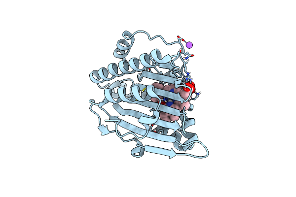
Neutron Structure Of Pcya I86D Mutant Complexed With Biliverdin At Room Temperature
Organism: Synechocystis sp. pcc 6803
Method: X-RAY DIFFRACTION, NEUTRON DIFFRACTION Resolution:1.90 Å, 2.0 Å Release Date: 2023-01-25 Classification: OXIDOREDUCTASE Ligands: BLR |
Organism: Synechocystis sp. pcc 6803
Method: X-RAY DIFFRACTION, NEUTRON DIFFRACTION
Release Date: 2023-01-25
Ligands: BLR
 |
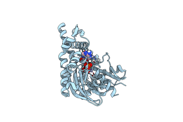
Neutron Structure Of Pcya D105N Mutant Complexed With Biliverdin At Room Temperature
Organism: Synechocystis sp. pcc 6803 substr. kazusa
Method: X-RAY DIFFRACTION, NEUTRON DIFFRACTION Resolution:1.38 Å, 2.10 Å Release Date: 2023-01-25 Classification: OXIDOREDUCTASE Ligands: BLR, NA |
Organism: Synechocystis sp. pcc 6803 substr. kazusa
Method: X-RAY DIFFRACTION, NEUTRON DIFFRACTION
Release Date: 2023-01-25
Ligands: BLR, NA
 |
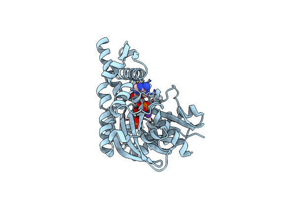
Joint Neutron And X-Ray Crystal Structure Of The Nucleotide-Binding Domain Of Hsp72 In Complex With Adp
Organism: Homo sapiens
Method: X-RAY DIFFRACTION, NEUTRON DIFFRACTION Resolution:1.600 Å, 2.199 Å Release Date: 2022-06-29 Classification: HYDROLASE Ligands: ADP, MG, NA |
Organism: Homo sapiens
Method: X-RAY DIFFRACTION, NEUTRON DIFFRACTION
Release Date: 2022-06-29
Ligands: ADP, MG, NA
 |
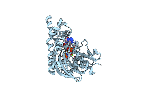
X-Ray Crystal Structure Of Y149A Mutated Hsp72-Nbd In Complex With Adp
Organism: Homo sapiens
Method: X-RAY DIFFRACTION Resolution:1.80 Å Release Date: 2022-06-29 Classification: HYDROLASE Ligands: ADP, MG, CL, K, PO4 |
Organism: Homo sapiens
Method: X-RAY DIFFRACTION
Release Date: 2022-06-29
Ligands: ADP, MG, CL, K, PO4
 |
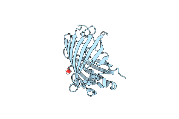
X-Ray Crystal Structure Of Y149A Mutated Hsp72-Nbd In Complex With Amppnp
Organism: Homo sapiens
Method: X-RAY DIFFRACTION Resolution:1.70 Å Release Date: 2022-06-29 Classification: HYDROLASE Ligands: MG, CL, ANP |
Organism: Homo sapiens
Method: X-RAY DIFFRACTION
Release Date: 2022-06-29
Ligands: MG, CL, ANP
 |
Neutron Crystal Structure Of The Mutant Green Fluorescent Protein (Egfp)
Organism: Aequorea victoria
Method: NEUTRON DIFFRACTION Resolution:1.44 Å Release Date: 2020-04-08 Classification: FLUORESCENT PROTEIN Ligands: D3O, DOD |
Organism: Aequorea victoria
Method: NEUTRON DIFFRACTION
Release Date: 2020-04-08
Ligands: D3O, DOD