
Search Count: 55
All
Selected
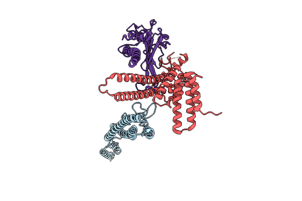 |
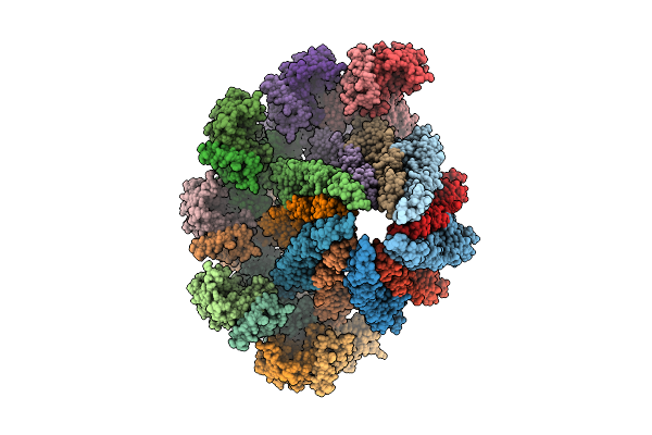
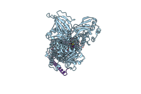
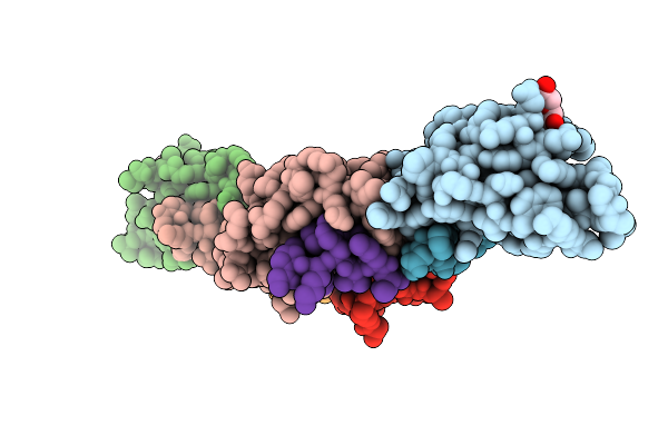
Straight And Symmetrical Filament Of The Spirochete Periplasmic Flagella Of Leptospira Biflexa
Organism: Leptospira biflexa
Method: ELECTRON MICROSCOPY Resolution:2.30 Å Release Date: 2025-12-24 Classification: MOTOR PROTEIN |
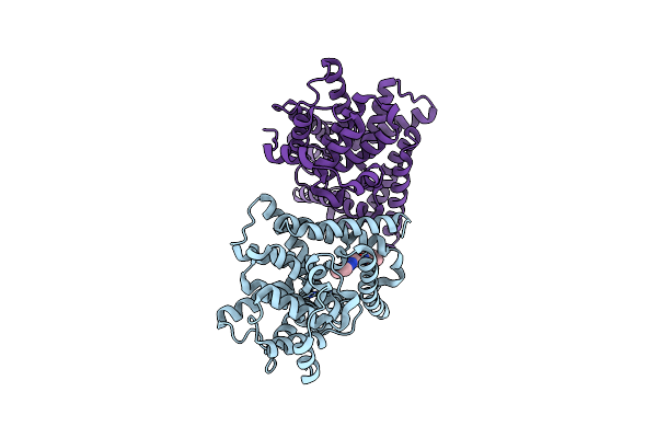
 |
Organism: Leptospira biflexa
Method: ELECTRON MICROSCOPY Resolution:2.40 Å Release Date: 2025-12-24 Classification: MOTOR PROTEIN |
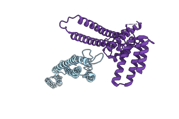 |
Straight And Symmetrical Filament Of The Spirochete Periplasmic Flagella Of Leptospira Biflexa Deleted Fcpb Strain
Organism: Leptospira biflexa
Method: ELECTRON MICROSCOPY Resolution:2.40 Å Release Date: 2025-12-24 Classification: MOTOR PROTEIN |
 |
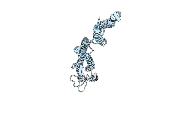
Core Filament Of The Spirochete Periplasmic Flagella Of Leptospira Biflexa Deleted Fcpb Strain
Organism: Leptospira biflexa
Method: ELECTRON MICROSCOPY Resolution:2.40 Å Release Date: 2025-12-24 Classification: MOTOR PROTEIN |
 |
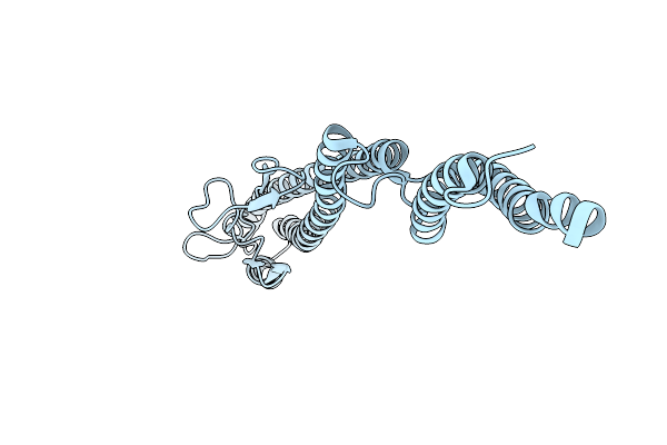
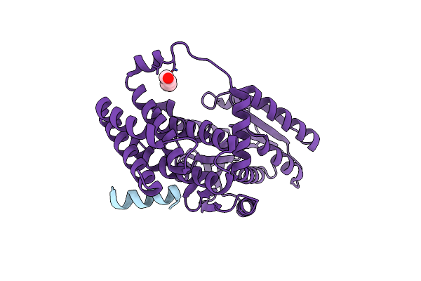
Core Filament Of The Spirochete Periplasmic Flagella Of Leptospira Biflexa Wild Type
Organism: Leptospira biflexa
Method: ELECTRON MICROSCOPY Release Date: 2025-12-24 Classification: MOTOR PROTEIN |
 |
Core Filament Of The Spirochete Periplasmic Flagella Of Leptospira Biflexa From The Flaa2-Complemented Stain
Organism: Leptospira biflexa
Method: ELECTRON MICROSCOPY Release Date: 2025-12-24 Classification: MOTOR PROTEIN |
 |
Core Filament Of The Spirochete Periplasmic Flagella Of Leptospira Biflexa From The Deleted Fcpb_Cl13 Strain
Organism: Leptospira biflexa
Method: ELECTRON MICROSCOPY Resolution:3.20 Å Release Date: 2025-12-24 Classification: MOTOR PROTEIN |
 |
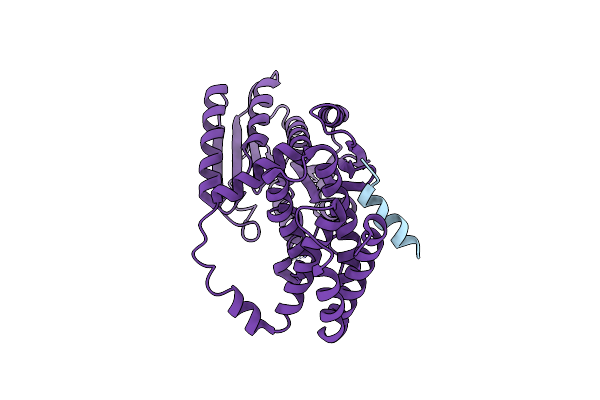
Sheathed Filament Of The Spirochete Periplasmic Flagella Of Leptospira Biflexa From The Flaa2-Complemented Stain
Organism: Leptospira biflexa
Method: ELECTRON MICROSCOPY Release Date: 2025-12-24 Classification: MOTOR PROTEIN |
 |
Sheathed Filament Of The Spirochete Periplasmic Flagella Of Leptospira Biflexa From The Deleted Fcpb_Cl13 Strain
Organism: Leptospira biflexa
Method: ELECTRON MICROSCOPY Release Date: 2025-12-24 Classification: MOTOR PROTEIN |
 |
Sheathed Filament Of The Spirochete Periplasmic Flagella Of Leptospira Biflexa Wild Type
Organism: Leptospira biflexa
Method: ELECTRON MICROSCOPY Release Date: 2025-12-24 Classification: MOTOR PROTEIN |
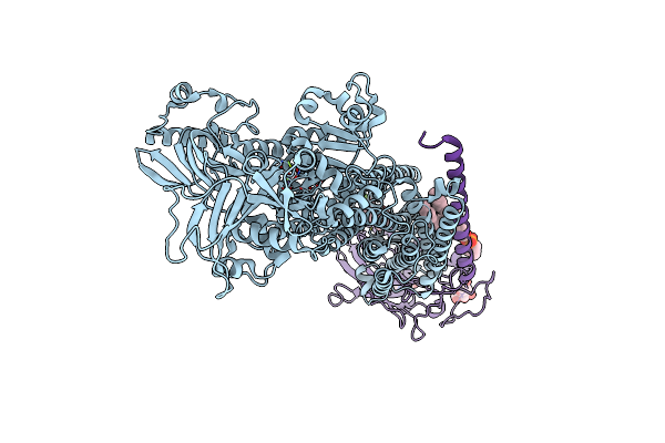 |
Organism: Sus scrofa
Method: ELECTRON MICROSCOPY Release Date: 2022-03-02 Classification: HYDROLASE Ligands: 8CK, NA, MG, NAG, PCW |
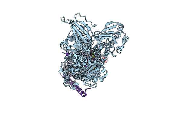 |
Organism: Sus scrofa
Method: X-RAY DIFFRACTION Resolution:3.00 Å Release Date: 2022-01-05 Classification: HYDROLASE Ligands: MG, RB, 8BN, NAG |
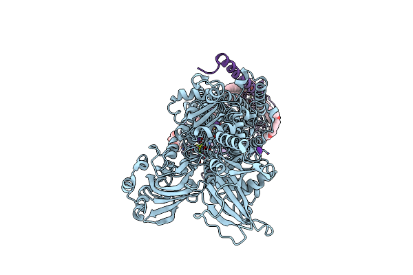 |
Organism: Sus scrofa
Method: X-RAY DIFFRACTION Resolution:3.50 Å Release Date: 2022-01-05 Classification: HYDROLASE Ligands: MG, CE1, RB, 8BZ, NAG, PCW |
 |
Organism: Sus scrofa
Method: X-RAY DIFFRACTION Resolution:3.10 Å Release Date: 2022-01-05 Classification: HYDROLASE Ligands: MG, RB, 8CE, NAG |
 |
Organism: Arabidopsis thaliana
Method: X-RAY DIFFRACTION Resolution:2.40 Å Release Date: 2020-09-02 Classification: TRANSCRIPTION Ligands: PEG |
 |
Organism: Arabidopsis thaliana
Method: X-RAY DIFFRACTION Resolution:3.20 Å Release Date: 2020-09-02 Classification: TRANSCRIPTION |
 |
Organism: Thermotoga maritima msb8
Method: X-RAY DIFFRACTION Resolution:1.70 Å Release Date: 2020-07-15 Classification: TRANSPORT PROTEIN Ligands: GAI, EDO |
 |
Organism: Arabidopsis thaliana
Method: X-RAY DIFFRACTION Resolution:1.35 Å Release Date: 2020-02-05 Classification: SIGNALING PROTEIN Ligands: PEG, EDO, PGE |
 |
Organism: Arabidopsis thaliana
Method: X-RAY DIFFRACTION Resolution:1.59 Å Release Date: 2020-02-05 Classification: SIGNALING PROTEIN Ligands: FLC, EDO |
 |
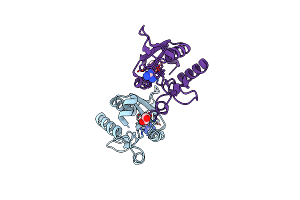
Organism: Homo sapiens
Method: X-RAY DIFFRACTION Resolution:2.44 Å Release Date: 2019-08-14 Classification: HYDROLASE Ligands: ZN, MG, D6X |