

Search Count: 21
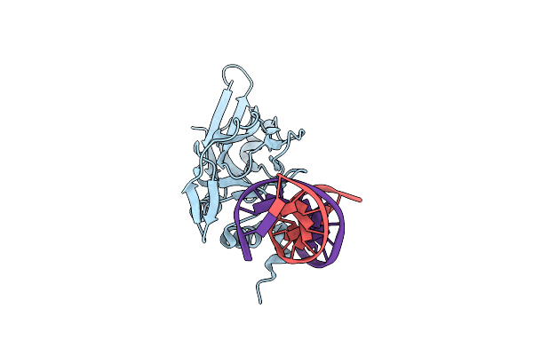 |
Crystal Structure Of Human Brachyury In Complex With A Single T Box Binding Element Dna
Organism: Homo sapiens
Method: X-RAY DIFFRACTION Resolution:2.55 Å Release Date: 2023-03-22 Classification: TRANSCRIPTION |
Organism: Homo sapiens
Method: X-RAY DIFFRACTION
Release Date: 2023-03-22
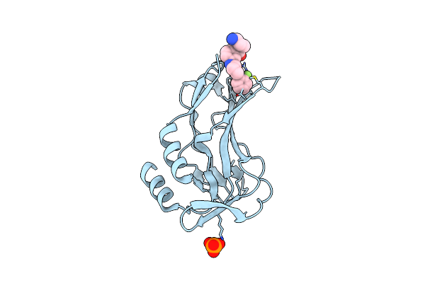 |
Crystal Structure Of Human Brachyury G177D Variant In Complex With (S)-N-(3-Aminopropyl)-3-((1-(2-Fluorophenyl)-2-Oxopyrrolidin-3-Yl)Amino)-N-Methylbenzamide (Cf-2-125)
Organism: Homo sapiens
Method: X-RAY DIFFRACTION Resolution:1.90 Å Release Date: 2022-10-05 Classification: TRANSCRIPTION Ligands: PO4, L9E |
Organism: Homo sapiens
Method: X-RAY DIFFRACTION
Release Date: 2022-10-05
Ligands: PO4, L9E
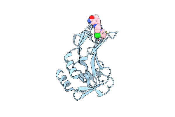 |
Crystal Structure Of Human Brachyury G177D Variant In Complex With Molpolrt-020-049-143
Organism: Homo sapiens
Method: X-RAY DIFFRACTION Resolution:1.88 Å Release Date: 2022-07-13 Classification: TRANSCRIPTION Ligands: A1IYK |
Organism: Homo sapiens
Method: X-RAY DIFFRACTION
Release Date: 2022-07-13
Ligands: A1IYK
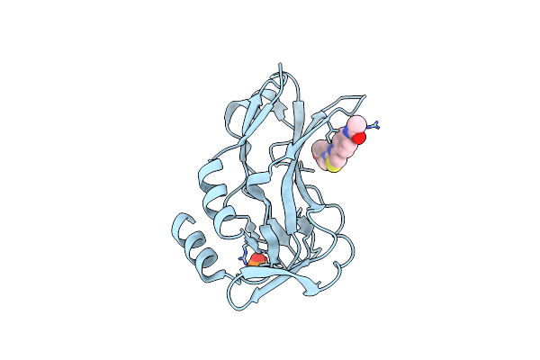 |
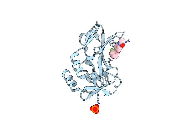
Crystal Structure Of Human Brachyury G177D Variant In Complex With Csc027898502
Organism: Homo sapiens
Method: X-RAY DIFFRACTION Resolution:1.60 Å Release Date: 2022-06-22 Classification: TRANSCRIPTION Ligands: PO4, IU4 |
Organism: Homo sapiens
Method: X-RAY DIFFRACTION
Release Date: 2022-06-22
Ligands: PO4, IU4
 |
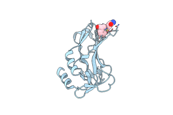
Crystal Structure Of Human Brachyury G177D Variant In Complex With Cf-5-86
Organism: Homo sapiens
Method: X-RAY DIFFRACTION Resolution:1.49 Å Release Date: 2022-06-22 Classification: TRANSCRIPTION Ligands: A1I1T, PO4 |
Organism: Homo sapiens
Method: X-RAY DIFFRACTION
Release Date: 2022-06-22
Ligands: A1I1T, PO4
 |
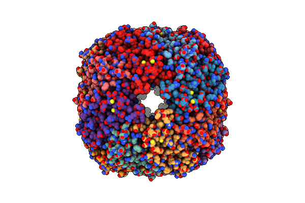
Crystal Structure Of Human Brachyury G177D Variant In Complex With Molpolrt-039-246-810
Organism: Homo sapiens
Method: X-RAY DIFFRACTION Resolution:1.80 Å Release Date: 2022-06-22 Classification: TRANSCRIPTION Ligands: IXB |
Organism: Homo sapiens
Method: X-RAY DIFFRACTION
Release Date: 2022-06-22
Ligands: IXB
 |
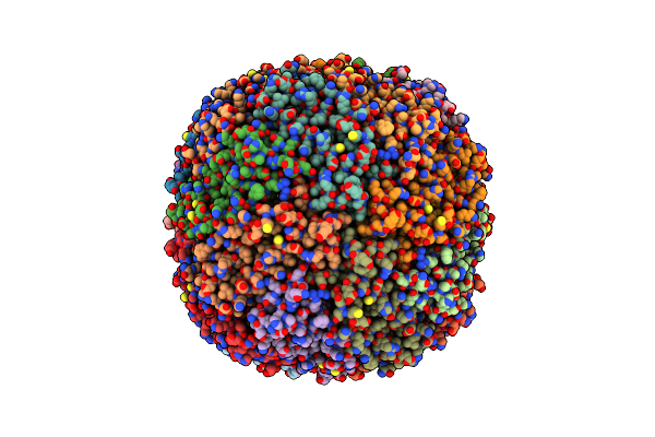
Human Ferritin Mutant - E-Helix Deletion
Organism: Homo sapiens
Method: ELECTRON MICROSCOPY Release Date: 2018-02-21 Classification: TRANSPORT PROTEIN |
Organism: Homo sapiens
Method: ELECTRON MICROSCOPY
Release Date: 2018-02-21
 |
Human Ferritin Mutant - E-Helix Deletion
Organism: Homo sapiens
Method: ELECTRON MICROSCOPY Release Date: 2018-02-21 Classification: TRANSPORT PROTEIN |
Organism: Homo sapiens
Method: ELECTRON MICROSCOPY
Release Date: 2018-02-21
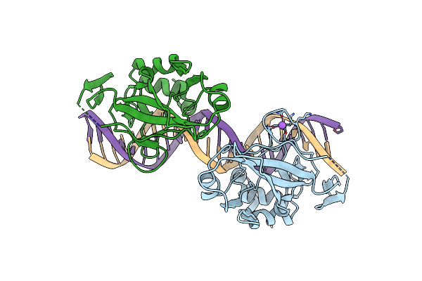 |
Crystal Structure Of Human Brachyury (T) In Complex With Dna
Organism: Homo sapiens
Method: X-RAY DIFFRACTION Resolution:2.25 Å Release Date: 2017-12-13 Classification: TRANSCRIPTION |
Organism: Homo sapiens
Method: X-RAY DIFFRACTION
Release Date: 2017-12-13
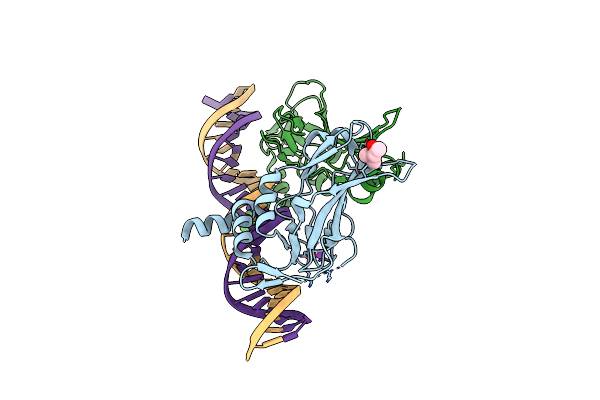 |
Crystal Structure Of Human Brachyury (T) G177D Variant In Complex With Dna
Organism: Homo sapiens
Method: X-RAY DIFFRACTION Resolution:2.15 Å Release Date: 2017-12-13 Classification: TRANSCRIPTION |
Organism: Homo sapiens
Method: X-RAY DIFFRACTION
Release Date: 2017-12-13
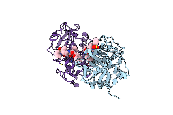 |
Crystal Structure Of Cathepsin K Complexed With 7-Methyl-Substituted Azepan-3-One Compound
Organism: Macaca mulatta
Method: X-RAY DIFFRACTION Resolution:2.55 Å Release Date: 2007-01-30 Classification: HYDROLASE Ligands: ILI |
Organism: Macaca mulatta
Method: X-RAY DIFFRACTION
Release Date: 2007-01-30
Ligands: ILI
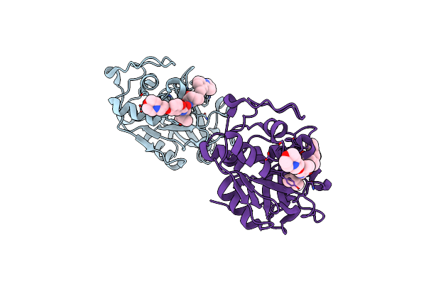 |
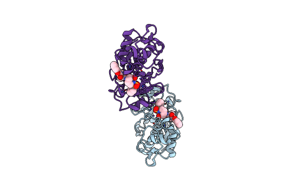
Crystal Structure Of The Cysteine Protease Human Cathepsin K In Complex With A Covalent Azepanone Inhibitor
Organism: Homo sapiens
Method: X-RAY DIFFRACTION Resolution:2.80 Å Release Date: 2003-01-14 Classification: HYDROLASE Ligands: 750 |
Organism: Homo sapiens
Method: X-RAY DIFFRACTION
Release Date: 2003-01-14
Ligands: 750
 |
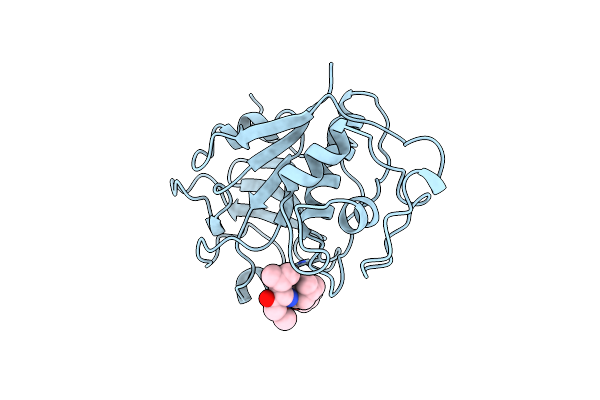
Crystal Structure Of The Cysteine Protease Human Cathepsin K In Complex With A Covalent Azepanone Inhibitor
Organism: Homo sapiens
Method: X-RAY DIFFRACTION Resolution:2.40 Å Release Date: 2003-01-14 Classification: HYDROLASE Ligands: 2CA |
Organism: Homo sapiens
Method: X-RAY DIFFRACTION
Release Date: 2003-01-14
Ligands: 2CA
 |
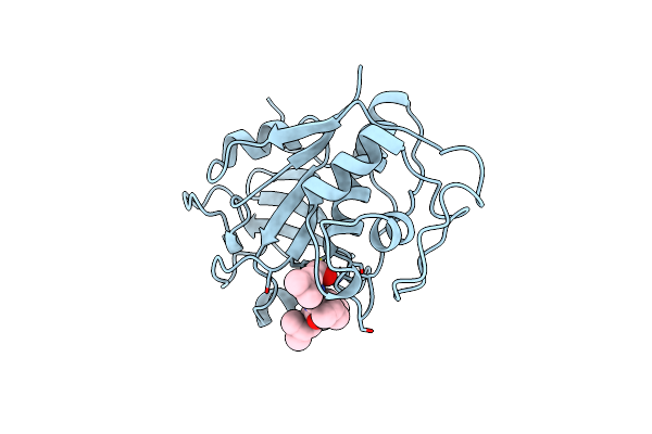
Use Of Papain As A Model For The Structure-Based Design Of Cathepsin K Inhibitors. Crystal Structures Of Two Papain Inhibitor Complexes Demonstrate Binding To S'-Subsites.
Organism: Carica papaya
Method: X-RAY DIFFRACTION Resolution:2.50 Å Release Date: 1999-08-16 Classification: HYDROLASE Ligands: SBA |
Organism: Carica papaya
Method: X-RAY DIFFRACTION
Release Date: 1999-08-16
Ligands: SBA
 |
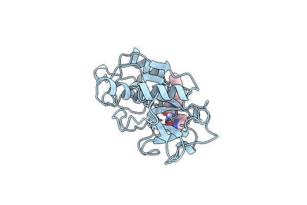
Use Of Papain As A Model For The Structure-Based Design Of Cathepsin K Inhibitors. Crystal Structures Of Two Papain Inhibitor Complexes Demonstrate Binding To S'-Subsites.
Organism: Carica papaya
Method: X-RAY DIFFRACTION Resolution:2.20 Å Release Date: 1999-08-12 Classification: HYDROLASE Ligands: ALD |
Organism: Carica papaya
Method: X-RAY DIFFRACTION
Release Date: 1999-08-12
Ligands: ALD
 |
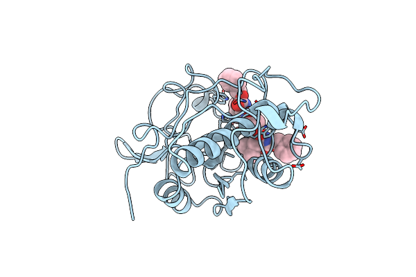
Crystal Structure Of Cysteine Protease Human Cathepsin K In Complex With A Covalent Peptidomimetic Inhibitor
Organism: Homo sapiens
Method: X-RAY DIFFRACTION Resolution:2.30 Å Release Date: 1999-06-08 Classification: HYDROLASE Ligands: I10 |
Organism: Homo sapiens
Method: X-RAY DIFFRACTION
Release Date: 1999-06-08
Ligands: I10
 |
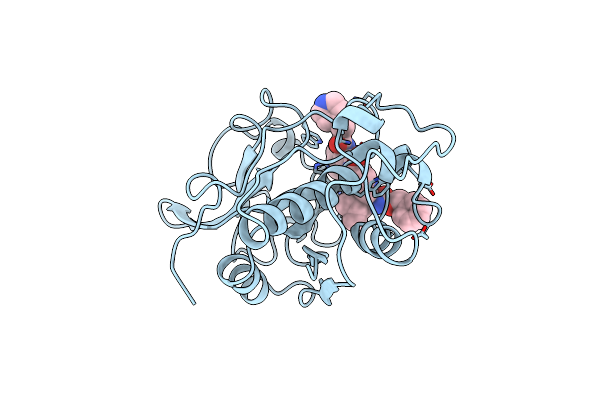
Crystal Structure Of The Cysteine Protease Human Cathepsin K In Complex With A Covalent Pyrrolidinone Inhibitor
Organism: Homo sapiens
Method: X-RAY DIFFRACTION Resolution:2.50 Å Release Date: 1998-10-14 Classification: HYDROLASE Ligands: PCM |
Organism: Homo sapiens
Method: X-RAY DIFFRACTION
Release Date: 1998-10-14
Ligands: PCM
 |
Crystal Structure Of The Cysteine Protease Human Cathepsin K In Complex With A Covalent Pyrrolidinone Inhibitor
Organism: Homo sapiens
Method: X-RAY DIFFRACTION Resolution:2.30 Å Release Date: 1998-10-14 Classification: HYDROLASE Ligands: INP |
Organism: Homo sapiens
Method: X-RAY DIFFRACTION
Release Date: 1998-10-14
Ligands: INP
 |
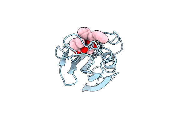
Design, Synthesis, And Kinetic Evaluation Of High-Affinity Fkbp Ligands, And The X-Ray Crystal Structures Of Their Complexes With Fkbp12
Organism: Homo sapiens
Method: X-RAY DIFFRACTION Resolution:2.00 Å Release Date: 1994-01-31 Classification: CIS-TRANS ISOMERASE Ligands: SB3 |
Organism: Homo sapiens
Method: X-RAY DIFFRACTION
Release Date: 1994-01-31
Ligands: SB3
 |
Design, Synthesis, And Kinetic Evaluation Of High-Affinity Fkbp Ligands, And The X-Ray Crystal Structures Of Their Complexes With Fkbp12
Organism: Homo sapiens
Method: X-RAY DIFFRACTION Resolution:1.95 Å Release Date: 1994-01-31 Classification: CIS-TRANS ISOMERASE Ligands: SBX |
Organism: Homo sapiens
Method: X-RAY DIFFRACTION
Release Date: 1994-01-31
Ligands: SBX