

Search Count: 20
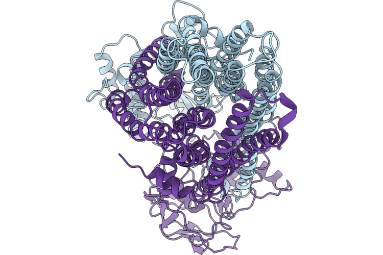 |
Apo State Of Cyddc In Nanodisc
Organism: Escherichia coli k-12
Method: ELECTRON MICROSCOPY Release Date: 2026-06-24 Classification: TRANSPORT PROTEIN |
Organism: Escherichia coli k-12
Method: ELECTRON MICROSCOPY
Release Date: 2026-06-24
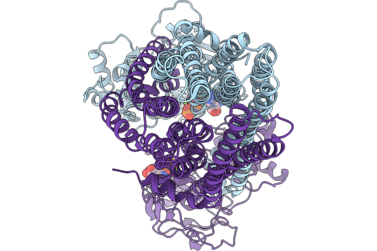 |
Cyddc In Nanodisc With Amp-Pnp-Bound
Organism: Escherichia coli k-12
Method: ELECTRON MICROSCOPY Release Date: 2026-06-24 Classification: TRANSPORT PROTEIN Ligands: MG, ANP |
Organism: Escherichia coli k-12
Method: ELECTRON MICROSCOPY
Release Date: 2026-06-24
Ligands: MG, ANP
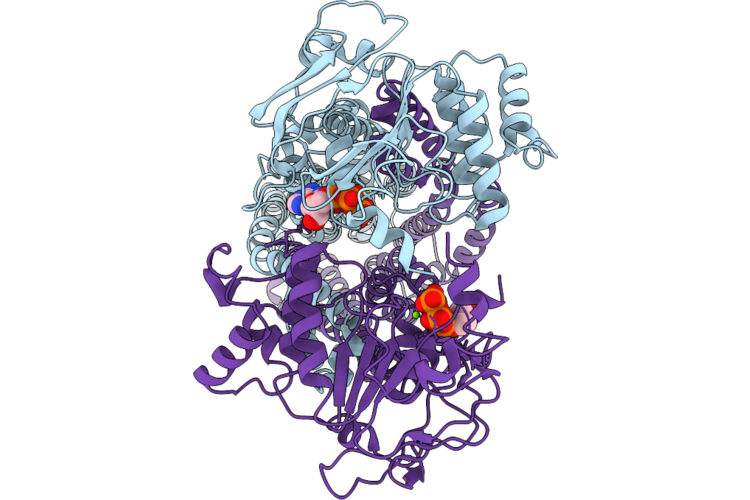 |
Cyddc In Nanodisc With Atp
Organism: Escherichia coli k-12
Method: ELECTRON MICROSCOPY Release Date: 2026-06-24 Classification: TRANSPORT PROTEIN Ligands: MG, ATP |
Organism: Escherichia coli k-12
Method: ELECTRON MICROSCOPY
Release Date: 2026-06-24
Ligands: MG, ATP
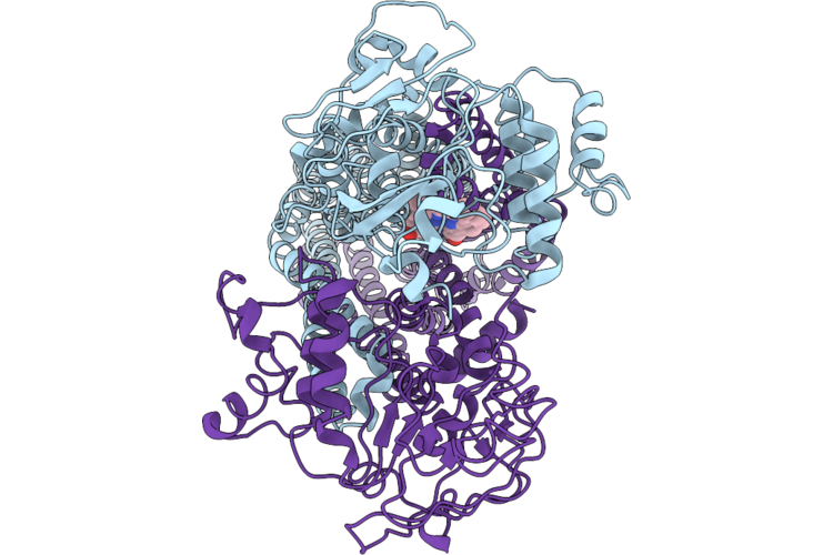 |
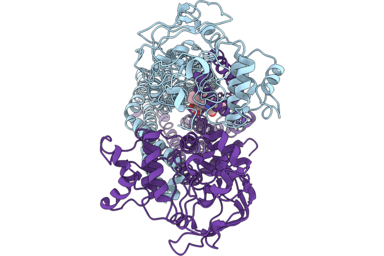
Cyddc In Nanodisc With Heme-Bound I
Organism: Escherichia coli k-12
Method: ELECTRON MICROSCOPY Release Date: 2026-06-24 Classification: TRANSPORT PROTEIN Ligands: HEM |
Organism: Escherichia coli k-12
Method: ELECTRON MICROSCOPY
Release Date: 2026-06-24
Ligands: HEM
 |
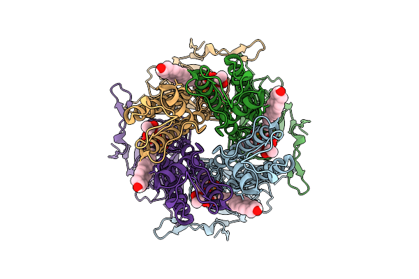
Cyddc In Nanodisc With Heme-Bound Ii
Organism: Escherichia coli k-12
Method: ELECTRON MICROSCOPY Release Date: 2026-06-24 Classification: TRANSPORT PROTEIN Ligands: HEM |
Organism: Escherichia coli k-12
Method: ELECTRON MICROSCOPY
Release Date: 2026-06-24
Ligands: HEM
 |
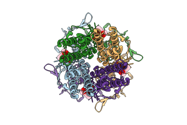
Cryo-Em Structure Of The Human Inward-Rectifier Potassium 7.1 Channel (Kir7.1) Extended State
Organism: Homo sapiens
Method: ELECTRON MICROSCOPY Resolution:3.30 Å Release Date: 2026-02-25 Classification: MEMBRANE PROTEIN Ligands: PIO, CLR |
Organism: Homo sapiens
Method: ELECTRON MICROSCOPY
Release Date: 2026-02-25
Ligands: PIO, CLR
 |
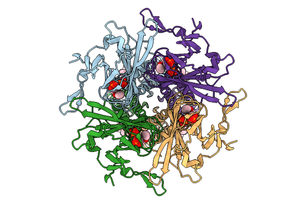
Cryo-Em Structure Of The Human Inward-Rectifier Potassium 7.1 Channel (Kir7.1) Docked State
Organism: Homo sapiens
Method: ELECTRON MICROSCOPY Resolution:3.90 Å Release Date: 2026-02-25 Classification: MEMBRANE PROTEIN Ligands: PIO |
Organism: Homo sapiens
Method: ELECTRON MICROSCOPY
Release Date: 2026-02-25
Ligands: PIO
 |
Cryo-Em Structure Of The Human Inward-Rectifier Potassium 7.1 Channel (Kir7.1) With Enantiomer Of 17-Hydroxyprogesterone Caproate
Organism: Homo sapiens
Method: ELECTRON MICROSCOPY Resolution:4.00 Å Release Date: 2026-02-25 Classification: MEMBRANE PROTEIN Ligands: PIO, A1CJG |
Organism: Homo sapiens
Method: ELECTRON MICROSCOPY
Release Date: 2026-02-25
Ligands: PIO, A1CJG
 |
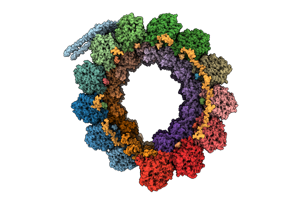
Cryo-Em Structure Of Conoid Fiber From Toxoplasma Gondii (24-Nm Repeat)
Organism: Toxoplasma gondii
Method: ELECTRON MICROSCOPY Release Date: 2025-12-03 Classification: STRUCTURAL PROTEIN Ligands: GTP, MG, GDP |
Organism: Toxoplasma gondii
Method: ELECTRON MICROSCOPY
Release Date: 2025-12-03
Ligands: GTP, MG, GDP
 |
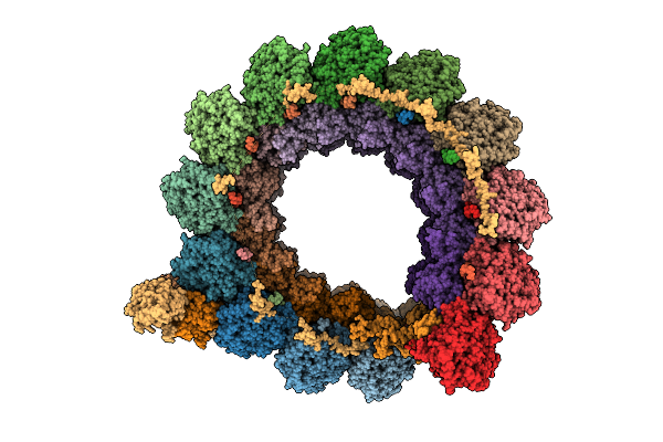
Cryo-Em Structure Of Intraconoidal Microtubule 2 (Icmt2) From Toxoplasma Gondii (8-Nm Repeat)
Organism: Toxoplasma gondii
Method: ELECTRON MICROSCOPY Release Date: 2025-12-03 Classification: STRUCTURAL PROTEIN Ligands: GTP, MG, GDP |
Organism: Toxoplasma gondii
Method: ELECTRON MICROSCOPY
Release Date: 2025-12-03
Ligands: GTP, MG, GDP
 |
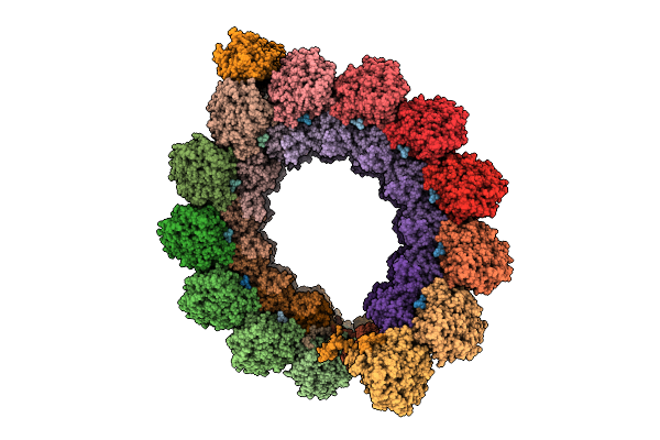
Cryo-Em Structure Of Intraconoidal Microtubule 1 (Icmt1) From Toxoplasma Gondii (8-Nm Repeat)
Organism: Toxoplasma gondii
Method: ELECTRON MICROSCOPY Release Date: 2025-12-03 Classification: STRUCTURAL PROTEIN Ligands: GTP, MG, GDP |
Organism: Toxoplasma gondii
Method: ELECTRON MICROSCOPY
Release Date: 2025-12-03
Ligands: GTP, MG, GDP
 |
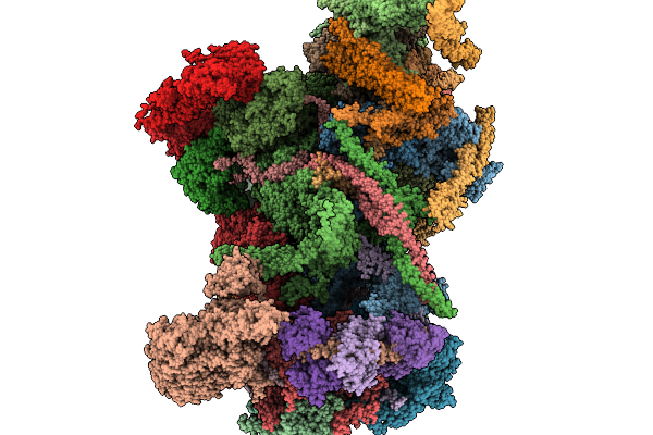
Cryo-Em Structure Of The Apical Region Of Subpellicular Microtubule (Spmt) From Toxoplasma Gondii (8-Nm Repeat)
Organism: Toxoplasma gondii
Method: ELECTRON MICROSCOPY Release Date: 2025-12-03 Classification: STRUCTURAL PROTEIN Ligands: GDP, GTP, MG |
Organism: Toxoplasma gondii
Method: ELECTRON MICROSCOPY
Release Date: 2025-12-03
Ligands: GDP, GTP, MG
 |
Cryo-Em Structure Of Pre-Conoid Ring 2 (Pcr P2 Ring)From Toxoplasma Gondii
Organism: Toxoplasma gondii
Method: ELECTRON MICROSCOPY Release Date: 2025-11-19 Classification: STRUCTURAL PROTEIN |
Organism: Toxoplasma gondii
Method: ELECTRON MICROSCOPY
Release Date: 2025-11-19
 |
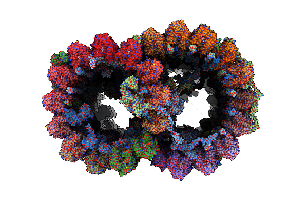
96-Nm Repeat Of Axonemal Doublet Microtubules From Bovine Sperm
Organism: Bos taurus
Method: ELECTRON MICROSCOPY Release Date: 2025-03-26 Classification: STRUCTURAL PROTEIN Ligands: GTP, MG, GDP |
Organism: Bos taurus
Method: ELECTRON MICROSCOPY
Release Date: 2025-03-26
Ligands: GTP, MG, GDP
 |
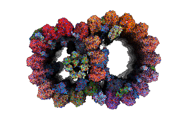
48-Nm Repeat Of The Leishmania Tarentolae Doublet Microtubule
Organism: Leishmania tarentolae
Method: ELECTRON MICROSCOPY Release Date: 2025-03-12 Classification: STRUCTURAL PROTEIN Ligands: GDP, MG, GTP, ZN, CA, ATP |
Organism: Leishmania tarentolae
Method: ELECTRON MICROSCOPY
Release Date: 2025-03-12
Ligands: GDP, MG, GTP, ZN, CA, ATP
 |
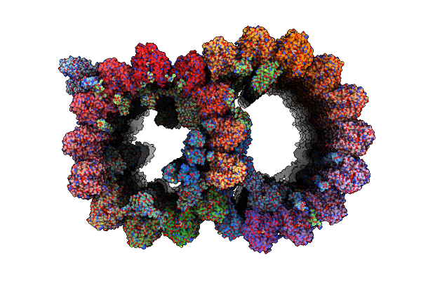
Atomic Model Of Bovine Fallopian Tube Cilia Doublet Microtubule (48-Nm Periodicity)
Organism: Bos taurus
Method: ELECTRON MICROSCOPY Release Date: 2024-10-30 Classification: STRUCTURAL PROTEIN Ligands: GDP, MG, GTP |
Organism: Bos taurus
Method: ELECTRON MICROSCOPY
Release Date: 2024-10-30
Ligands: GDP, MG, GTP
 |
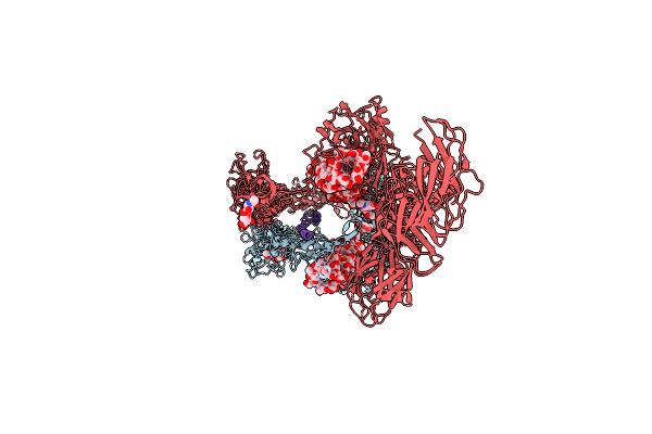
Atomic Model Of Porcine Brain Ventricles Cilia Doublet Microtubule (48-Nm Periodicity)
Organism: Sus scrofa
Method: ELECTRON MICROSCOPY Release Date: 2024-10-23 Classification: STRUCTURAL PROTEIN Ligands: GTP, GDP |
Organism: Sus scrofa
Method: ELECTRON MICROSCOPY
Release Date: 2024-10-23
Ligands: GTP, GDP
 |
Chlamydomonas Reinhardtii Mastigoneme Filament
Organism: Chlamydomonas reinhardtii
Method: ELECTRON MICROSCOPY Release Date: 2024-04-10 Classification: STRUCTURAL PROTEIN Ligands: A1AIO, GLA |
Organism: Chlamydomonas reinhardtii
Method: ELECTRON MICROSCOPY
Release Date: 2024-04-10
Ligands: A1AIO, GLA
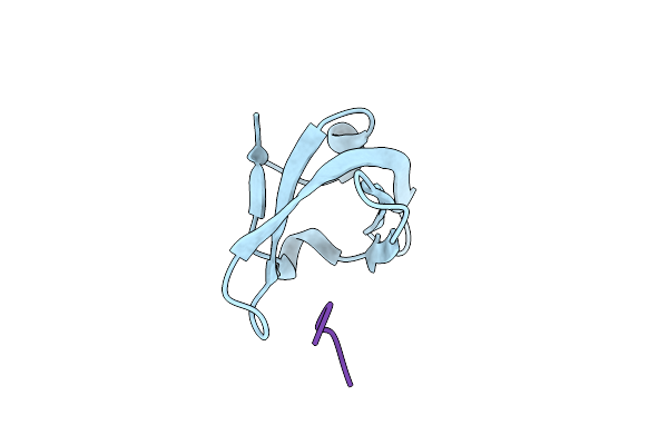 |
Crystal Structure Of Arabidopsis Rdm15 Tudor Domain In Complex With An H3K4Me1 Peptide
Organism: Arabidopsis thaliana
Method: X-RAY DIFFRACTION Resolution:1.71 Å Release Date: 2021-04-07 Classification: GENE REGULATION |
Organism: Arabidopsis thaliana
Method: X-RAY DIFFRACTION
Release Date: 2021-04-07
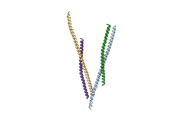 |
Crystal Structure Of Huntingtin-Interacting Protein 1 (Hip1) Coiled-Coil Domain With A Basic Surface Suitable For Hip-Protein Interactor (Hippi)
Organism: Homo sapiens
Method: X-RAY DIFFRACTION Resolution:2.80 Å Release Date: 2008-07-08 Classification: Actin Binding |
Organism: Homo sapiens
Method: X-RAY DIFFRACTION
Release Date: 2008-07-08