

Search Count: 223
 |
Crystal Structure Of Umpk From S. Aureus In Complex With Atp And Gtp
Organism: Staphylococcus aureus subsp. aureus nctc 8325
Method: X-RAY DIFFRACTION Resolution:2.88 Å Release Date: 2026-05-27 Classification: TRANSFERASE Ligands: GTP, GOL, ADP, ATP |
Organism: Staphylococcus aureus subsp. aureus nctc 8325
Method: X-RAY DIFFRACTION
Release Date: 2026-05-27
Ligands: GTP, GOL, ADP, ATP
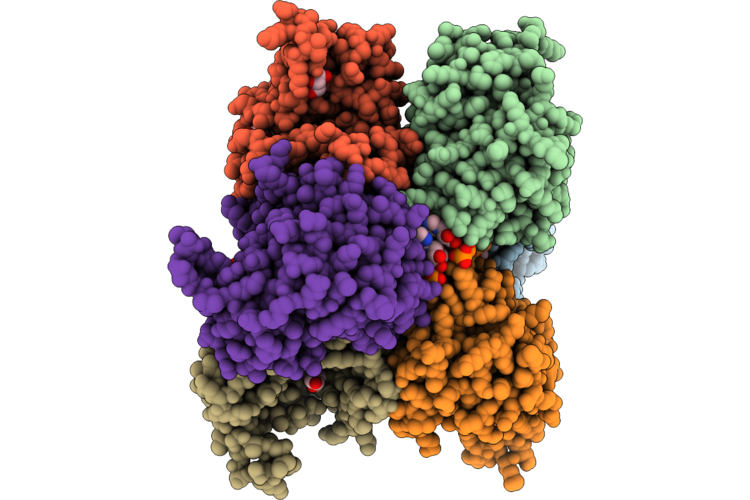 |
Crystal Structure Of Umpk From S. Aureus In Complex With Atp
Organism: Staphylococcus aureus subsp. aureus nctc 8325
Method: X-RAY DIFFRACTION Resolution:2.82 Å Release Date: 2026-05-27 Classification: TRANSFERASE Ligands: GOL, ATP |
Organism: Staphylococcus aureus subsp. aureus nctc 8325
Method: X-RAY DIFFRACTION
Release Date: 2026-05-27
Ligands: GOL, ATP
 |
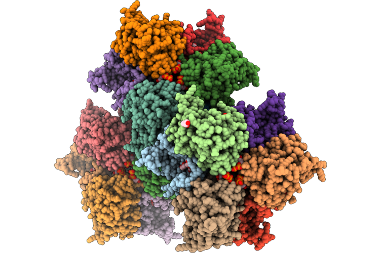
Crystal Structure Of Umpk From S. Aureus In Complex With Utp
Organism: Staphylococcus aureus subsp. aureus nctc 8325
Method: X-RAY DIFFRACTION Resolution:2.30 Å Release Date: 2026-05-27 Classification: TRANSFERASE Ligands: GOL, UTP, UDP, PO4 |
Organism: Staphylococcus aureus subsp. aureus nctc 8325
Method: X-RAY DIFFRACTION
Release Date: 2026-05-27
Ligands: GOL, UTP, UDP, PO4
 |
Crystal Structure Of Umpk From S. Aureus In Complex With Gtp
Organism: Staphylococcus aureus subsp. aureus nctc 8325
Method: X-RAY DIFFRACTION Resolution:2.88 Å Release Date: 2026-05-13 Classification: TRANSFERASE Ligands: GTP, GOL |
Organism: Staphylococcus aureus subsp. aureus nctc 8325
Method: X-RAY DIFFRACTION
Release Date: 2026-05-13
Ligands: GTP, GOL
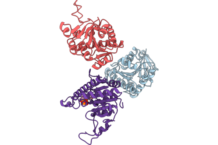 |
Crystal Structure Of Umpk From S. Aureus In Complex With U5P
Organism: Staphylococcus aureus subsp. aureus nctc 8325
Method: X-RAY DIFFRACTION Resolution:3.26 Å Release Date: 2026-05-13 Classification: TRANSFERASE Ligands: U5P |
Organism: Staphylococcus aureus subsp. aureus nctc 8325
Method: X-RAY DIFFRACTION
Release Date: 2026-05-13
Ligands: U5P
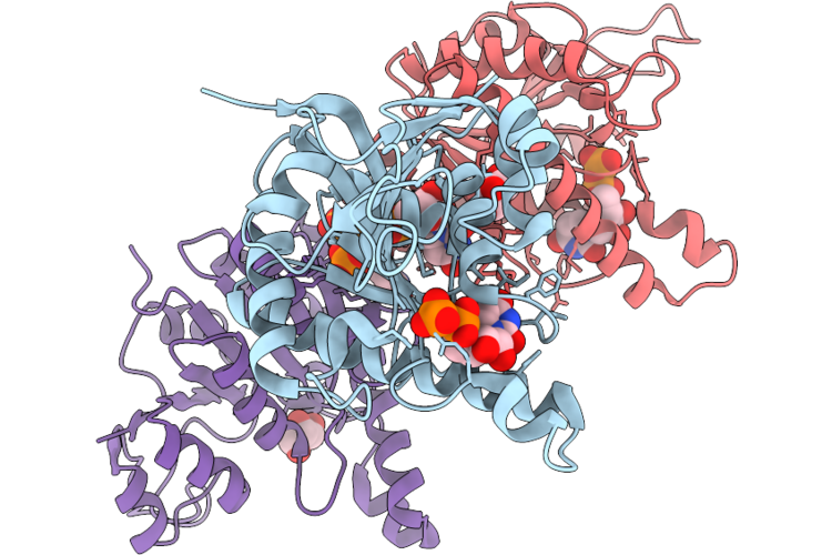 |
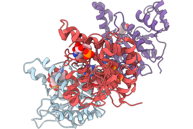
Crystal Structure Of Umpk From S. Aureus In Complex With Udp
Organism: Staphylococcus aureus subsp. aureus nctc 8325
Method: X-RAY DIFFRACTION Resolution:2.75 Å Release Date: 2026-05-13 Classification: TRANSFERASE Ligands: UDP, GOL, PO4 |
Organism: Staphylococcus aureus subsp. aureus nctc 8325
Method: X-RAY DIFFRACTION
Release Date: 2026-05-13
Ligands: UDP, GOL, PO4
 |
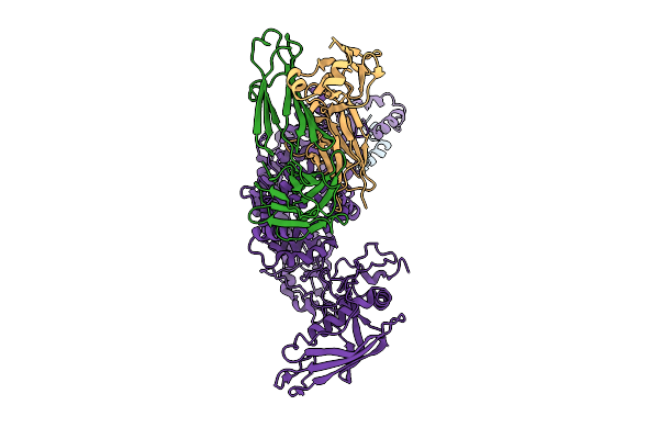
Crystal Structure Of Umpk From S. Aureus In Complex With Atp And U5P
Organism: Staphylococcus aureus subsp. aureus nctc 8325
Method: X-RAY DIFFRACTION Resolution:2.57 Å Release Date: 2026-05-13 Classification: TRANSFERASE Ligands: U5P, ATP, PO4, GOL |
Organism: Staphylococcus aureus subsp. aureus nctc 8325
Method: X-RAY DIFFRACTION
Release Date: 2026-05-13
Ligands: U5P, ATP, PO4, GOL
 |
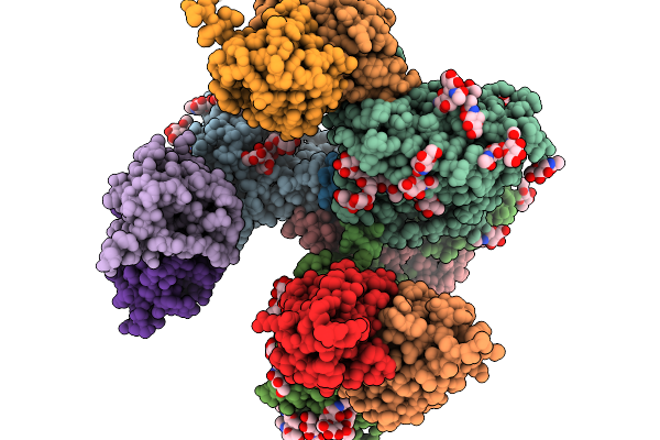
P116 From Mycoplasma Pneumoniae In Complex With Mild Growth Suppressor Monoclonal Antibody
Organism: Mus musculus, Mycoplasmoides pneumoniae m129
Method: ELECTRON MICROSCOPY Release Date: 2025-12-24 Classification: PROTEIN TRANSPORT |
Organism: Mus musculus, Mycoplasmoides pneumoniae m129
Method: ELECTRON MICROSCOPY
Release Date: 2025-12-24
 |
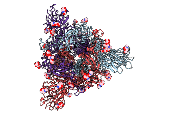
Structure Of Cd4 Mimetic Cjf-Iii-288 In Complex With Bg505 Sosip.664 Hiv-1Env Trimer And 17B Fab
Organism: Human immunodeficiency virus 1, Homo sapiens
Method: ELECTRON MICROSCOPY Release Date: 2025-11-19 Classification: VIRAL PROTEIN/IMMUNE SYSTEM Ligands: NAG, Y26 |
Organism: Human immunodeficiency virus 1, Homo sapiens
Method: ELECTRON MICROSCOPY
Release Date: 2025-11-19
Ligands: NAG, Y26
 |
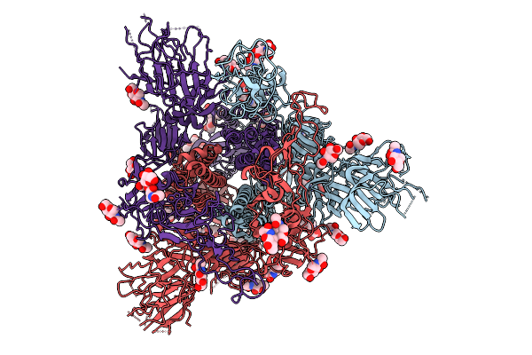
Sars-Cov-2-6P-Mut7 S Protein-Dy-Iii-281 Complex Closed Conformation
Organism: Severe acute respiratory syndrome coronavirus 2
Method: ELECTRON MICROSCOPY Resolution:3.56 Å Release Date: 2025-10-01 Classification: VIRAL PROTEIN Ligands: NAG |
Organism: Severe acute respiratory syndrome coronavirus 2
Method: ELECTRON MICROSCOPY
Release Date: 2025-10-01
Ligands: NAG
 |
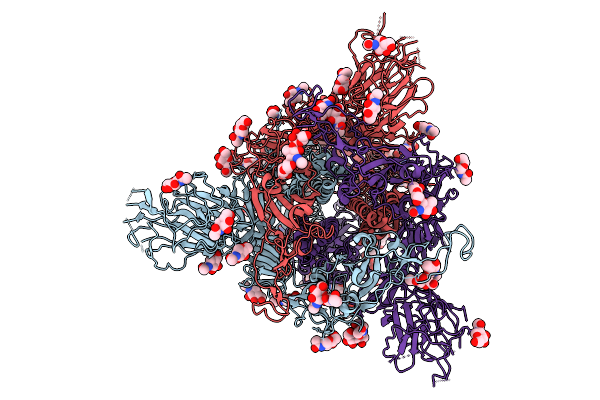
Sars-Cov-2-6P-Mut7 S Protein-Dy-Iii-281 Complex 1 Rbd Up Conformation
Organism: Severe acute respiratory syndrome coronavirus 2
Method: ELECTRON MICROSCOPY Resolution:3.30 Å Release Date: 2025-10-01 Classification: VIRAL PROTEIN Ligands: NAG |
Organism: Severe acute respiratory syndrome coronavirus 2
Method: ELECTRON MICROSCOPY
Release Date: 2025-10-01
Ligands: NAG
 |
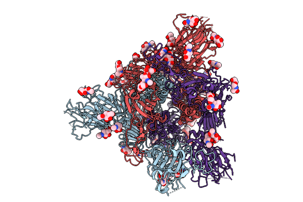
Apo Sars-Cov-2-6P-Mut7 S Protein Closed Conformation
Organism: Severe acute respiratory syndrome coronavirus 2
Method: ELECTRON MICROSCOPY Resolution:3.14 Å Release Date: 2025-10-01 Classification: VIRAL PROTEIN Ligands: NAG |
Organism: Severe acute respiratory syndrome coronavirus 2
Method: ELECTRON MICROSCOPY
Release Date: 2025-10-01
Ligands: NAG
 |
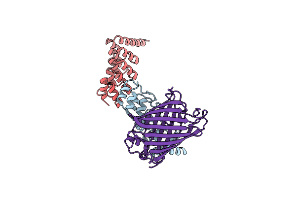
Apo Sars-Cov-2-6P-Mut7 S Protein 1 Rbd Up Conformation
Organism: Severe acute respiratory syndrome coronavirus 2
Method: ELECTRON MICROSCOPY Resolution:3.09 Å Release Date: 2025-10-01 Classification: VIRAL PROTEIN Ligands: NAG |
Organism: Severe acute respiratory syndrome coronavirus 2
Method: ELECTRON MICROSCOPY
Release Date: 2025-10-01
Ligands: NAG
 |
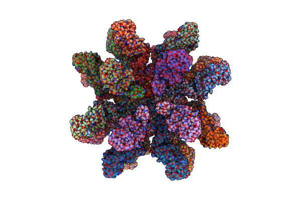
Multibody Refinement Of Dimeric Darpin And Its Bound Gfp On A Symmetric Scaffold
Organism: Synthetic construct, Aequorea victoria
Method: ELECTRON MICROSCOPY Release Date: 2025-06-04 Classification: BIOSYNTHETIC PROTEIN |
Organism: Synthetic construct, Aequorea victoria
Method: ELECTRON MICROSCOPY
Release Date: 2025-06-04
 |
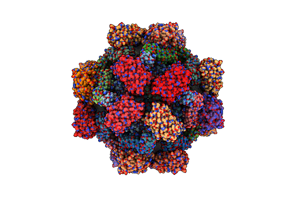
24-Mer Darpin-Apoferritin Scaffold In Complex With The Maltose Binding Protein
Organism: Synthetic construct, Homo sapiens, Escherichia coli
Method: ELECTRON MICROSCOPY Release Date: 2025-06-04 Classification: BIOSYNTHETIC PROTEIN |
Organism: Synthetic construct, Homo sapiens, Escherichia coli
Method: ELECTRON MICROSCOPY
Release Date: 2025-06-04
 |
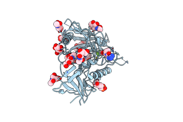
Gfp Bound To 24-Mer Darpin-Apoferritin Model 6C
Organism: Homo sapiens, Aequorea victoria
Method: ELECTRON MICROSCOPY Release Date: 2025-06-04 Classification: METAL BINDING PROTEIN/LUMINESCENT PROTEIN |
Organism: Homo sapiens, Aequorea victoria
Method: ELECTRON MICROSCOPY
Release Date: 2025-06-04
 |
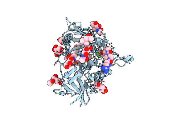
Crystal Structure Of Hiv-1 Lm/Hs Clade A/E Crf01 Gp120 Core In Complex With Dl-Iii-14
Organism: Human immunodeficiency virus 1
Method: X-RAY DIFFRACTION Resolution:2.50 Å Release Date: 2025-04-02 Classification: VIRAL PROTEIN Ligands: NAG, EPE, A1AS5 |
Organism: Human immunodeficiency virus 1
Method: X-RAY DIFFRACTION
Release Date: 2025-04-02
Ligands: NAG, EPE, A1AS5
 |
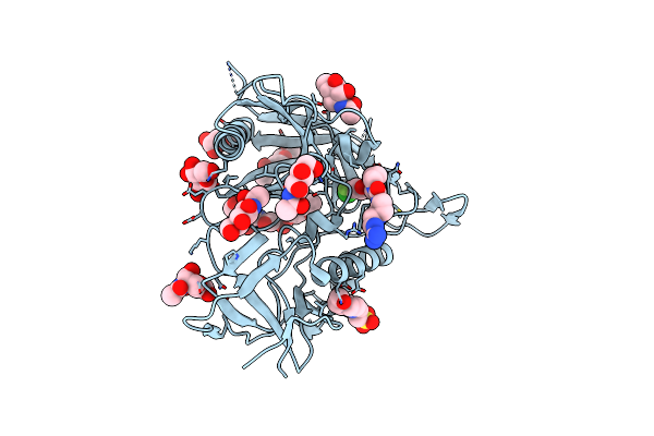
Crystal Structure Of Hiv-1 Lm/Hs Clade A/E Crf01 Gp120 Core In Complex With Hz-Iv-188
Organism: Human immunodeficiency virus 1
Method: X-RAY DIFFRACTION Resolution:1.85 Å Release Date: 2025-04-02 Classification: VIRAL PROTEIN Ligands: NAG, EPE, A1ATK, MPD |
Organism: Human immunodeficiency virus 1
Method: X-RAY DIFFRACTION
Release Date: 2025-04-02
Ligands: NAG, EPE, A1ATK, MPD
 |
Crystal Structure Of Hiv-1 Lm/Hs Clade A/E Crf01 Gp120 Core In Complex With Hz-Iv-236
Organism: Human immunodeficiency virus 1
Method: X-RAY DIFFRACTION Resolution:2.69 Å Release Date: 2025-04-02 Classification: VIRAL PROTEIN Ligands: NAG, EPE, A1AS8 |
Organism: Human immunodeficiency virus 1
Method: X-RAY DIFFRACTION
Release Date: 2025-04-02
Ligands: NAG, EPE, A1AS8
 |
Crystal Structure Of Hiv-1 Lm/Hs Clade A/E Crf01 Gp120 Core In Complex With Hz-Iv-242
Organism: Human immunodeficiency virus 1
Method: X-RAY DIFFRACTION Resolution:1.79 Å Release Date: 2025-04-02 Classification: VIRAL PROTEIN Ligands: NAG, EPE, A1ATA |
Organism: Human immunodeficiency virus 1
Method: X-RAY DIFFRACTION
Release Date: 2025-04-02
Ligands: NAG, EPE, A1ATA