

Search Count: 139
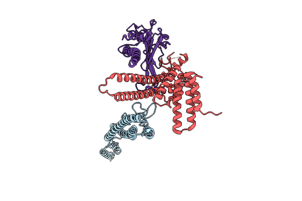 |
Straight And Symmetrical Filament Of The Spirochete Periplasmic Flagella Of Leptospira Biflexa
Organism: Leptospira biflexa
Method: ELECTRON MICROSCOPY Resolution:2.30 Å Release Date: 2025-12-24 Classification: MOTOR PROTEIN |
Organism: Leptospira biflexa
Method: ELECTRON MICROSCOPY
Release Date: 2025-12-24
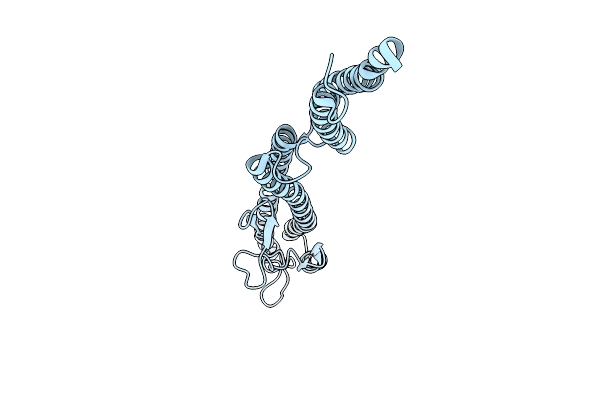 |
Core Filament Of The Spirochete Periplasmic Flagella Of Leptospira Biflexa
Organism: Leptospira biflexa
Method: ELECTRON MICROSCOPY Resolution:2.40 Å Release Date: 2025-12-24 Classification: MOTOR PROTEIN |
Organism: Leptospira biflexa
Method: ELECTRON MICROSCOPY
Release Date: 2025-12-24
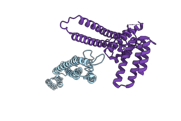 |
Straight And Symmetrical Filament Of The Spirochete Periplasmic Flagella Of Leptospira Biflexa Deleted Fcpb Strain
Organism: Leptospira biflexa
Method: ELECTRON MICROSCOPY Resolution:2.40 Å Release Date: 2025-12-24 Classification: MOTOR PROTEIN |
Organism: Leptospira biflexa
Method: ELECTRON MICROSCOPY
Release Date: 2025-12-24
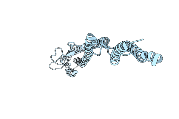 |
Core Filament Of The Spirochete Periplasmic Flagella Of Leptospira Biflexa Deleted Fcpb Strain
Organism: Leptospira biflexa
Method: ELECTRON MICROSCOPY Resolution:2.40 Å Release Date: 2025-12-24 Classification: MOTOR PROTEIN |
Organism: Leptospira biflexa
Method: ELECTRON MICROSCOPY
Release Date: 2025-12-24
 |
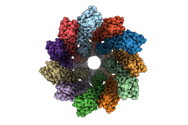
Core Filament Of The Spirochete Periplasmic Flagella Of Leptospira Biflexa Wild Type
Organism: Leptospira biflexa
Method: ELECTRON MICROSCOPY Release Date: 2025-12-24 Classification: MOTOR PROTEIN |
Organism: Leptospira biflexa
Method: ELECTRON MICROSCOPY
Release Date: 2025-12-24
 |
Core Filament Of The Spirochete Periplasmic Flagella Of Leptospira Biflexa From The Flaa2-Complemented Stain
Organism: Leptospira biflexa
Method: ELECTRON MICROSCOPY Release Date: 2025-12-24 Classification: MOTOR PROTEIN |
Organism: Leptospira biflexa
Method: ELECTRON MICROSCOPY
Release Date: 2025-12-24
 |
Core Filament Of The Spirochete Periplasmic Flagella Of Leptospira Biflexa From The Deleted Fcpb_Cl13 Strain
Organism: Leptospira biflexa
Method: ELECTRON MICROSCOPY Resolution:3.20 Å Release Date: 2025-12-24 Classification: MOTOR PROTEIN |
Organism: Leptospira biflexa
Method: ELECTRON MICROSCOPY
Release Date: 2025-12-24
 |
Sheathed Filament Of The Spirochete Periplasmic Flagella Of Leptospira Biflexa From The Flaa2-Complemented Stain
Organism: Leptospira biflexa
Method: ELECTRON MICROSCOPY Release Date: 2025-12-24 Classification: MOTOR PROTEIN |
Organism: Leptospira biflexa
Method: ELECTRON MICROSCOPY
Release Date: 2025-12-24
 |
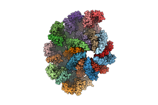
Sheathed Filament Of The Spirochete Periplasmic Flagella Of Leptospira Biflexa From The Deleted Fcpb_Cl13 Strain
Organism: Leptospira biflexa
Method: ELECTRON MICROSCOPY Release Date: 2025-12-24 Classification: MOTOR PROTEIN |
Organism: Leptospira biflexa
Method: ELECTRON MICROSCOPY
Release Date: 2025-12-24
 |
Sheathed Filament Of The Spirochete Periplasmic Flagella Of Leptospira Biflexa Wild Type
Organism: Leptospira biflexa
Method: ELECTRON MICROSCOPY Release Date: 2025-12-24 Classification: MOTOR PROTEIN |
Organism: Leptospira biflexa
Method: ELECTRON MICROSCOPY
Release Date: 2025-12-24
 |
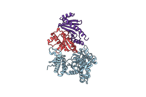
Crystal Structure Of The Type Iii Secretion Chaperone Veca From Vibrio Parahaemolyticus
Organism: Vibrio parahaemolyticus
Method: X-RAY DIFFRACTION Resolution:2.20 Å Release Date: 2025-06-25 Classification: CHAPERONE Ligands: PO4, TBR |
Organism: Vibrio parahaemolyticus
Method: X-RAY DIFFRACTION
Release Date: 2025-06-25
Ligands: PO4, TBR
 |
Crystal Structure Of The Virulence Effector Vepa In Complex With Its Secretion Chaperone Veca
Organism: Vibrio parahaemolyticus
Method: X-RAY DIFFRACTION Resolution:2.49 Å Release Date: 2025-06-25 Classification: TOXIN |
Organism: Vibrio parahaemolyticus
Method: X-RAY DIFFRACTION
Release Date: 2025-06-25
 |
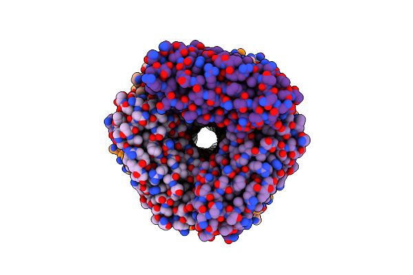
Cryo-Em Structure Of The Type Ivb Pilus From Enterotoxigenic Escherichia Coli
Organism: Escherichia coli
Method: ELECTRON MICROSCOPY Release Date: 2025-05-28 Classification: CELL ADHESION |
Organism: Escherichia coli
Method: ELECTRON MICROSCOPY
Release Date: 2025-05-28
 |
Cryo-Em Structure Of The Type I Pilus From Enterotoxigenic Escherichia Coli
Organism: Escherichia coli
Method: ELECTRON MICROSCOPY Release Date: 2025-05-28 Classification: CELL ADHESION |
Organism: Escherichia coli
Method: ELECTRON MICROSCOPY
Release Date: 2025-05-28
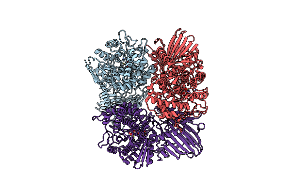 |
Crystal Structure Of Gh65 Alpha-1,2-Glucosidase From Flavobacterium Johnsoniae In Complex With 1-Deoxynojirimycin
Organism: Flavobacterium johnsoniae uw101
Method: X-RAY DIFFRACTION Resolution:1.90 Å Release Date: 2025-03-19 Classification: HYDROLASE Ligands: NOJ, EDO |
Organism: Flavobacterium johnsoniae uw101
Method: X-RAY DIFFRACTION
Release Date: 2025-03-19
Ligands: NOJ, EDO
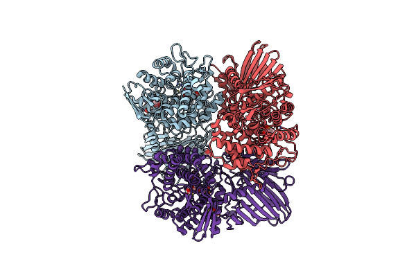 |
Crystal Structure Of Gh65 Alpha-1,2-Glucosidase From Flavobacterium Johnsoniae In Complex With Castanospermine
Organism: Flavobacterium johnsoniae uw101
Method: X-RAY DIFFRACTION Resolution:1.60 Å Release Date: 2025-03-19 Classification: HYDROLASE Ligands: CTS, EDO |
Organism: Flavobacterium johnsoniae uw101
Method: X-RAY DIFFRACTION
Release Date: 2025-03-19
Ligands: CTS, EDO
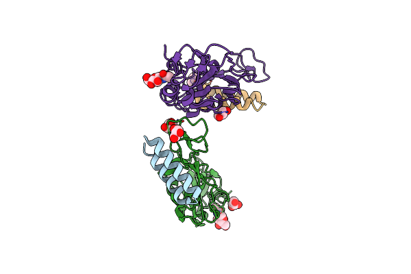 |
Crystal Structure Of The Receptor Binding Domain Of Sars-Cov-2 Spike Protein In Complex With Ce9
Organism: Severe acute respiratory syndrome coronavirus 2, Synthetic construct
Method: X-RAY DIFFRACTION Resolution:2.30 Å Release Date: 2025-01-15 Classification: VIRAL PROTEIN/INHIBITOR Ligands: NAG, GOL |
Organism: Severe acute respiratory syndrome coronavirus 2, Synthetic construct
Method: X-RAY DIFFRACTION
Release Date: 2025-01-15
Ligands: NAG, GOL
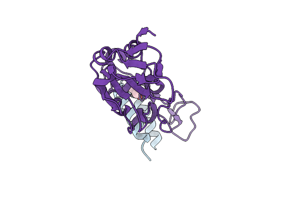 |
Crystal Structure Of The Receptor Binding Domain Of Sars-Cov-2 Omicron Ba.2 Variant Spike Protein In Complex With Ce149
Organism: Severe acute respiratory syndrome coronavirus 2, Synthetic construct
Method: X-RAY DIFFRACTION Resolution:1.90 Å Release Date: 2025-01-15 Classification: VIRAL PROTEIN/INHIBITOR Ligands: GOL |
Organism: Severe acute respiratory syndrome coronavirus 2, Synthetic construct
Method: X-RAY DIFFRACTION
Release Date: 2025-01-15
Ligands: GOL
 |
Crystal Structure Of The Receptor Binding Domain Of Sars-Cov-2 Alpha Variant Spike Protein In Complex With Ce59
Organism: Severe acute respiratory syndrome coronavirus 2, Synthetic construct
Method: X-RAY DIFFRACTION Resolution:2.30 Å Release Date: 2025-01-15 Classification: VIRAL PROTEIN/INHIBITOR Ligands: NAG |
Organism: Severe acute respiratory syndrome coronavirus 2, Synthetic construct
Method: X-RAY DIFFRACTION
Release Date: 2025-01-15
Ligands: NAG
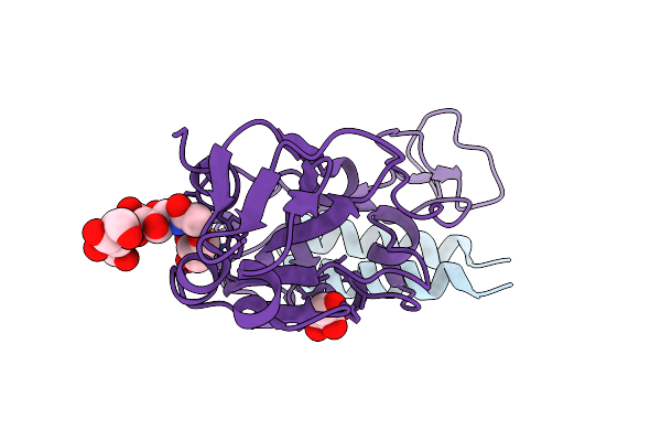 |
Crystal Structure Of The Receptor Binding Domain Of Sars-Cov-2 Delta Variant Spike Protein In Complex With Ce59
Organism: Severe acute respiratory syndrome coronavirus 2, Synthetic construct
Method: X-RAY DIFFRACTION Resolution:1.90 Å Release Date: 2025-01-15 Classification: VIRAL PROTEIN/INHIBITOR Ligands: GOL |
Organism: Severe acute respiratory syndrome coronavirus 2, Synthetic construct
Method: X-RAY DIFFRACTION
Release Date: 2025-01-15
Ligands: GOL