

Search Count: 16
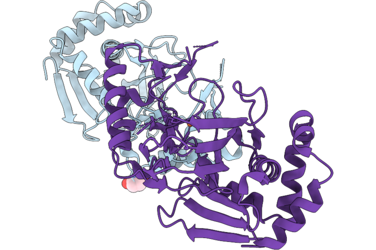 |
Glutarate L-2-Hydroxylase K270C Mutant-5'-Mal-C2-Aaattt Dna Conjugate In P 4 21 2 Space Group
Organism: Escherichia coli
Method: X-RAY DIFFRACTION Resolution:1.65 Å Release Date: 2026-07-29 Classification: METAL BINDING PROTEIN Ligands: NEN, FE2 |
Organism: Escherichia coli
Method: X-RAY DIFFRACTION
Release Date: 2026-07-29
Ligands: NEN, FE2
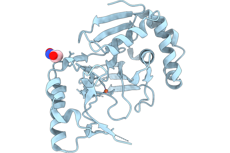 |
Glutarate L-2-Hydroxylase K270C Mutant-5'-Mal-C2-Aaattt Dna Conjugate In I422 Space Group
Organism: Escherichia coli
Method: X-RAY DIFFRACTION Resolution:1.64 Å Release Date: 2026-07-29 Classification: METAL BINDING PROTEIN Ligands: NEN, FE2 |
Organism: Escherichia coli
Method: X-RAY DIFFRACTION
Release Date: 2026-07-29
Ligands: NEN, FE2
 |
Human 20S Proteasome In Complex With Peptide Activator Peptide Blm42
Organism: Synthetic construct, Homo sapiens
Method: X-RAY DIFFRACTION Resolution:2.14 Å Release Date: 2023-12-27 Classification: HYDROLASE Ligands: CL, MG, 1PE, NEN, K |
Organism: Synthetic construct, Homo sapiens
Method: X-RAY DIFFRACTION
Release Date: 2023-12-27
Ligands: CL, MG, 1PE, NEN, K
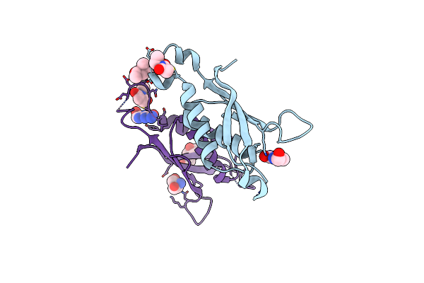 |
Dihydroneopterin Aldolase (Dhna) From Yersinia Pestis With Alkylated Cys50 Co-Crystallized With 7,8-Dihydroneopterin
Organism: Yersinia pestis
Method: X-RAY DIFFRACTION Resolution:2.65 Å Release Date: 2022-12-28 Classification: BIOSYNTHETIC PROTEIN Ligands: NEN, PH2, MPD, TRS |
Organism: Yersinia pestis
Method: X-RAY DIFFRACTION
Release Date: 2022-12-28
Ligands: NEN, PH2, MPD, TRS
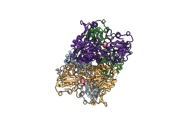 |
Structure Of The Short Lor Domain Of Human Aass Bound To N-Ethylsuccinimide
Organism: Homo sapiens
Method: X-RAY DIFFRACTION Resolution:2.45 Å Release Date: 2022-10-05 Classification: OXIDOREDUCTASE Ligands: NEN |
Organism: Homo sapiens
Method: X-RAY DIFFRACTION
Release Date: 2022-10-05
Ligands: NEN
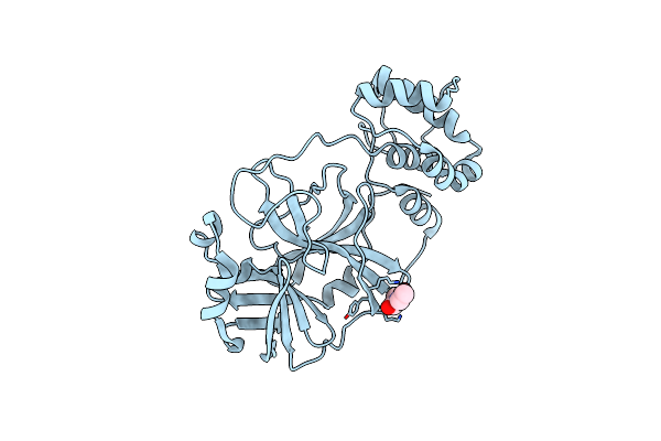 |
Room Temperature X-Ray Crystallography Reveals Catalytic Cysteine In The Sars-Cov-2 3Cl Mpro Is Highly Reactive: Insights For Enzyme Mechanism And Drug Design
Organism: Severe acute respiratory syndrome coronavirus 2
Method: X-RAY DIFFRACTION Resolution:1.80 Å Release Date: 2020-06-17 Classification: VIRAL PROTEIN, HYDROLASE Ligands: NEN |
Organism: Severe acute respiratory syndrome coronavirus 2
Method: X-RAY DIFFRACTION
Release Date: 2020-06-17
Ligands: NEN
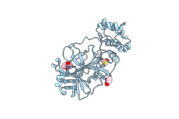 |
Room Temperature X-Ray Crystallography Reveals Catalytic Cysteine In The Sars-Cov-2 3Cl Mpro Is Highly Reactive: Insights For Enzyme Mechanism And Drug Design
Organism: Severe acute respiratory syndrome coronavirus 2
Method: X-RAY DIFFRACTION Resolution:2.10 Å Release Date: 2020-06-17 Classification: VIRAL PROTEIN, HYDROLASE Ligands: NEN, DMS |
Organism: Severe acute respiratory syndrome coronavirus 2
Method: X-RAY DIFFRACTION
Release Date: 2020-06-17
Ligands: NEN, DMS
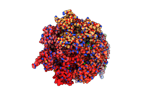 |
Human 20S Proteasome Complex With Z-Lly-Ketoaldehyde At 2.1 Angstrom
Organism: Homo sapiens
Method: X-RAY DIFFRACTION Resolution:2.07 Å Release Date: 2016-08-17 Classification: HYDROLASE Ligands: CL, NEN, K, MG, 1PE |
Organism: Homo sapiens
Method: X-RAY DIFFRACTION
Release Date: 2016-08-17
Ligands: CL, NEN, K, MG, 1PE
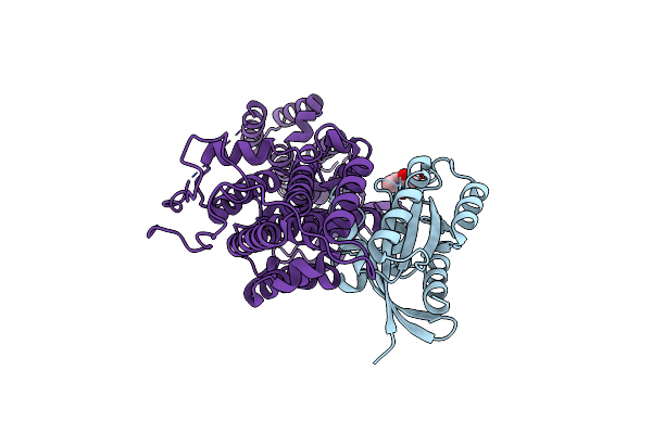 |
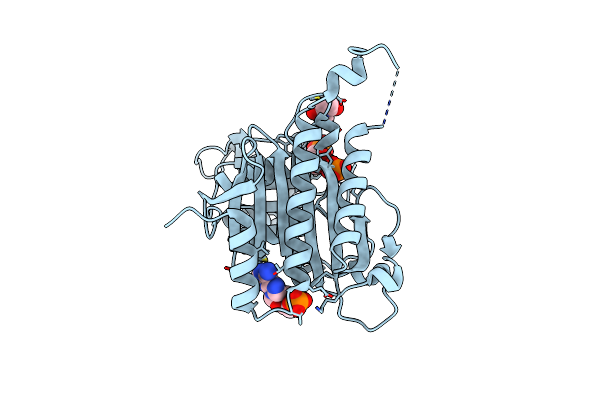
The Crystal Structure Of H-Ras And Sos In Complex With Ligands
Organism: Homo sapiens
Method: X-RAY DIFFRACTION Resolution:2.17 Å Release Date: 2015-03-04 Classification: SIGNALING PROTEIN Ligands: NEN |
Organism: Homo sapiens
Method: X-RAY DIFFRACTION
Release Date: 2015-03-04
Ligands: NEN
 |
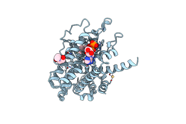
Crystal Structure Of Amp Complexes Of Nem Modified Porcine Liver Fructose-1,6-Bisphosphatase
Organism: Sus scrofa
Method: X-RAY DIFFRACTION Resolution:2.50 Å Release Date: 2013-09-18 Classification: HYDROLASE Ligands: F6P, MG, PO4, AMP, NEN |
Organism: Sus scrofa
Method: X-RAY DIFFRACTION
Release Date: 2013-09-18
Ligands: F6P, MG, PO4, AMP, NEN
 |
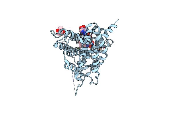
Structure Of The C150A/C295A Mutant Of S. Cerevisiae Ero1P
Organism: Saccharomyces cerevisiae
Method: X-RAY DIFFRACTION Resolution:1.85 Å Release Date: 2010-11-03 Classification: OXIDOREDUCTASE Ligands: NEN, FAD, CD |
Organism: Saccharomyces cerevisiae
Method: X-RAY DIFFRACTION
Release Date: 2010-11-03
Ligands: NEN, FAD, CD
 |
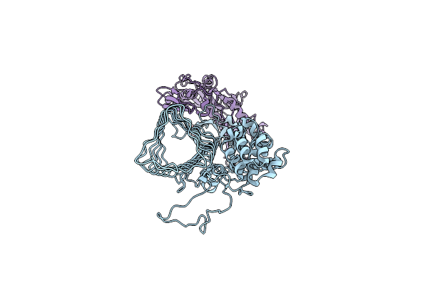
Crystal Structure Of The C143A/C166A Mutant Of Ero1P
Organism: Saccharomyces cerevisiae
Method: X-RAY DIFFRACTION Resolution:3.20 Å Release Date: 2010-11-03 Classification: OXIDOREDUCTASE Ligands: NEN, FAD, CD |
Organism: Saccharomyces cerevisiae
Method: X-RAY DIFFRACTION
Release Date: 2010-11-03
Ligands: NEN, FAD, CD
 |
Crystal Structure Analysis Of (Gp27-Gp5)3 Conjugated With Fe(Iii) Protoporphyrin
Organism: Enterobacteria phage t4
Method: X-RAY DIFFRACTION Resolution:3.11 Å Release Date: 2008-04-15 Classification: HYDROLASE/STRUCTURAL PROTEIN Ligands: NEN |
Organism: Enterobacteria phage t4
Method: X-RAY DIFFRACTION
Release Date: 2008-04-15
Ligands: NEN
 |
Structure Of Ero1P, Source Of Disulfide Bonds For Oxidative Protein Folding In The Cell
Organism: Saccharomyces cerevisiae
Method: X-RAY DIFFRACTION Resolution:2.20 Å Release Date: 2004-06-08 Classification: OXIDOREDUCTASE Ligands: CD, NEN, FAD |
Organism: Saccharomyces cerevisiae
Method: X-RAY DIFFRACTION
Release Date: 2004-06-08
Ligands: CD, NEN, FAD
 |
Structure Of Ero1P, Source Of Disulfide Bonds For Oxidative Protein Folding In The Cell
Organism: Saccharomyces cerevisiae
Method: X-RAY DIFFRACTION Resolution:2.80 Å Release Date: 2004-06-08 Classification: OXIDOREDUCTASE Ligands: CD, NEN, FAD |
Organism: Saccharomyces cerevisiae
Method: X-RAY DIFFRACTION
Release Date: 2004-06-08
Ligands: CD, NEN, FAD
 |
N-Terminal Domain Of Sec18P
Organism: Saccharomyces cerevisiae
Method: X-RAY DIFFRACTION Resolution:2.30 Å Release Date: 1999-12-22 Classification: ENDOCYTOSIS/EXOCYTOSIS Ligands: NEN |
Organism: Saccharomyces cerevisiae
Method: X-RAY DIFFRACTION
Release Date: 1999-12-22
Ligands: NEN