

Search Count: 57
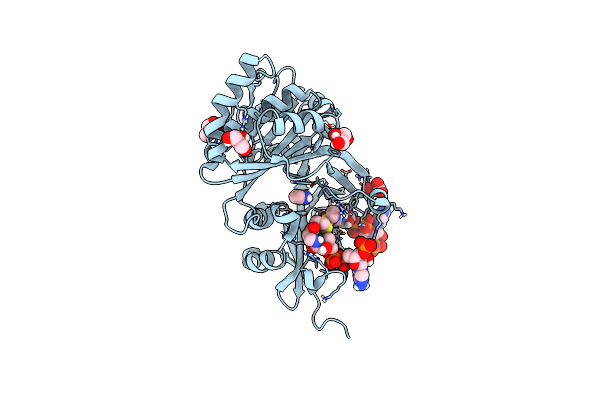 |
Crystal Structure Of Histidine Acetyltransferase With Imidazole And Coenzyme A Disulfide
Organism: Homo sapiens
Method: X-RAY DIFFRACTION Resolution:1.40 Å Release Date: 2025-03-26 Classification: TRANSFERASE Ligands: 5NG, IMD, GOL, MLI |
Organism: Homo sapiens
Method: X-RAY DIFFRACTION
Release Date: 2025-03-26
Ligands: 5NG, IMD, GOL, MLI
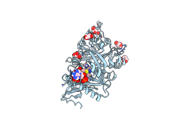 |
Crystal Structure Of Histidine Acetyltransferase With L-Histidine And S-Ethyl-Coenzyme A
Organism: Homo sapiens
Method: X-RAY DIFFRACTION Resolution:1.40 Å Release Date: 2025-03-26 Classification: TRANSFERASE Ligands: GOL, HIS, A2U, CL |
Organism: Homo sapiens
Method: X-RAY DIFFRACTION
Release Date: 2025-03-26
Ligands: GOL, HIS, A2U, CL
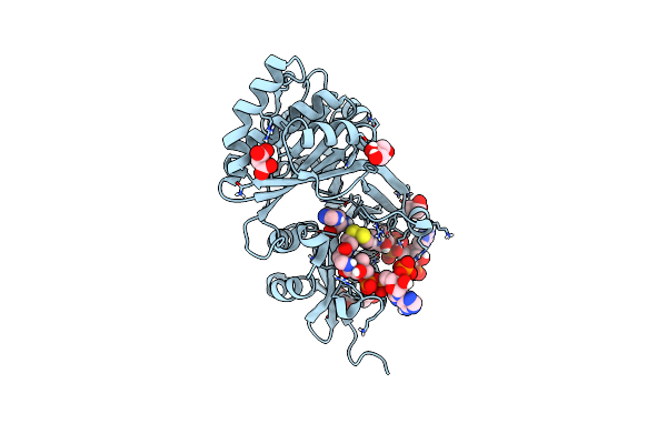 |
Crystal Structure Of Histidine Acetyltransferase With L-Histidine And Coenzyme A Disulfide
Organism: Homo sapiens
Method: X-RAY DIFFRACTION Resolution:1.60 Å Release Date: 2025-03-19 Classification: TRANSFERASE Ligands: GOL, 5NG, MLI, HIS, CL |
Organism: Homo sapiens
Method: X-RAY DIFFRACTION
Release Date: 2025-03-19
Ligands: GOL, 5NG, MLI, HIS, CL
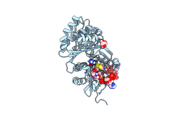 |
Crystal Structure Of Histidine Acetyltransferase With L-Arginine And Coenzyme A Disulfide
Organism: Homo sapiens
Method: X-RAY DIFFRACTION Resolution:1.90 Å Release Date: 2025-03-19 Classification: TRANSFERASE Ligands: 5NG, GOL, ARG |
Organism: Homo sapiens
Method: X-RAY DIFFRACTION
Release Date: 2025-03-19
Ligands: 5NG, GOL, ARG
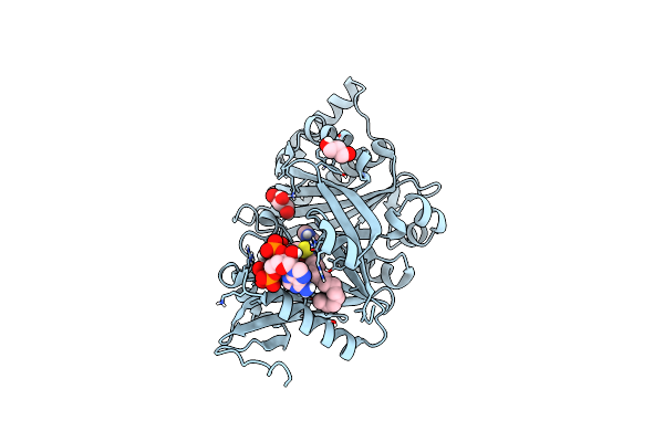 |
Crystal Structure Of Histidine Acetyltransferase With N-Myristoyl Histidine And Coenzyme A
Organism: Homo sapiens
Method: X-RAY DIFFRACTION Resolution:1.45 Å Release Date: 2025-03-19 Classification: TRANSFERASE Ligands: COA, A1H51, GOL |
Organism: Homo sapiens
Method: X-RAY DIFFRACTION
Release Date: 2025-03-19
Ligands: COA, A1H51, GOL
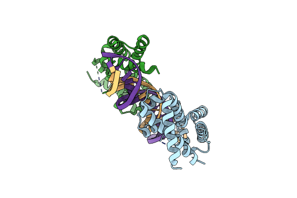 |
Dna Binding Domain Of Hnf-1A Bound To P2-Hnf4A Promoter Dna Variant (P2 -169C>T)
Organism: Homo sapiens
Method: X-RAY DIFFRACTION Resolution:3.20 Å Release Date: 2024-06-19 Classification: DNA BINDING PROTEIN Ligands: CL |
Organism: Homo sapiens
Method: X-RAY DIFFRACTION
Release Date: 2024-06-19
Ligands: CL
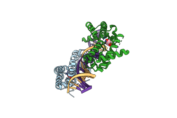 |
Dna Binding Domain Of Hnf-1A Bound To P2-Hnf4A Promoter Dna
Organism: Homo sapiens
Method: X-RAY DIFFRACTION Resolution:2.30 Å Release Date: 2024-06-19 Classification: DNA BINDING PROTEIN Ligands: GOL |
Organism: Homo sapiens
Method: X-RAY DIFFRACTION
Release Date: 2024-06-19
Ligands: GOL
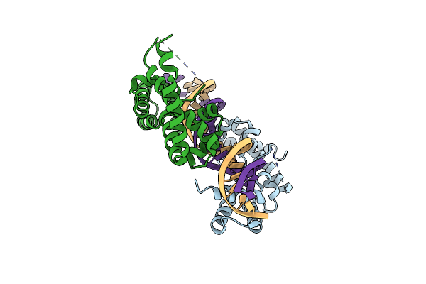 |
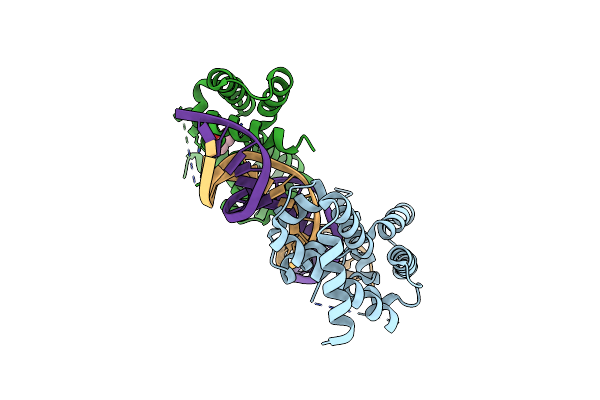
Dna Binding Domain Of Hnf-1A Bound To P2-Hnf4A Promoter Dna Variant (P2 -181G>A)
Organism: Homo sapiens
Method: X-RAY DIFFRACTION Resolution:2.80 Å Release Date: 2024-06-19 Classification: DNA BINDING PROTEIN |
Organism: Homo sapiens
Method: X-RAY DIFFRACTION
Release Date: 2024-06-19
 |
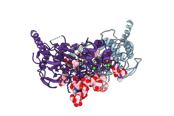
Dna Binding Domain Of Hnf-1A Bound To P2-Hnf4A Promoter Dna Variant (P2 -181G>T)
Organism: Homo sapiens
Method: X-RAY DIFFRACTION Resolution:2.80 Å Release Date: 2024-06-19 Classification: DNA BINDING PROTEIN Ligands: GOL |
Organism: Homo sapiens
Method: X-RAY DIFFRACTION
Release Date: 2024-06-19
Ligands: GOL
 |
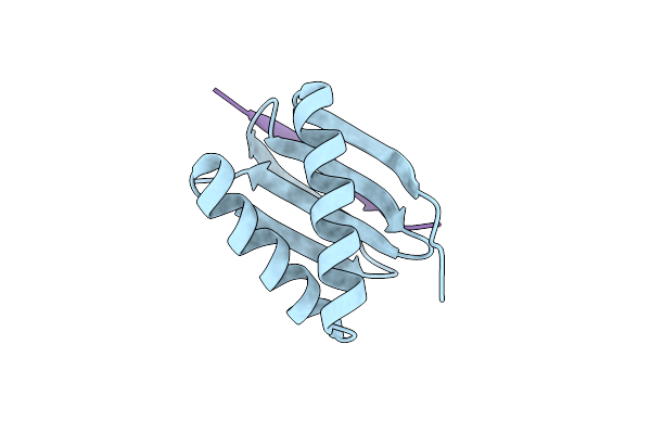
Crystal Structure Of Human Transmembrane Prolyl 4-Hydroxylase
Organism: Homo sapiens
Method: X-RAY DIFFRACTION Resolution:2.25 Å Release Date: 2020-12-23 Classification: OXIDOREDUCTASE Ligands: OGA, FE2, TBU, GLY, CA, CL |
Organism: Homo sapiens
Method: X-RAY DIFFRACTION
Release Date: 2020-12-23
Ligands: OGA, FE2, TBU, GLY, CA, CL
 |
Complex Between The Dynein Light Chain Dynll1/Dlc8 And The Specific Domain Of Large Myelin-Associated Glycoprotein L-Mag
Organism: Homo sapiens, Mus musculus
Method: X-RAY DIFFRACTION Resolution:1.98 Å Release Date: 2018-10-10 Classification: PROTEIN BINDING Ligands: CL |
Organism: Homo sapiens, Mus musculus
Method: X-RAY DIFFRACTION
Release Date: 2018-10-10
Ligands: CL
 |
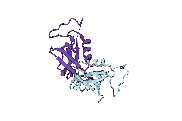
Complex Between The Dynein Light Chain Dynll1/Dlc8 And A Peptide From The Large Myelin-Associated Glycoprotein L-Mag
Organism: Homo sapiens, Mus musculus
Method: X-RAY DIFFRACTION Resolution:1.95 Å Release Date: 2018-10-10 Classification: PROTEIN BINDING Ligands: CL |
Organism: Homo sapiens, Mus musculus
Method: X-RAY DIFFRACTION
Release Date: 2018-10-10
Ligands: CL
 |
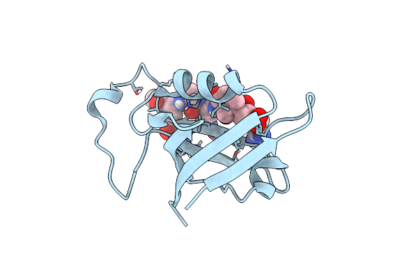
Apo Pdz Domain From Rat Shank3
Organism: Rattus norvegicus
Method: X-RAY DIFFRACTION Resolution:2.30 Å Release Date: 2018-03-07 Classification: PROTEIN BINDING |
Organism: Rattus norvegicus
Method: X-RAY DIFFRACTION
Release Date: 2018-03-07
 |
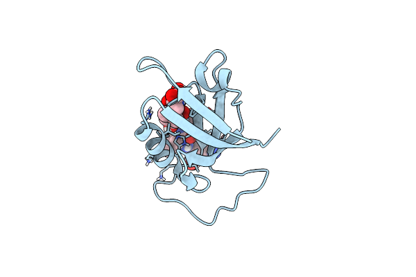
Pdz Domain From Rat Shank3 Bound To The C Terminus Of Gkap
Organism: Rattus norvegicus
Method: X-RAY DIFFRACTION Resolution:1.55 Å Release Date: 2018-03-07 Classification: PROTEIN BINDING Ligands: CL |
Organism: Rattus norvegicus
Method: X-RAY DIFFRACTION
Release Date: 2018-03-07
Ligands: CL
 |
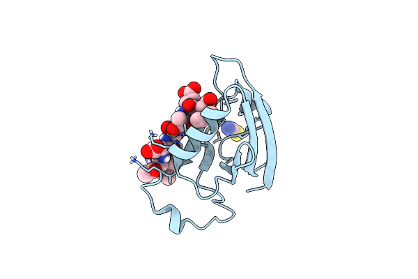
Pdz Domain From Rat Shank3 Bound To The C Terminus Of Cirl
Organism: Rattus norvegicus
Method: X-RAY DIFFRACTION Resolution:1.50 Å Release Date: 2018-03-07 Classification: PROTEIN BINDING |
Organism: Rattus norvegicus
Method: X-RAY DIFFRACTION
Release Date: 2018-03-07
 |
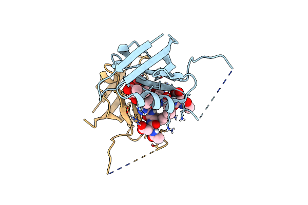
Pdz Domain From Rat Shank3 Bound To The C Terminus Of Prosapip1
Organism: Rattus norvegicus
Method: X-RAY DIFFRACTION Resolution:1.40 Å Release Date: 2018-03-07 Classification: PROTEIN BINDING Ligands: SCN |
Organism: Rattus norvegicus
Method: X-RAY DIFFRACTION
Release Date: 2018-03-07
Ligands: SCN
 |
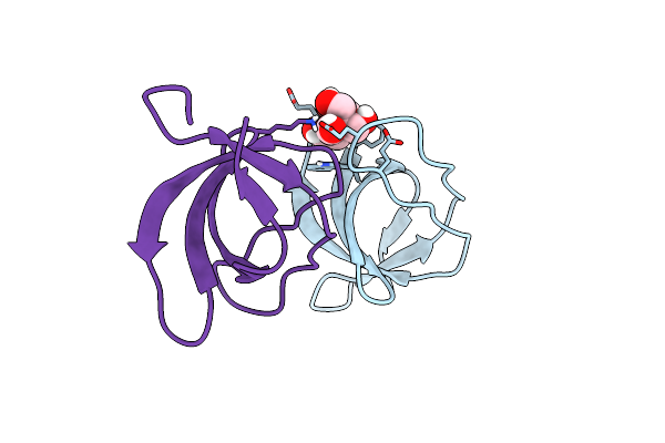
Pdz Domain From Rat Shank3 Bound To The C Terminus Of Somatostatin Receptor Subtype 2
Organism: Rattus norvegicus
Method: X-RAY DIFFRACTION Resolution:1.80 Å Release Date: 2018-03-07 Classification: PROTEIN BINDING |
Organism: Rattus norvegicus
Method: X-RAY DIFFRACTION
Release Date: 2018-03-07
 |
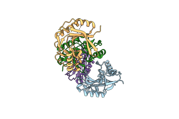
Unconventional Sh3 Domain From The Postsynaptic Density Scaffold Protein Shank3
Organism: Rattus norvegicus
Method: X-RAY DIFFRACTION Resolution:0.87 Å Release Date: 2017-07-05 Classification: PROTEIN BINDING Ligands: BTB |
Organism: Rattus norvegicus
Method: X-RAY DIFFRACTION
Release Date: 2017-07-05
Ligands: BTB
 |
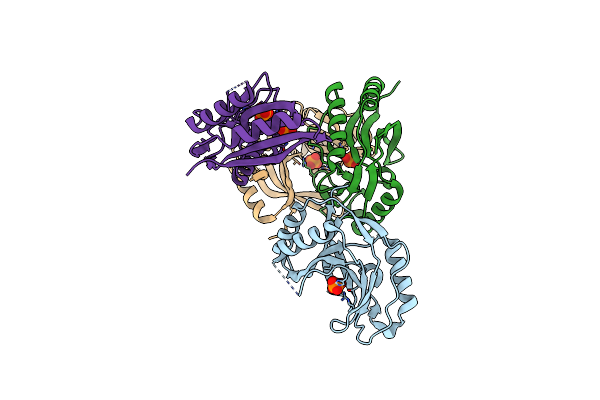
Crystal Structure Of E.Coli Ligt In Apo Form
Organism: Escherichia coli bl21(de3)
Method: X-RAY DIFFRACTION Resolution:2.10 Å Release Date: 2017-01-18 Classification: HYDROLASE Ligands: CL |
Organism: Escherichia coli bl21(de3)
Method: X-RAY DIFFRACTION
Release Date: 2017-01-18
Ligands: CL
 |
Crystal Structure Of E.Coli Ligt Complexed With Phosphate
Organism: Escherichia coli bl21(de3)
Method: X-RAY DIFFRACTION Resolution:2.80 Å Release Date: 2017-01-18 Classification: HYDROLASE Ligands: PO4 |
Organism: Escherichia coli bl21(de3)
Method: X-RAY DIFFRACTION
Release Date: 2017-01-18
Ligands: PO4