

Search Count: 44
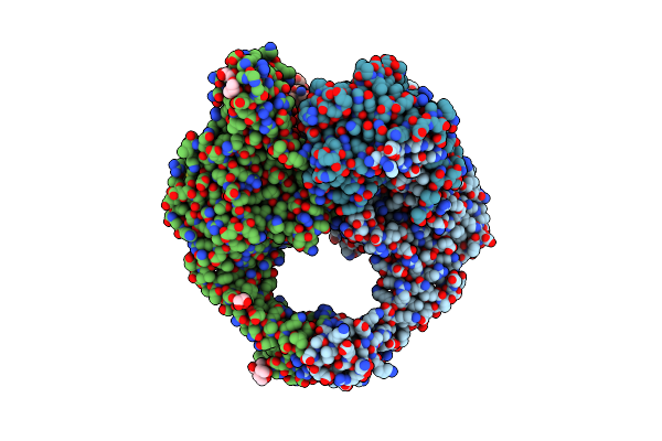 |
A 2.58A Crystal Structure Of S. Aureus Dna Gyrase And Dna With Metals Identified Through Anomalous Scattering
Organism: Staphylococcus aureus, Dna molecule
Method: X-RAY DIFFRACTION Resolution:2.58 Å Release Date: 2024-10-23 Classification: ISOMERASE Ligands: GOL, MN, BTB |
Organism: Staphylococcus aureus, Dna molecule
Method: X-RAY DIFFRACTION
Release Date: 2024-10-23
Ligands: GOL, MN, BTB
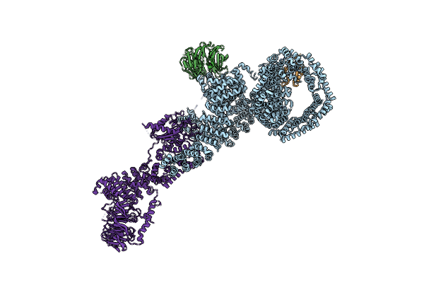 |
Human Deptor In A Complex With Mutant Human Mtorc1 A1459P
Organism: Homo sapiens
Method: ELECTRON MICROSCOPY Resolution:4.70 Å Release Date: 2021-09-08 Classification: SIGNALING PROTEIN |
Organism: Homo sapiens
Method: ELECTRON MICROSCOPY
Release Date: 2021-09-08
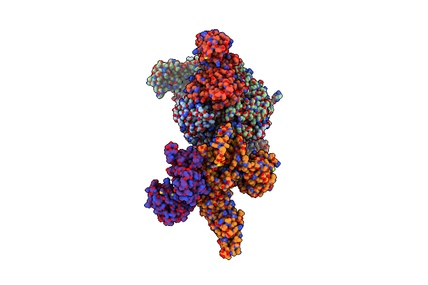 |
Muts In Complex With The N-Terminal Domain Of Mutl - Crystal Form 1
Organism: Escherichia coli k-12
Method: X-RAY DIFFRACTION Resolution:4.71 Å Release Date: 2015-07-22 Classification: DNA BINDING PROTEIN Ligands: ANP |
Organism: Escherichia coli k-12
Method: X-RAY DIFFRACTION
Release Date: 2015-07-22
Ligands: ANP
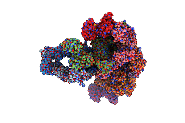 |
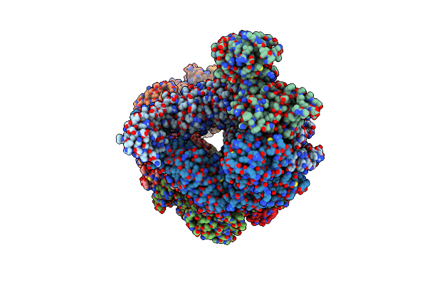
Muts In Complex With The N-Terminal Domain Of Mutl - Crystal Form 2
Organism: Escherichia coli k-12
Method: X-RAY DIFFRACTION Resolution:6.60 Å Release Date: 2015-07-22 Classification: HYDROLASE Ligands: ANP |
Organism: Escherichia coli k-12
Method: X-RAY DIFFRACTION
Release Date: 2015-07-22
Ligands: ANP
 |
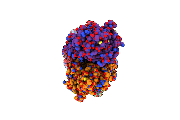
Muts In Complex With The N-Terminal Domain Of Mutl - Crystal Form 3
Organism: Escherichia coli
Method: X-RAY DIFFRACTION Resolution:7.60 Å Release Date: 2015-07-22 Classification: DNA BINDING PROTEIN Ligands: ANP |
Organism: Escherichia coli
Method: X-RAY DIFFRACTION
Release Date: 2015-07-22
Ligands: ANP
 |
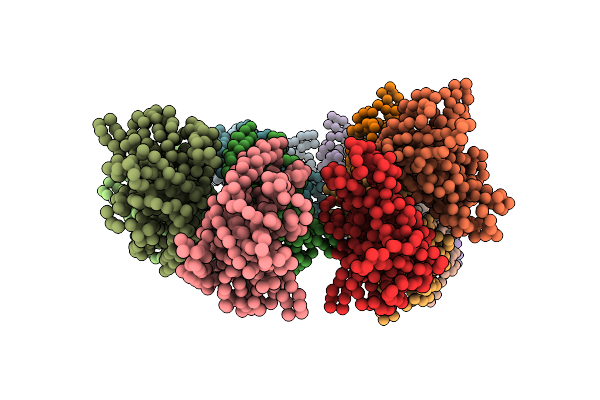
Actin-Like Parm Protein Bound To Amppnp
Organism: Escherichia coli
Method: ELECTRON MICROSCOPY Resolution:4.30 Å Release Date: 2015-04-22 Classification: STRUCTURAL PROTEIN Ligands: ANP |
Organism: Escherichia coli
Method: ELECTRON MICROSCOPY
Release Date: 2015-04-22
Ligands: ANP
 |
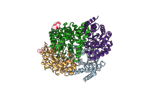
Parm Doublet Model
Organism: Escherichia coli
Method: ELECTRON MICROSCOPY Release Date: 2015-04-22 Classification: MOTOR PROTEIN |
Organism: Escherichia coli
Method: ELECTRON MICROSCOPY
Release Date: 2015-04-22
 |
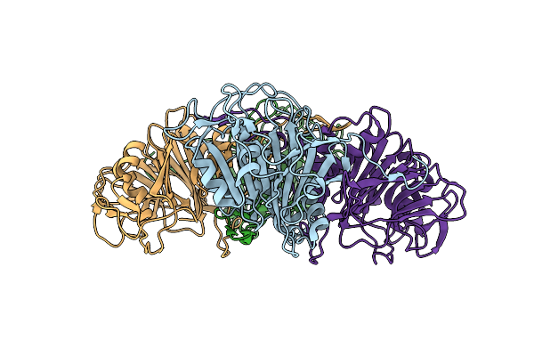
Structure Of Wild Type E. Coli N-Acetylneuraminic Acid Lyase In Space Group P21 Crystal Form Ii
Organism: Escherichia coli
Method: X-RAY DIFFRACTION Resolution:1.90 Å Release Date: 2012-04-04 Classification: LYASE Ligands: CL, 1PE |
Organism: Escherichia coli
Method: X-RAY DIFFRACTION
Release Date: 2012-04-04
Ligands: CL, 1PE
 |
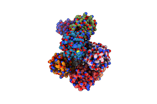
Activation Of Catalytic Cysteine Without A Base In A Mutant Penicillin Acylase Precursor
Organism: Bacillus sphaericus
Method: X-RAY DIFFRACTION Resolution:2.50 Å Release Date: 2011-07-20 Classification: HYDROLASE |
Organism: Bacillus sphaericus
Method: X-RAY DIFFRACTION
Release Date: 2011-07-20
 |
1.9 Angstrom Crystal Structure Of Clec5A
Organism: Homo sapiens
Method: X-RAY DIFFRACTION Resolution:1.90 Å Release Date: 2011-05-11 Classification: IMMUNE SYSTEM |
Organism: Homo sapiens
Method: X-RAY DIFFRACTION
Release Date: 2011-05-11
 |
Crystal Structure Of Dethiobiotin Synthetase (Biod) From Helicobacter Pylori Complexed With Atp
Organism: Helicobacter pylori
Method: X-RAY DIFFRACTION Resolution:1.34 Å Release Date: 2011-03-30 Classification: LIGASE Ligands: ATP, MG, NO3, EDO, GOL, PEG |
Organism: Helicobacter pylori
Method: X-RAY DIFFRACTION
Release Date: 2011-03-30
Ligands: ATP, MG, NO3, EDO, GOL, PEG
 |
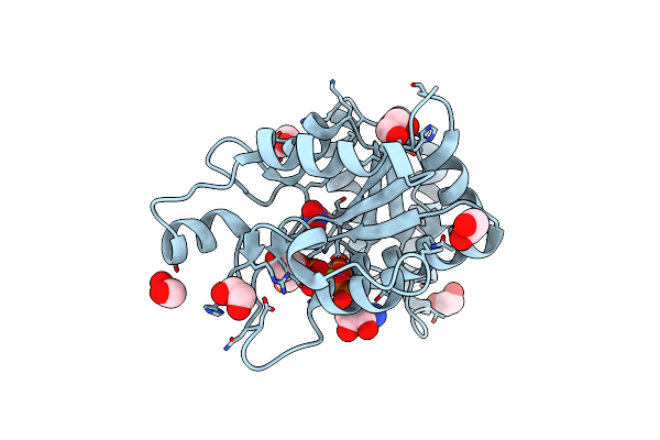
Crystal Structure Of Dethiobiotin Synthetase (Biod) From Helicobacter Pylori Complexed With Adp And 8-Aminocaprylic Acid
Organism: Helicobacter pylori
Method: X-RAY DIFFRACTION Resolution:1.36 Å Release Date: 2011-03-30 Classification: LIGASE Ligands: 8AC, ADP, MG, PO4, EDO |
Organism: Helicobacter pylori
Method: X-RAY DIFFRACTION
Release Date: 2011-03-30
Ligands: 8AC, ADP, MG, PO4, EDO
 |
Crystal Structure Of Dethiobiotin Synthetase (Biod) From Helicobacter Pylori Complexed With Gtp
Organism: Helicobacter pylori
Method: X-RAY DIFFRACTION Resolution:1.38 Å Release Date: 2011-03-30 Classification: LIGASE Ligands: GTP, EDO, MG, NO3 |
Organism: Helicobacter pylori
Method: X-RAY DIFFRACTION
Release Date: 2011-03-30
Ligands: GTP, EDO, MG, NO3
 |
Crystal Structure Of Dethiobiotin Synthetase (Biod) From Helicobacter Pylori Complexed With Anp
Organism: Helicobacter pylori
Method: X-RAY DIFFRACTION Resolution:1.35 Å Release Date: 2011-03-30 Classification: LIGASE Ligands: EDO, ANP, MG, NO3 |
Organism: Helicobacter pylori
Method: X-RAY DIFFRACTION
Release Date: 2011-03-30
Ligands: EDO, ANP, MG, NO3
 |
Crystal Structure Of Dethiobiotin Synthetase (Biod) From Helicobacter Pylori Complexed With Gdp And 8-Aminocaprylic Acid
Organism: Helicobacter pylori
Method: X-RAY DIFFRACTION Resolution:1.36 Å Release Date: 2011-03-30 Classification: LIGASE Ligands: 8AC, GDP, MG, PO4, EDO, GOL |
Organism: Helicobacter pylori
Method: X-RAY DIFFRACTION
Release Date: 2011-03-30
Ligands: 8AC, GDP, MG, PO4, EDO, GOL
 |
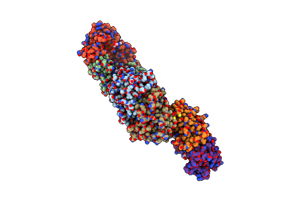
Crystal Structure Of Dethiobiotin Synthetase (Biod) From Helicobacter Pylori Complexed With Gdp
Organism: Helicobacter pylori
Method: X-RAY DIFFRACTION Resolution:1.60 Å Release Date: 2011-03-30 Classification: LIGASE Ligands: GDP, PO4, MG, EDO |
Organism: Helicobacter pylori
Method: X-RAY DIFFRACTION
Release Date: 2011-03-30
Ligands: GDP, PO4, MG, EDO
 |
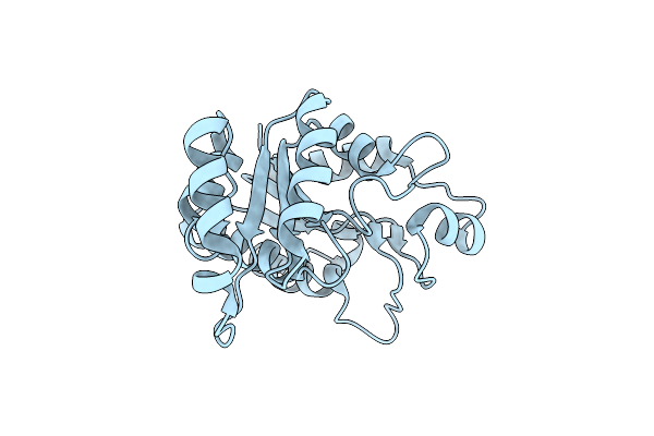
Crystal Structure Of Dethiobiotin Synthetase (Biod) From Helicobacter Pylori Cocrystallized With Atp
Organism: Helicobacter pylori
Method: X-RAY DIFFRACTION Resolution:2.80 Å Release Date: 2010-05-19 Classification: LIGASE Ligands: PO4, MG, NO3, CL, 8AC, ADP |
Organism: Helicobacter pylori
Method: X-RAY DIFFRACTION
Release Date: 2010-05-19
Ligands: PO4, MG, NO3, CL, 8AC, ADP
 |
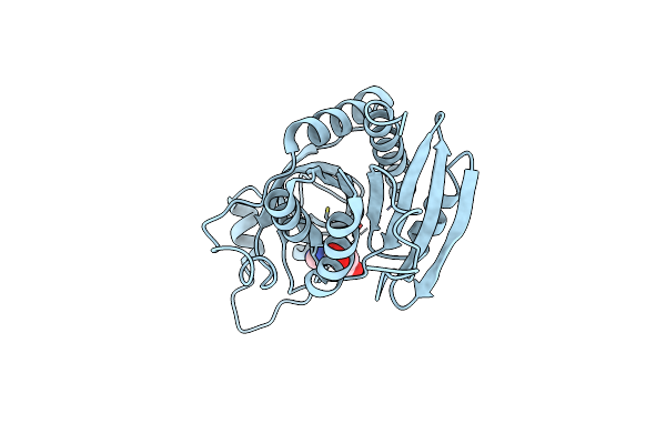
Crystal Structure Of Dethiobiotin Synthetase (Biod) From Helicobacter Pylori
Organism: Helicobacter pylori
Method: X-RAY DIFFRACTION Resolution:1.47 Å Release Date: 2007-07-31 Classification: LIGASE Ligands: CL |
Organism: Helicobacter pylori
Method: X-RAY DIFFRACTION
Release Date: 2007-07-31
Ligands: CL
 |
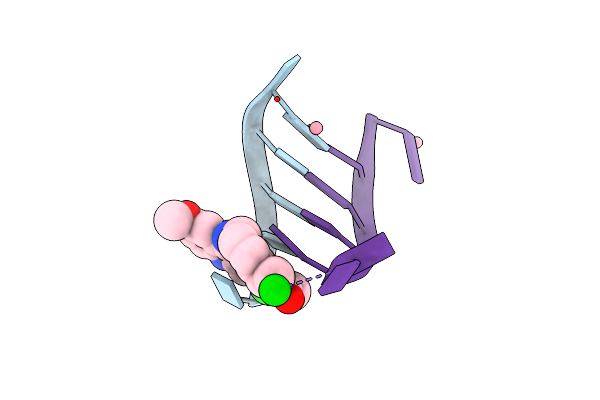
Crystal Structure Of The Plasmodium Falciparum Purine Nucleoside Phosphorylase Complexed With Inosine
Organism: Plasmodium falciparum
Method: X-RAY DIFFRACTION Resolution:2.00 Å Release Date: 2005-08-18 Classification: TRANSFERASE Ligands: NOS |
Organism: Plasmodium falciparum
Method: X-RAY DIFFRACTION
Release Date: 2005-08-18
Ligands: NOS
 |
Structure Of Oligonucleotide/Drug Complex
Method: X-RAY DIFFRACTION
Resolution:1.40 Å Release Date: 2005-07-19 Classification: DNA Ligands: CO, A4L, BA |
Method: X-RAY DIFFRACTION
Release Date: 2005-07-19
Ligands: CO, A4L, BA