

Search Count: 27
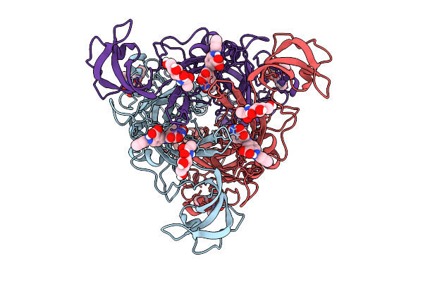 |
Cryo Em Structure Of The E307T Mutant Of The Human P2X4 Receptor In Complex With The Anthraquinone Derivative Psb-0704
Organism: Homo sapiens
Method: ELECTRON MICROSCOPY Release Date: 2025-11-26 Classification: MEMBRANE PROTEIN Ligands: A1INJ, NAG |
Organism: Homo sapiens
Method: ELECTRON MICROSCOPY
Release Date: 2025-11-26
Ligands: A1INJ, NAG
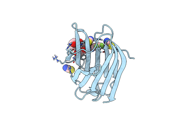 |
Co-Crystal Structure Of Galectin-3 With An Inhibitor
Organism: Homo sapiens
Method: X-RAY DIFFRACTION Resolution:1.45 Å Release Date: 2025-03-26 Classification: SUGAR BINDING PROTEIN Ligands: SCN, A1IB3 |
Organism: Homo sapiens
Method: X-RAY DIFFRACTION
Release Date: 2025-03-26
Ligands: SCN, A1IB3
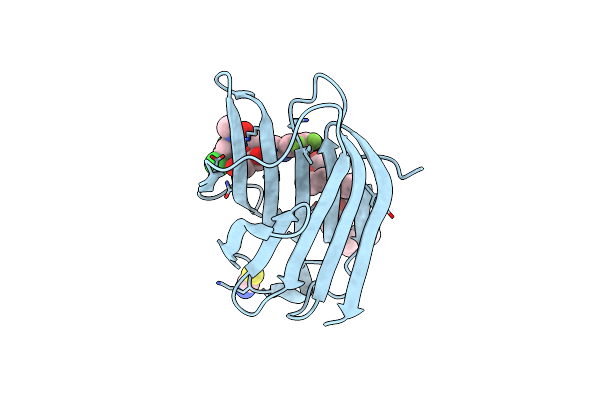 |
Co-Crystal Structure Of Galectin-3 With An Inhibitor
Organism: Homo sapiens
Method: X-RAY DIFFRACTION Resolution:1.78 Å Release Date: 2025-03-26 Classification: SUGAR BINDING PROTEIN Ligands: 2PE, A1IB4, SCN |
Organism: Homo sapiens
Method: X-RAY DIFFRACTION
Release Date: 2025-03-26
Ligands: 2PE, A1IB4, SCN
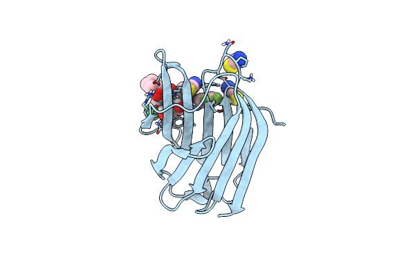 |
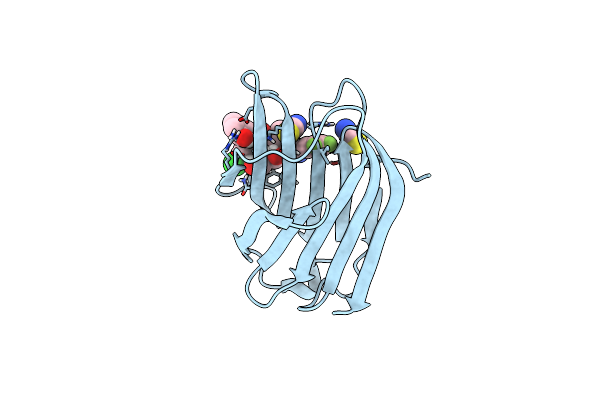
Galectin-3 With A Bound Inhibitor
Organism: Homo sapiens
Method: X-RAY DIFFRACTION Resolution:1.25 Å Release Date: 2025-01-29 Classification: SUGAR BINDING PROTEIN Ligands: A1H1W, SCN |
Organism: Homo sapiens
Method: X-RAY DIFFRACTION
Release Date: 2025-01-29
Ligands: A1H1W, SCN
 |
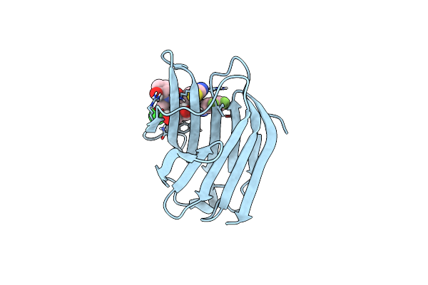
Galectin-3 With A Bound Inhibitor
Organism: Homo sapiens
Method: X-RAY DIFFRACTION Resolution:1.20 Å Release Date: 2025-01-29 Classification: SUGAR BINDING PROTEIN Ligands: A1H1Y, SCN |
Organism: Homo sapiens
Method: X-RAY DIFFRACTION
Release Date: 2025-01-29
Ligands: A1H1Y, SCN
 |
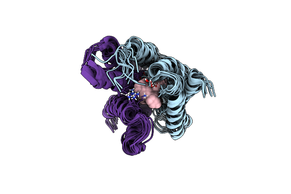
Galectin-3 With A Bound Inhibitor
Organism: Homo sapiens
Method: X-RAY DIFFRACTION Resolution:1.35 Å Release Date: 2025-01-29 Classification: SUGAR BINDING PROTEIN Ligands: A1H1X, SCN |
Organism: Homo sapiens
Method: X-RAY DIFFRACTION
Release Date: 2025-01-29
Ligands: A1H1X, SCN
 |
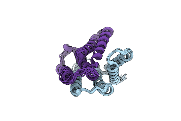
Emre Structure In The Tpp-Bound State (Wt/E14Q Heterodimer)
Organism: Escherichia coli
Method: SOLUTION NMR, SOLID-STATE NMR Release Date: 2024-05-29 Classification: MEMBRANE PROTEIN Ligands: P4P |
Organism: Escherichia coli
Method: SOLUTION NMR, SOLID-STATE NMR
Release Date: 2024-05-29
Ligands: P4P
 |
Emre Structure In The Proton-Bound State (Wt/L51I Heterodimer)
Organism: Escherichia coli
Method: SOLUTION NMR, SOLID-STATE NMR Release Date: 2024-05-29 Classification: MEMBRANE PROTEIN |
Organism: Escherichia coli
Method: SOLUTION NMR, SOLID-STATE NMR
Release Date: 2024-05-29
 |
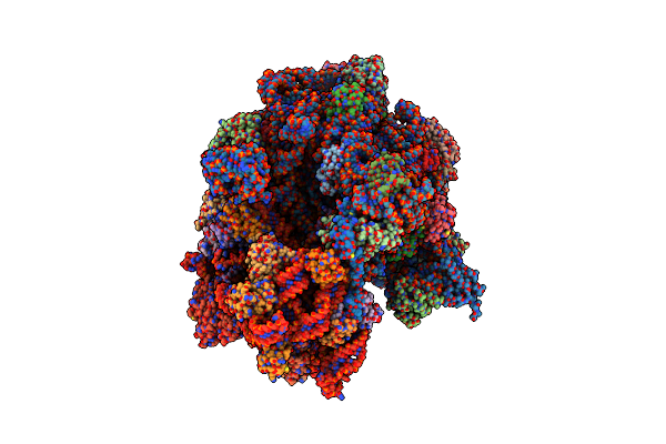
Structure Of The P+9 Stalled Ribosome Complex
Organism: Escherichia coli
Method: ELECTRON MICROSCOPY Release Date: 2020-08-19 Classification: RIBOSOME Ligands: MG, ZN, CL, NA |
Organism: Escherichia coli
Method: ELECTRON MICROSCOPY
Release Date: 2020-08-19
Ligands: MG, ZN, CL, NA
 |
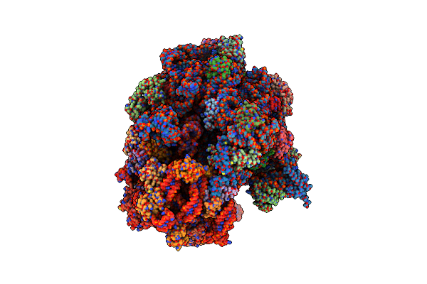
Structure Of The P+9 Arfb-Ribosome Complex In The Post-Hydrolysis State
Organism: Apis mellifera, Escherichia coli
Method: ELECTRON MICROSCOPY Release Date: 2020-08-19 Classification: RIBOSOME Ligands: MG, ZN, NA |
Organism: Apis mellifera, Escherichia coli
Method: ELECTRON MICROSCOPY
Release Date: 2020-08-19
Ligands: MG, ZN, NA
 |
Structure Of The P+9 Arfb-Ribosome Complex With P/E Hybrid Trna In The Post-Hydrolysis State
Organism: Apis mellifera, Escherichia coli
Method: ELECTRON MICROSCOPY Release Date: 2020-08-19 Classification: RIBOSOME Ligands: MG, ZN, NA |
Organism: Apis mellifera, Escherichia coli
Method: ELECTRON MICROSCOPY
Release Date: 2020-08-19
Ligands: MG, ZN, NA
 |
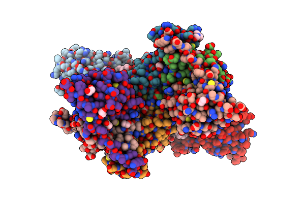
Structure Of The P+0 Arfb-Ribosome Complex In The Post-Hydrolysis State
Organism: Apis mellifera, Escherichia coli
Method: ELECTRON MICROSCOPY Release Date: 2020-08-19 Classification: RIBOSOME Ligands: MG, ZN, NA |
Organism: Apis mellifera, Escherichia coli
Method: ELECTRON MICROSCOPY
Release Date: 2020-08-19
Ligands: MG, ZN, NA
 |
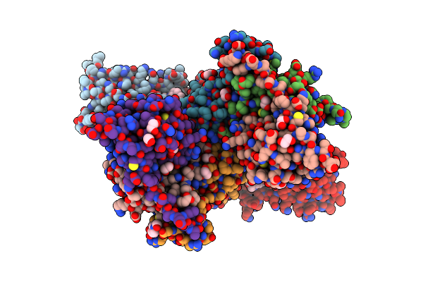
Crystal Structure Of Protac 2 In Complex With The Bromodomain Of Human Smarca2 And Pvhl:Elonginc:Elonginb
Organism: Homo sapiens
Method: X-RAY DIFFRACTION Resolution:2.35 Å Release Date: 2019-06-12 Classification: GENE REGULATION Ligands: EDO, FWZ, EPE |
Organism: Homo sapiens
Method: X-RAY DIFFRACTION
Release Date: 2019-06-12
Ligands: EDO, FWZ, EPE
 |
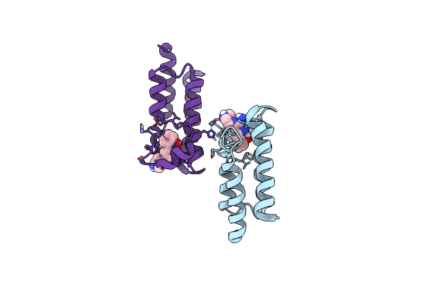
Crystal Structure Of Protac 1 In Complex With The Bromodomain Of Human Smarca2 And Pvhl:Elonginc:Elonginb
Organism: Homo sapiens
Method: X-RAY DIFFRACTION Resolution:2.24 Å Release Date: 2019-06-12 Classification: GENE REGULATION Ligands: FX8, EDO, FMT, EPE |
Organism: Homo sapiens
Method: X-RAY DIFFRACTION
Release Date: 2019-06-12
Ligands: FX8, EDO, FMT, EPE
 |
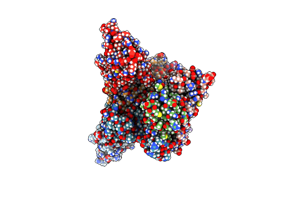
Crystal Structure Of The Bromodomain Of Human Smarca2 In Complex With Smarca-Bd Ligand
Organism: Homo sapiens
Method: X-RAY DIFFRACTION Resolution:1.31 Å Release Date: 2019-06-12 Classification: GENE REGULATION Ligands: FX5, ZN |
Organism: Homo sapiens
Method: X-RAY DIFFRACTION
Release Date: 2019-06-12
Ligands: FX5, ZN
 |
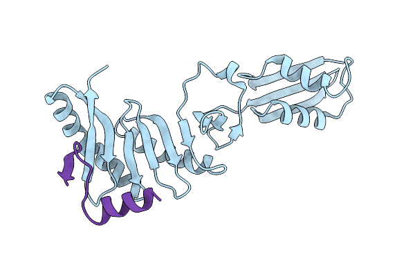
Crystal Structure Of Protac 2 In Complex With The Bromodomain Of Human Smarca4 And Pvhl:Elonginc:Elonginb
Organism: Homo sapiens
Method: X-RAY DIFFRACTION Resolution:1.76 Å Release Date: 2019-06-12 Classification: TRANSCRIPTION Ligands: FWZ, DMS, EDO |
Organism: Homo sapiens
Method: X-RAY DIFFRACTION
Release Date: 2019-06-12
Ligands: FWZ, DMS, EDO
 |
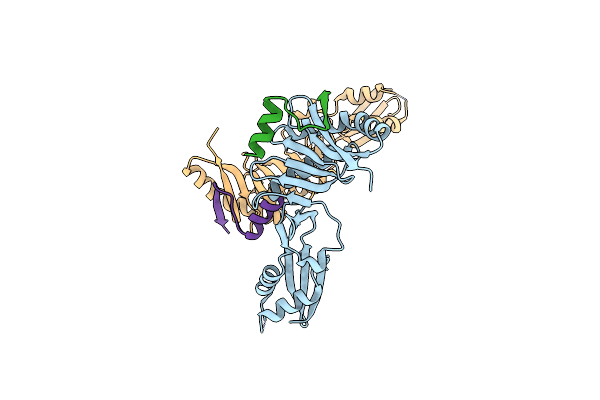
Complex Of The Periplasmic Domains Of Bacterial Cell Division Proteins Ftsq And Ftsb
Organism: Escherichia coli k-12, Escherichia coli 55989
Method: X-RAY DIFFRACTION Resolution:2.60 Å Release Date: 2018-09-05 Classification: CELL CYCLE |
Organism: Escherichia coli k-12, Escherichia coli 55989
Method: X-RAY DIFFRACTION
Release Date: 2018-09-05
 |
Complex Of The Periplasmic Domains Of Bacterial Cell Division Proteins Ftsq And Ftsb
Organism: Escherichia coli, Escherichia coli s88
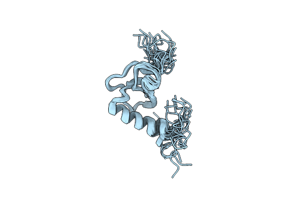
Method: X-RAY DIFFRACTION Resolution:2.80 Å Release Date: 2018-09-05 Classification: CELL CYCLE |
Organism: Escherichia coli, Escherichia coli s88
Method: X-RAY DIFFRACTION
Release Date: 2018-09-05
 |
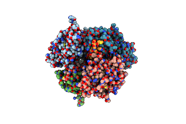
Solution Structure Of The Human Chemokine Ccl19
Organism: Homo sapiens
Method: SOLUTION NMR Release Date: 2015-06-10 Classification: SIGNALING PROTEIN |
Organism: Homo sapiens
Method: SOLUTION NMR
Release Date: 2015-06-10
 |
Crystal Structure Of The Minimal Pho-Sfmbt Complex (P21 Spacegroup)
Organism: Drosophila melanogaster
Method: X-RAY DIFFRACTION Resolution:1.95 Å Release Date: 2013-11-13 Classification: TRANSCRIPTION |
Organism: Drosophila melanogaster
Method: X-RAY DIFFRACTION
Release Date: 2013-11-13