

Search Count: 26
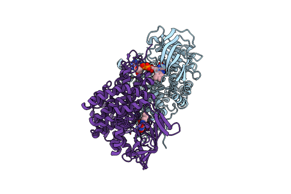 |
Bd2924 Apo-Form
Organism: Bdellovibrio bacteriovorus (strain atcc 15356 / dsm 50701 / ncib 9529 / hd100)
Method: X-RAY DIFFRACTION Resolution:1.51 Å Release Date: 2019-09-11 Classification: OXIDOREDUCTASE Ligands: FAD |
Organism: Bdellovibrio bacteriovorus (strain atcc 15356 / dsm 50701 / ncib 9529 / hd100)
Method: X-RAY DIFFRACTION
Release Date: 2019-09-11
Ligands: FAD
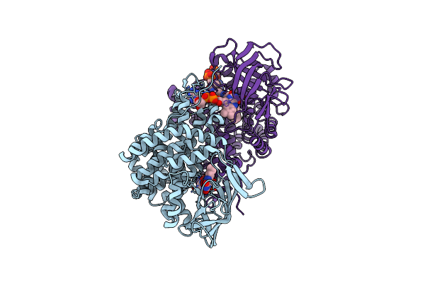 |
Bd2924 C10 Acyl-Coenzymea Bound Form
Organism: Bdellovibrio bacteriovorus hd100
Method: X-RAY DIFFRACTION Resolution:1.87 Å Release Date: 2019-09-11 Classification: OXIDOREDUCTASE Ligands: FAD, MFK |
Organism: Bdellovibrio bacteriovorus hd100
Method: X-RAY DIFFRACTION
Release Date: 2019-09-11
Ligands: FAD, MFK
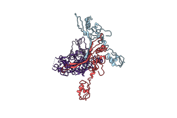 |
Cryo-Em Structure Of The Anti-Feeding Prophage (Afp) Baseplate In Extended State, 3-Fold Symmetrised
Organism: Serratia entomophila
Method: ELECTRON MICROSCOPY Release Date: 2019-04-24 Classification: VIRUS LIKE PARTICLE |
Organism: Serratia entomophila
Method: ELECTRON MICROSCOPY
Release Date: 2019-04-24
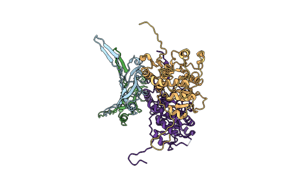 |
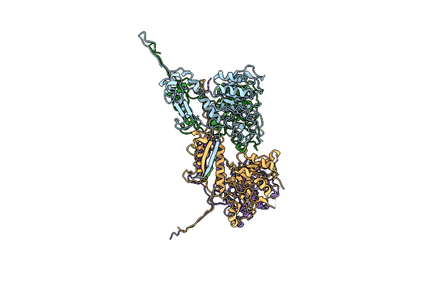
Cryo-Em Structure Of The Anti-Feeding Prophage (Afp) Helical Sheath-Tube Complex In Extended State
Organism: Serratia entomophila
Method: ELECTRON MICROSCOPY Release Date: 2019-04-24 Classification: VIRUS LIKE PARTICLE |
Organism: Serratia entomophila
Method: ELECTRON MICROSCOPY
Release Date: 2019-04-24
 |
Cryo-Em Structure Of The Anti-Feeding Prophage (Afp) Baseplate In Contracted State
Organism: Serratia entomophila
Method: ELECTRON MICROSCOPY Release Date: 2019-04-24 Classification: VIRUS LIKE PARTICLE |
Organism: Serratia entomophila
Method: ELECTRON MICROSCOPY
Release Date: 2019-04-24
 |
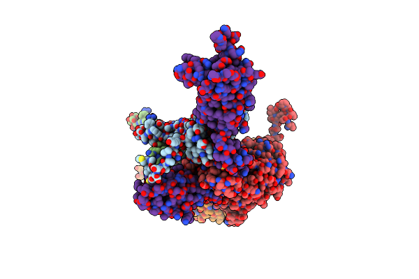
Cryo-Em Structure Of The Anti-Feeding Prophage (Afp) Baseplate, 6-Fold Symmetrised
Organism: Serratia entomophila
Method: ELECTRON MICROSCOPY Release Date: 2019-04-17 Classification: VIRUS LIKE PARTICLE |
Organism: Serratia entomophila
Method: ELECTRON MICROSCOPY
Release Date: 2019-04-17
 |
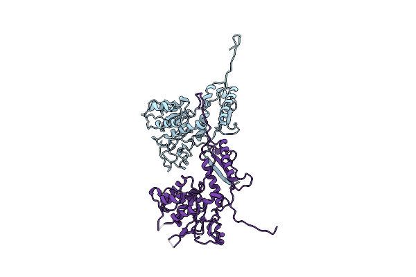
Cryo-Em Structure Of The Anti-Feeding Prophage Cap (Afp Tube Terminating Cap)
Organism: Serratia entomophila
Method: ELECTRON MICROSCOPY Release Date: 2019-04-17 Classification: VIRUS LIKE PARTICLE |
Organism: Serratia entomophila
Method: ELECTRON MICROSCOPY
Release Date: 2019-04-17
 |
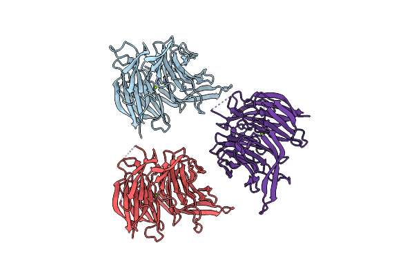
Cryo-Em Structure Of The Anti-Feeding Prophage (Afp) Helical Sheath In Contracted State
Organism: Serratia entomophila
Method: ELECTRON MICROSCOPY Release Date: 2019-04-17 Classification: VIRUS LIKE PARTICLE |
Organism: Serratia entomophila
Method: ELECTRON MICROSCOPY
Release Date: 2019-04-17
 |
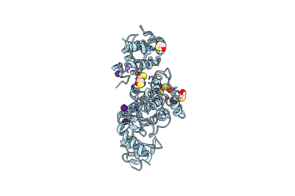
Crystal Structure Of The Wd40-Repeat Of Rfwd3
Organism: Homo sapiens
Method: X-RAY DIFFRACTION Resolution:1.80 Å Release Date: 2018-06-27 Classification: TRANSFERASE Ligands: MG |
Organism: Homo sapiens
Method: X-RAY DIFFRACTION
Release Date: 2018-06-27
Ligands: MG
 |
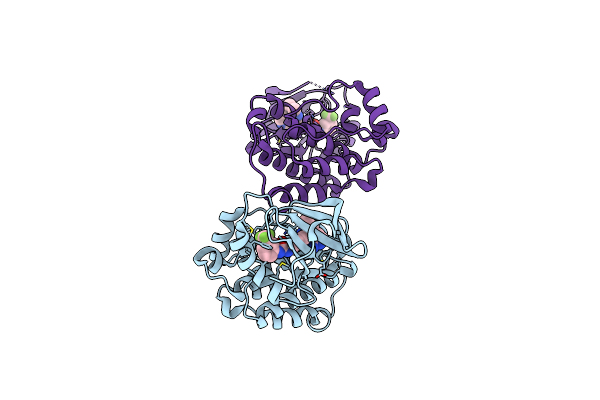
Crystal Structure Of Apo Human Ire1 Alpha
Organism: Homo sapiens
Method: X-RAY DIFFRACTION Resolution:2.58 Å Release Date: 2016-06-08 Classification: Transferase/Oxidoreductase Ligands: CS, NA, GOL, DTT, BME, CL |
Organism: Homo sapiens
Method: X-RAY DIFFRACTION
Release Date: 2016-06-08
Ligands: CS, NA, GOL, DTT, BME, CL
 |
Src Kinase In Complex With A Sulfonamide Inhibitor
Organism: Gallus gallus
Method: X-RAY DIFFRACTION Resolution:2.15 Å Release Date: 2016-06-01 Classification: Transferase/Transferase Inhibitor Ligands: 6G3 |
Organism: Gallus gallus
Method: X-RAY DIFFRACTION
Release Date: 2016-06-01
Ligands: 6G3
 |
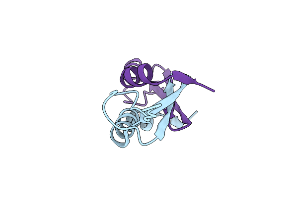
Structure Of Human Peroxiredoxin 3 As Three Stacked Rings
Organism: Homo sapiens
Method: X-RAY DIFFRACTION Resolution:2.80 Å Release Date: 2016-05-25 Classification: OXIDOREDUCTASE |
Organism: Homo sapiens
Method: X-RAY DIFFRACTION
Release Date: 2016-05-25
 |
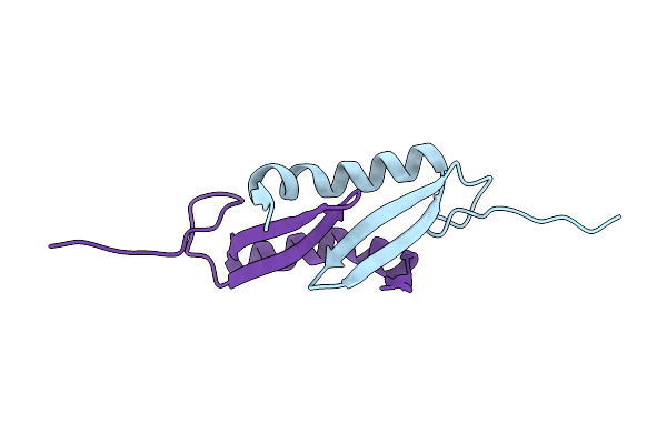
Crystal Structure Of The Dimerization Domain Of Lsr2 From Mycobacterium Tuberculosis In The P 1 21 1 Space Group
Organism: Mycobacterium tuberculosis
Method: X-RAY DIFFRACTION Resolution:1.73 Å Release Date: 2012-06-20 Classification: DNA BINDING PROTEIN |
Organism: Mycobacterium tuberculosis
Method: X-RAY DIFFRACTION
Release Date: 2012-06-20
 |
Crystal Structure Of The Dimerization Domain Of Lsr2 From Mycobacterium Tuberculosis In The P 31 2 1 Space Group
Organism: Mycobacterium tuberculosis
Method: X-RAY DIFFRACTION Resolution:2.04 Å Release Date: 2012-06-20 Classification: DNA BINDING PROTEIN |
Organism: Mycobacterium tuberculosis
Method: X-RAY DIFFRACTION
Release Date: 2012-06-20
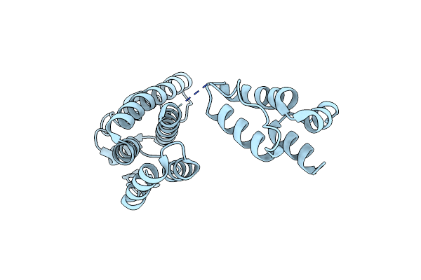 |
Pseudo-Atomic Model Of The Rous Sarcoma Virus Capsid Hexamer
Organism: Rous sarcoma virus
Method: X-RAY DIFFRACTION Resolution:4.10 Å Release Date: 2012-04-04 Classification: VIRAL PROTEIN |
Organism: Rous sarcoma virus
Method: X-RAY DIFFRACTION
Release Date: 2012-04-04
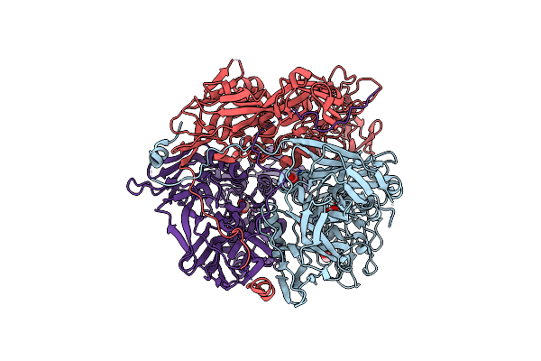 |
Structure Of D13, The Scaffolding Protein Of Vaccinia Virus (Mutant D513G)
Organism: Vaccinia virus
Method: X-RAY DIFFRACTION Resolution:2.55 Å Release Date: 2011-06-22 Classification: VIRAL PROTEIN Ligands: FMT |
Organism: Vaccinia virus
Method: X-RAY DIFFRACTION
Release Date: 2011-06-22
Ligands: FMT
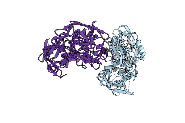 |
Structure Of D13, The Scaffolding Protein Of Vaccinia Virus
Organism: Vaccinia virus
Method: X-RAY DIFFRACTION Resolution:3.51 Å Release Date: 2011-06-22 Classification: VIRAL PROTEIN |
Organism: Vaccinia virus
Method: X-RAY DIFFRACTION
Release Date: 2011-06-22
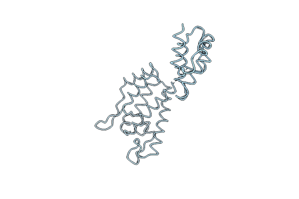 |
Cryo-Em 3D Model Of The Icosahedral Particle Composed Of Rous Sarcoma Virus Capsid Protein Pentamers
Organism: Rous sarcoma virus - prague c
Method: ELECTRON MICROSCOPY Resolution:18.30 Å Release Date: 2010-05-19 Classification: VIRUS |
Organism: Rous sarcoma virus - prague c
Method: ELECTRON MICROSCOPY
Release Date: 2010-05-19
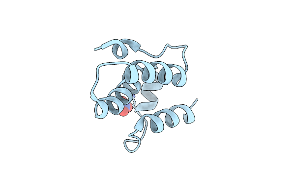 |
Crystal Structure Of The C-Terminal Domain From The Rous Sarcoma Virus Capsid Protein: Mutant D179A
Organism: Rous sarcoma virus
Method: X-RAY DIFFRACTION Resolution:2.00 Å Release Date: 2009-06-02 Classification: VIRAL PROTEIN Ligands: NO3 |
Organism: Rous sarcoma virus
Method: X-RAY DIFFRACTION
Release Date: 2009-06-02
Ligands: NO3
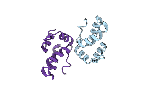 |
Crystal Structure Of The C-Terminal Domain From The Rous Sarcoma Virus Capsid Protein: High Ph
Organism: Rous sarcoma virus
Method: X-RAY DIFFRACTION Resolution:2.01 Å Release Date: 2009-06-02 Classification: VIRAL PROTEIN |
Organism: Rous sarcoma virus
Method: X-RAY DIFFRACTION
Release Date: 2009-06-02