

Search Count: 10
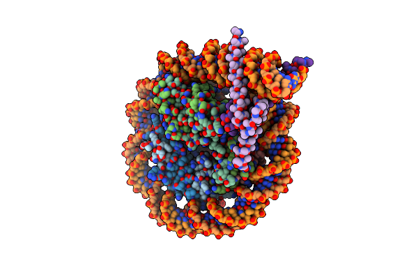 |
Cryo-Em Structure Of Clock-Bmal1 Bound To A Nucleosomal E-Box At Position Shl-6.2 (Dna Conformation 1)
Organism: Homo sapiens, Mus musculus, Synthetic construct
Method: ELECTRON MICROSCOPY Release Date: 2023-05-24 Classification: GENE REGULATION |
Organism: Homo sapiens, Mus musculus, Synthetic construct
Method: ELECTRON MICROSCOPY
Release Date: 2023-05-24
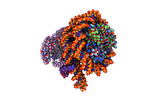 |
Cryo-Em Structure Of Clock-Bmal1 Bound To A Nucleosomal E-Box At Position Shl+5.8 (Composite Map)
Organism: Homo sapiens, Mus musculus, Synthetic construct
Method: ELECTRON MICROSCOPY Release Date: 2023-05-24 Classification: GENE REGULATION |
Organism: Homo sapiens, Mus musculus, Synthetic construct
Method: ELECTRON MICROSCOPY
Release Date: 2023-05-24
 |
Cryo-Em Structure Of Clock-Bmal1 Bound To The Native Por Enhancer Nucleosome (Map 2, Additional 3D Classification And Flexible Refinement)
Organism: Homo sapiens, Mus musculus
Method: ELECTRON MICROSCOPY Release Date: 2023-05-24 Classification: GENE REGULATION |
Organism: Homo sapiens, Mus musculus
Method: ELECTRON MICROSCOPY
Release Date: 2023-05-24
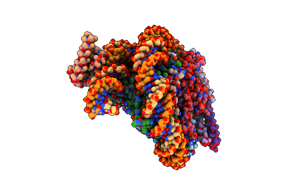 |
Oct4 And Myc-Max Co-Bound To A Nucleosome
Organism: Homo sapiens, Aequorea victoria, Synthetic construct
Method: ELECTRON MICROSCOPY Release Date: 2023-05-24 Classification: TRANSCRIPTION Ligands: PTD |
Organism: Homo sapiens, Aequorea victoria, Synthetic construct
Method: ELECTRON MICROSCOPY
Release Date: 2023-05-24
Ligands: PTD
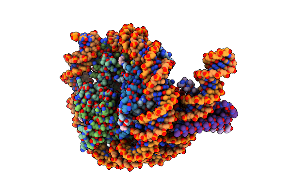 |
Myc-Max Bound To A Nucleosome At Shl+5.8
Organism: Homo sapiens, Synthetic construct
Method: ELECTRON MICROSCOPY Release Date: 2023-05-24 Classification: TRANSCRIPTION Ligands: PTD |
Organism: Homo sapiens, Synthetic construct
Method: ELECTRON MICROSCOPY
Release Date: 2023-05-24
Ligands: PTD
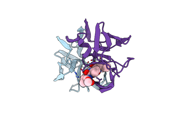 |
Hydroxyethylene Isostere Inhibitors Of Human Immunodeficiency Virus-1 Protease: Structure-Activity Analysis Using Enzyme Kinetics, X-Ray Crystallography, And Infected T-Cell Assays
Organism: Human immunodeficiency virus 1
Method: X-RAY DIFFRACTION Resolution:2.50 Å Release Date: 1994-06-22 Classification: HYDROLASE/HYDROLASE INHIBITOR Ligands: PSI |
Organism: Human immunodeficiency virus 1
Method: X-RAY DIFFRACTION
Release Date: 1994-06-22
Ligands: PSI
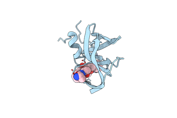 |
The Crystal Structures At 2.2 Angstroms Resolution Of Hydroxyethylene-Based Inhibitors Bound To Human Immunodeficiency Virus Type 1 Protease Show That The Inhibitors Are Present In Two Distinct Orientations
Organism: Human immunodeficiency virus 1
Method: X-RAY DIFFRACTION Resolution:2.20 Å Release Date: 1994-05-31 Classification: HYDROLASE/HYDROLASE INHIBITOR |
Organism: Human immunodeficiency virus 1
Method: X-RAY DIFFRACTION
Release Date: 1994-05-31
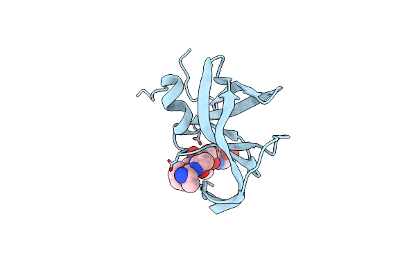 |
The Crystal Structures At 2.2 Angstroms Resolution Of Hydroxyethylene-Based Inhibitors Bound To Human Immunodeficiency Virus Type 1 Protease Show That The Inhibitors Are Present In Two Distinct Orientations
Organism: Human immunodeficiency virus 1
Method: X-RAY DIFFRACTION Resolution:2.20 Å Release Date: 1994-05-31 Classification: HYDROLASE/HYDROLASE INHIBITOR Ligands: PSI |
Organism: Human immunodeficiency virus 1
Method: X-RAY DIFFRACTION
Release Date: 1994-05-31
Ligands: PSI
 |
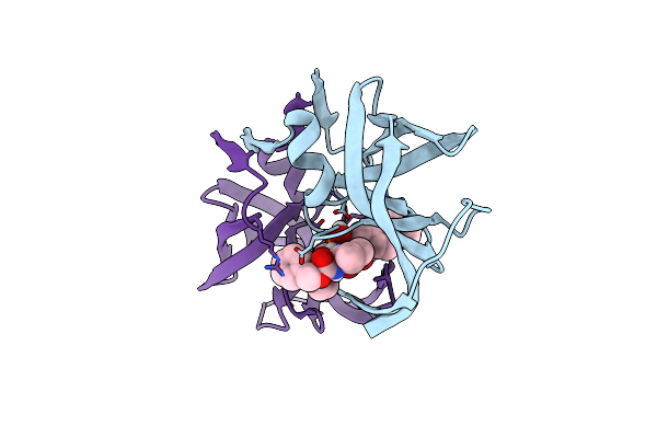
Three-Dimensional Structure Of A Siv Protease(Slash)Inhibitor Complex. Implications For The Design Of Hiv-1 And Hiv-2 Protease Inhibitors
Organism: Simian immunodeficiency virus
Method: X-RAY DIFFRACTION Resolution:2.50 Å Release Date: 1994-01-31 Classification: HYDROLASE/HYDROLASE INHIBITOR Ligands: PSI |
Organism: Simian immunodeficiency virus
Method: X-RAY DIFFRACTION
Release Date: 1994-01-31
Ligands: PSI
 |
Inhibition Of Human Immunodeficiency Virus-1 Protease By A C2-Symmetric Phosphinate Synthesis And Crystallographic Analysis
Organism: Human immunodeficiency virus 1
Method: X-RAY DIFFRACTION Resolution:2.30 Å Release Date: 1993-10-31 Classification: HYDROLASE(ACID PROTEINASE) Ligands: PHP |
Organism: Human immunodeficiency virus 1
Method: X-RAY DIFFRACTION
Release Date: 1993-10-31
Ligands: PHP