

Search Count: 11
 |
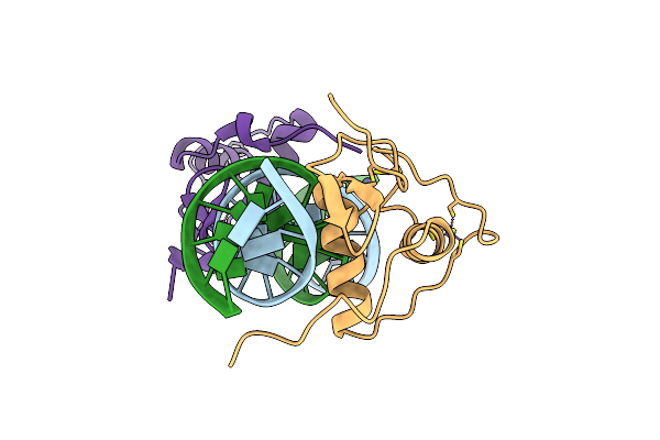
Crystal Structure Of The Human Rxr/Rar Dna-Binding Domain Heterodimer Bound To The Human Rarb2 Dr5 Response Element
Organism: Homo sapiens
Method: X-RAY DIFFRACTION Resolution:2.40 Å Release Date: 2020-09-09 Classification: TRANSCRIPTION Ligands: ZN, GOL, CL |
Organism: Homo sapiens
Method: X-RAY DIFFRACTION
Release Date: 2020-09-09
Ligands: ZN, GOL, CL
 |
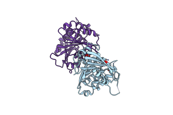
Crystal Structure Of The Human Rxr Dna-Binding Domain Homodimer Bound To The Human Hoxb13 Dr0 Response Element
Organism: Homo sapiens
Method: X-RAY DIFFRACTION Resolution:2.10 Å Release Date: 2020-09-09 Classification: TRANSCRIPTION Ligands: ZN |
Organism: Homo sapiens
Method: X-RAY DIFFRACTION
Release Date: 2020-09-09
Ligands: ZN
 |
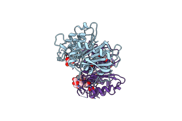
Crystal Structure Of The Ferric Enterobactin Esterase (Pfee) From Pseudomonas Aeruginosa
Organism: Pseudomonas aeruginosa
Method: X-RAY DIFFRACTION Resolution:2.00 Å Release Date: 2018-06-20 Classification: HYDROLASE Ligands: NO3 |
Organism: Pseudomonas aeruginosa
Method: X-RAY DIFFRACTION
Release Date: 2018-06-20
Ligands: NO3
 |
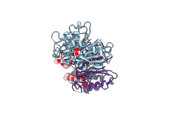
Crystal Structure Of The Ferric Enterobactin Esterase (Pfee) Mutant(S157A) From Pseudomonas Aeruginosa In Presence Of Enterobactin
Organism: Pseudomonas aeruginosa pao1
Method: X-RAY DIFFRACTION Resolution:1.66 Å Release Date: 2018-06-20 Classification: HYDROLASE Ligands: FE, DBS, NO3, EDO, EB4 |
Organism: Pseudomonas aeruginosa pao1
Method: X-RAY DIFFRACTION
Release Date: 2018-06-20
Ligands: FE, DBS, NO3, EDO, EB4
 |
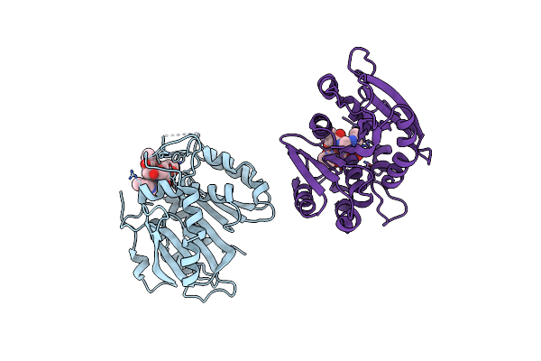
Crystal Structure Of The Ferric Enterobactin Esterase (Pfee) Mutant(S157A) From Pseudomonas Aeruginosa In Complex With Tris-Catechol Vector
Organism: Pseudomonas aeruginosa
Method: X-RAY DIFFRACTION Resolution:1.49 Å Release Date: 2018-06-20 Classification: HYDROLASE Ligands: FE, 8SW, NO3, EDO |
Organism: Pseudomonas aeruginosa
Method: X-RAY DIFFRACTION
Release Date: 2018-06-20
Ligands: FE, 8SW, NO3, EDO
 |
Crystal Structure Of The Ferric Enterobactin Esterase (Pfee) From Pseudomonas Aeruginosa In Complex With The Tris-Catechol Vector
Organism: Pseudomonas aeruginosa
Method: X-RAY DIFFRACTION Resolution:3.11 Å Release Date: 2018-06-20 Classification: HYDROLASE Ligands: FE, 8SW |
Organism: Pseudomonas aeruginosa
Method: X-RAY DIFFRACTION
Release Date: 2018-06-20
Ligands: FE, 8SW
 |
Structural Insights Into The Ctar Dna Recognition By The Hiv-1 Nucleocapsid Protein: Role Of Sugar Deoxyriboses In The Binding Polarity Of Nc
Organism: Human immunodeficiency virus 1
Method: SOLUTION NMR Release Date: 2010-12-08 Classification: VIRAL PROTEIN/DNA Ligands: ZN |
Organism: Human immunodeficiency virus 1
Method: SOLUTION NMR
Release Date: 2010-12-08
Ligands: ZN
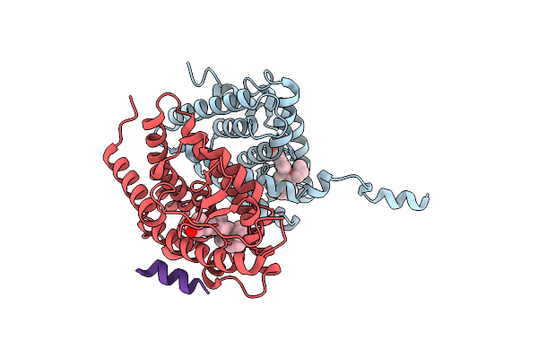 |
Crystal Structure Of A Mixed Agonist-Bound Rar-Alpha And Antagonist-Bound Rxr-Alpha Heterodimer Ligand Binding Domains
Organism: Mus musculus, Homo sapiens, Homo sapiens
Method: X-RAY DIFFRACTION Resolution:2.75 Å Release Date: 2010-10-06 Classification: TRANSCRIPTION Ligands: 754, REA |
Organism: Mus musculus, Homo sapiens, Homo sapiens
Method: X-RAY DIFFRACTION
Release Date: 2010-10-06
Ligands: 754, REA
 |
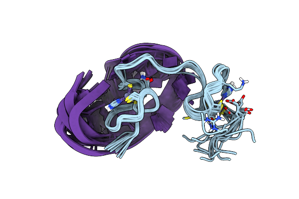
How The Hiv-1 Nucleocapsid Protein Binds And Destabilises The (-)Primer Binding Site During Reverse Transcription
|
Method: SOLUTION NMR
Release Date: 2009-01-13
Ligands: ZN
 |
Solution Structure Of The Hiv-1 Nucleocapsid (Ncp7(12-55)) Complexed With The Dna (-) Primer Binding Site
|
Method: SOLUTION NMR
Release Date: 2007-04-24
Ligands: ZN
 |
Dimeric Structure Of The C-Terminal Domain Of Vpr
|
Method: SOLUTION NMR
Release Date: 2005-06-14