

Search Count: 83
 |
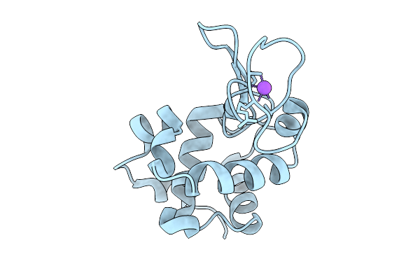
5 Micrometer Hewl Crystals Solved At Room-Temperature Using Fixed-Target Serial Crystallography.
Organism: Gallus gallus
Method: X-RAY DIFFRACTION Resolution:1.55 Å Release Date: 2025-11-12 Classification: HYDROLASE Ligands: CL, NA |
Organism: Gallus gallus
Method: X-RAY DIFFRACTION
Release Date: 2025-11-12
Ligands: CL, NA
 |
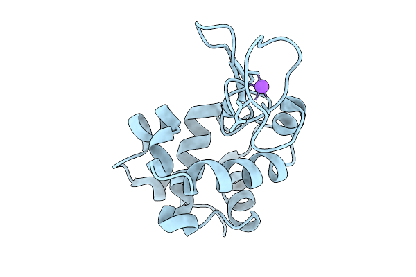
10 Micrometer Hewl Crystals Solved At Room-Temperature Using Fixed-Target Serial Crystallography.
Organism: Gallus gallus
Method: X-RAY DIFFRACTION Resolution:1.47 Å Release Date: 2025-09-10 Classification: HYDROLASE Ligands: CL, NA |
Organism: Gallus gallus
Method: X-RAY DIFFRACTION
Release Date: 2025-09-10
Ligands: CL, NA
 |
25 Micrometer Hewl Crystals Solved At Room-Temperature Using Fixed-Target Serial Crystallography.
Organism: Gallus gallus
Method: X-RAY DIFFRACTION Resolution:1.33 Å Release Date: 2025-09-10 Classification: HYDROLASE Ligands: CL, NA |
Organism: Gallus gallus
Method: X-RAY DIFFRACTION
Release Date: 2025-09-10
Ligands: CL, NA
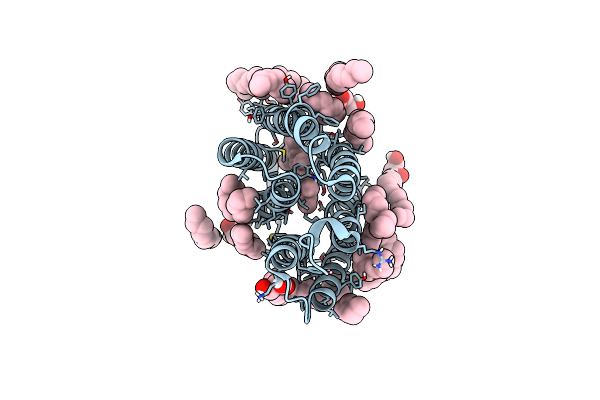 |
Laser Excitation Effects On Br: Reprocessed Dark Dataset Recorded From Nango Et Al.
Organism: Halobacterium salinarum
Method: X-RAY DIFFRACTION Resolution:2.00 Å Release Date: 2025-01-22 Classification: PROTON TRANSPORT Ligands: LI1, OLC, RET |
Organism: Halobacterium salinarum
Method: X-RAY DIFFRACTION
Release Date: 2025-01-22
Ligands: LI1, OLC, RET
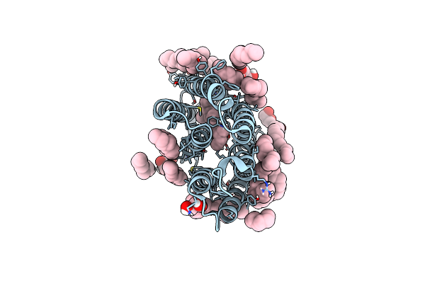 |
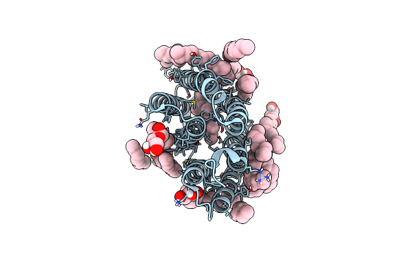
Laser Excitation Effects On Br: Reprocessed Extrapolated 16 Ns Light Dataset Recorded At 0.04 Gw/Cm2 From Nango Et Al.
Organism: Halobacterium salinarum
Method: X-RAY DIFFRACTION Resolution:2.00 Å Release Date: 2025-01-22 Classification: PROTON TRANSPORT Ligands: LI1, OLC, RET |
Organism: Halobacterium salinarum
Method: X-RAY DIFFRACTION
Release Date: 2025-01-22
Ligands: LI1, OLC, RET
 |
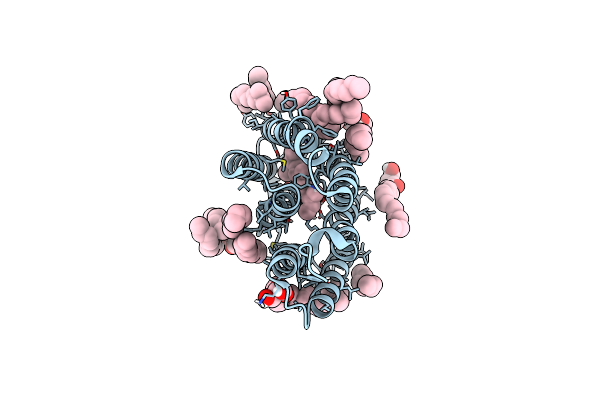
Laser Excitation Effects On Br: Dark Dataset Recorded At Swissfel
Organism: Halobacterium salinarum
Method: X-RAY DIFFRACTION Resolution:1.70 Å Release Date: 2025-01-22 Classification: PROTON TRANSPORT Ligands: LI1, OLC, RET |
Organism: Halobacterium salinarum
Method: X-RAY DIFFRACTION
Release Date: 2025-01-22
Ligands: LI1, OLC, RET
 |
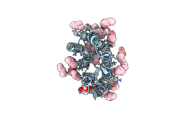
Laser Excitation Effects On Br: Extrapolated 6 Ps Light Dataset Recorded At 342 Gw/Cm2 At Swissfel
Organism: Halobacterium salinarum
Method: X-RAY DIFFRACTION Resolution:1.80 Å Release Date: 2025-01-22 Classification: PROTON TRANSPORT Ligands: LI1, OLC, RET |
Organism: Halobacterium salinarum
Method: X-RAY DIFFRACTION
Release Date: 2025-01-22
Ligands: LI1, OLC, RET
 |
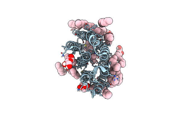
Laser Excitation Effects On Br: Extrapolated 6 Ps Light Dataset Recorded At 2493 Gw/Cm2 At Swissfel
Organism: Halobacterium salinarum
Method: X-RAY DIFFRACTION Resolution:1.80 Å Release Date: 2025-01-22 Classification: PROTON TRANSPORT Ligands: LI1, OLC, RET |
Organism: Halobacterium salinarum
Method: X-RAY DIFFRACTION
Release Date: 2025-01-22
Ligands: LI1, OLC, RET
 |
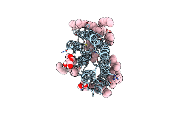
Laser Excitation Effects On Br: Reprocessed Dark From Nogly Et Al.
Organism: Halobacterium salinarum
Method: X-RAY DIFFRACTION Resolution:1.44 Å Release Date: 2025-01-22 Classification: PROTON TRANSPORT Ligands: LI1, OLC, RET |
Organism: Halobacterium salinarum
Method: X-RAY DIFFRACTION
Release Date: 2025-01-22
Ligands: LI1, OLC, RET
 |
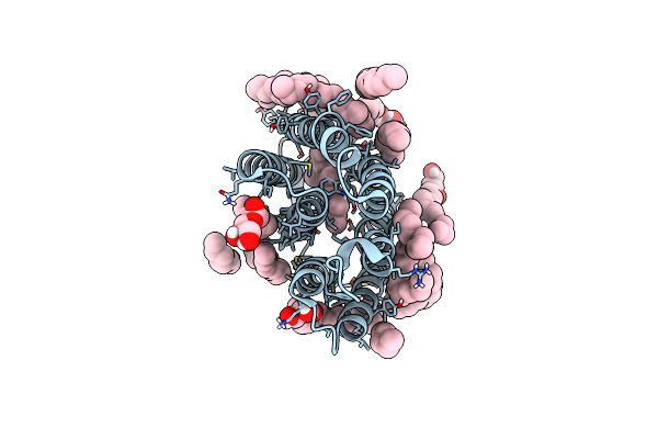
Laser Excitation Effects On Br: Reprocessed Extrapolated 10Ps Light Dataset Recorded At 525 Gw/Cm2 From Nogly Et Al.
Organism: Halobacterium salinarum
Method: X-RAY DIFFRACTION Resolution:1.66 Å Release Date: 2025-01-22 Classification: PROTON TRANSPORT Ligands: LI1, OLC, RET |
Organism: Halobacterium salinarum
Method: X-RAY DIFFRACTION
Release Date: 2025-01-22
Ligands: LI1, OLC, RET
 |
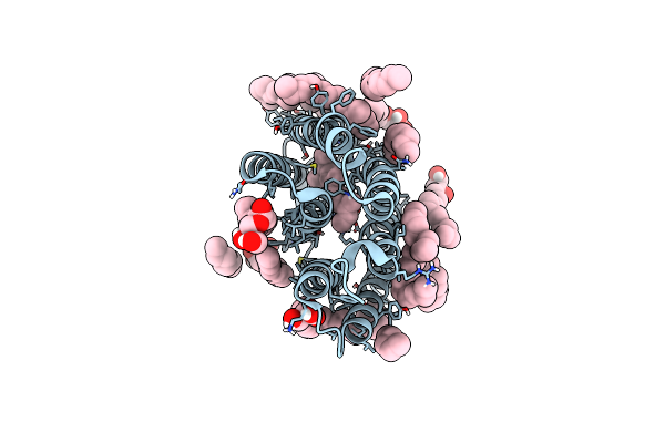
Laser Excitation Effects On Br: Dark Dataset Recorded At Sacla
Organism: Halobacterium salinarum
Method: X-RAY DIFFRACTION Resolution:1.60 Å Release Date: 2025-01-22 Classification: PROTON TRANSPORT Ligands: LI1, OLC, RET |
Organism: Halobacterium salinarum
Method: X-RAY DIFFRACTION
Release Date: 2025-01-22
Ligands: LI1, OLC, RET
 |
Laser Excitation Effects On Br: Extrapolated 10Ps Light Dataset Recorded At 1281 Gw/Cm2 At Sacla (+100Ps, 1Ns, 10Ns)
Organism: Halobacterium salinarum
Method: X-RAY DIFFRACTION Resolution:1.76 Å Release Date: 2025-01-22 Classification: PROTON TRANSPORT Ligands: LI1, OLC, RET |
Organism: Halobacterium salinarum
Method: X-RAY DIFFRACTION
Release Date: 2025-01-22
Ligands: LI1, OLC, RET
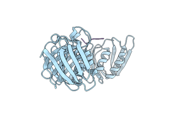 |
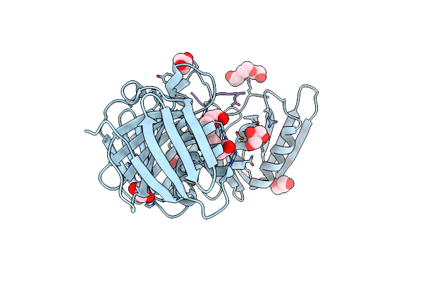
Structure Of The E.Coli Dna Polymerase Sliding Clamp With A Covalently Bound Peptide 3.
Organism: Escherichia coli
Method: X-RAY DIFFRACTION Resolution:1.45 Å Release Date: 2024-03-13 Classification: REPLICATION |
Organism: Escherichia coli
Method: X-RAY DIFFRACTION
Release Date: 2024-03-13
 |
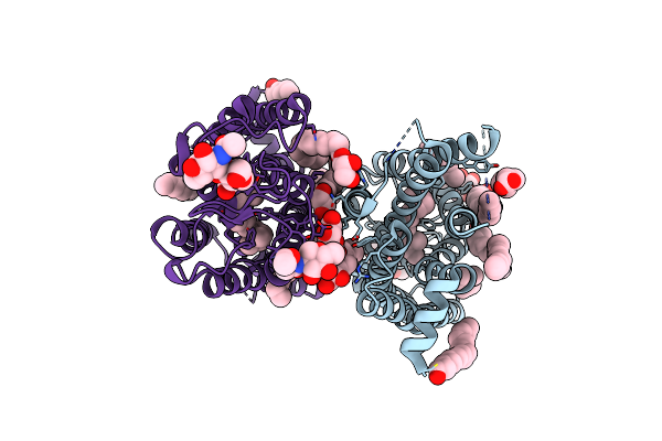
Structure Of The E.Coli Dna Polymerase Sliding Clamp With A Covalently Bound Peptide 2.
Organism: Escherichia coli
Method: X-RAY DIFFRACTION Resolution:1.21 Å Release Date: 2024-03-13 Classification: REPLICATION Ligands: GOL, 1PE |
Organism: Escherichia coli
Method: X-RAY DIFFRACTION
Release Date: 2024-03-13
Ligands: GOL, 1PE
 |
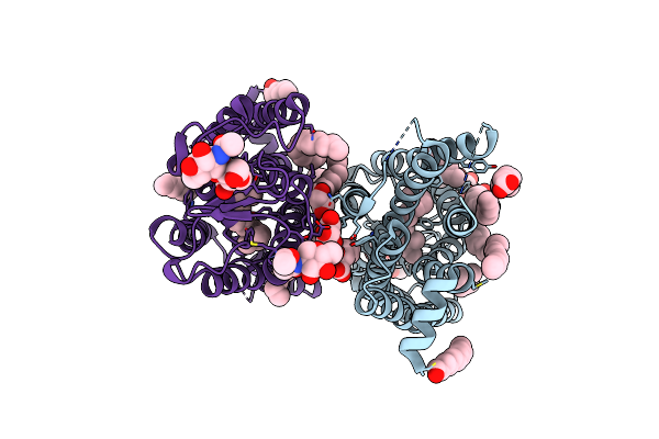
Dark State Crystal Structure Of Bovine Rhodopsin In Lipidic Cubic Phase (Sacla)
Organism: Bos taurus
Method: X-RAY DIFFRACTION Resolution:1.80 Å Release Date: 2023-03-29 Classification: MEMBRANE PROTEIN Ligands: ACE, RET, NAG, DAO, OLC, PLM |
Organism: Bos taurus
Method: X-RAY DIFFRACTION
Release Date: 2023-03-29
Ligands: ACE, RET, NAG, DAO, OLC, PLM
 |
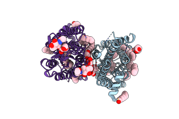
Dark State Crystal Structure Of Bovine Rhodopsin In Lipidic Cubic Phase (Swissfel)
Organism: Bos taurus
Method: X-RAY DIFFRACTION Resolution:1.80 Å Release Date: 2023-03-29 Classification: MEMBRANE PROTEIN Ligands: ACE, RET, NAG, DAO, OLC, PLM |
Organism: Bos taurus
Method: X-RAY DIFFRACTION
Release Date: 2023-03-29
Ligands: ACE, RET, NAG, DAO, OLC, PLM
 |
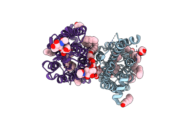
1 Picosecond Light Activated Crystal Structure Of Bovine Rhodopsin In Lipidic Cubic Phase
Organism: Bos taurus
Method: X-RAY DIFFRACTION Resolution:1.80 Å Release Date: 2023-03-29 Classification: MEMBRANE PROTEIN Ligands: ACE, RET, NAG, DAO, OLC, PLM |
Organism: Bos taurus
Method: X-RAY DIFFRACTION
Release Date: 2023-03-29
Ligands: ACE, RET, NAG, DAO, OLC, PLM
 |
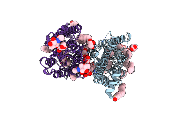
10 Picosecond Light Activated Crystal Structure Of Bovine Rhodopsin In Lipidic Cubic Phase
Organism: Bos taurus
Method: X-RAY DIFFRACTION Resolution:1.80 Å Release Date: 2023-03-29 Classification: MEMBRANE PROTEIN Ligands: ACE, RET, NAG, DAO, OLC, PLM |
Organism: Bos taurus
Method: X-RAY DIFFRACTION
Release Date: 2023-03-29
Ligands: ACE, RET, NAG, DAO, OLC, PLM
 |
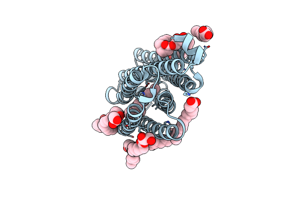
100 Picosecond Light Activated Crystal Structure Of Bovine Rhodopsin In Lipidic Cubic Phase (Sacla)
Organism: Bos taurus
Method: X-RAY DIFFRACTION Resolution:1.80 Å Release Date: 2023-03-29 Classification: MEMBRANE PROTEIN Ligands: RET, NAG, DAO, OLC, PLM |
Organism: Bos taurus
Method: X-RAY DIFFRACTION
Release Date: 2023-03-29
Ligands: RET, NAG, DAO, OLC, PLM
 |
Nmhr Dark State Structure Determined By Serial Femtosecond Crystallography
Organism: Nonlabens marinus s1-08
Method: X-RAY DIFFRACTION Resolution:1.45 Å Release Date: 2022-02-09 Classification: MEMBRANE PROTEIN Ligands: RET, CL, OLC, OLA |
Organism: Nonlabens marinus s1-08
Method: X-RAY DIFFRACTION
Release Date: 2022-02-09
Ligands: RET, CL, OLC, OLA