

Search Count: 17
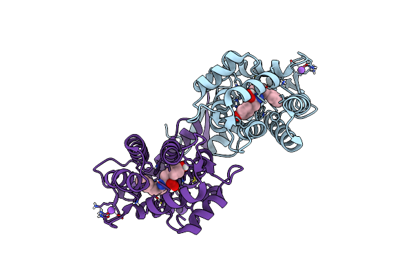 |
Crystal Structure Of H64F Obelin Mutant From Obelia Longissima At 2.2 Angstrom Resolution
Organism: Obelia longissima
Method: X-RAY DIFFRACTION Resolution:2.20 Å Release Date: 2023-03-29 Classification: LUMINESCENT PROTEIN Ligands: CZH, NA |
Organism: Obelia longissima
Method: X-RAY DIFFRACTION
Release Date: 2023-03-29
Ligands: CZH, NA
 |
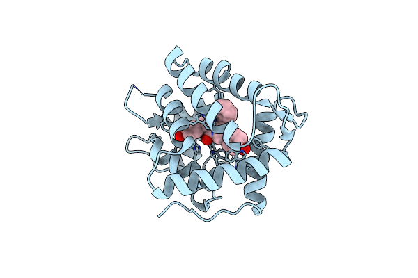
Crystal Structure Of Ca(2+)-Regulated Photoprotein Mitrocomin From Jellyfish Mitrocoma Cellularia At 1.3 Angstrom Resolution
Organism: Mitrocoma cellularia
Method: X-RAY DIFFRACTION Resolution:1.30 Å Release Date: 2014-11-26 Classification: LUMINESCENT PROTEIN Ligands: CZH |
Organism: Mitrocoma cellularia
Method: X-RAY DIFFRACTION
Release Date: 2014-11-26
Ligands: CZH
 |
Crystal Structure Of Y138F Obelin Mutant From Obelia Longissima At 1.72 Angstrom Resolution
Organism: Obelia longissima
Method: X-RAY DIFFRACTION Resolution:1.72 Å Release Date: 2014-03-12 Classification: LUMINESCENT PROTEIN Ligands: CZH |
Organism: Obelia longissima
Method: X-RAY DIFFRACTION
Release Date: 2014-03-12
Ligands: CZH
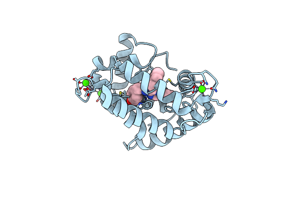 |
Crystal Structure Of Ca(2+)- Discharged Y138F Obelin Mutant From Obelia Longissima At 1.30 Angstrom Resolution
Organism: Obelia longissima
Method: X-RAY DIFFRACTION Resolution:1.30 Å Release Date: 2014-03-12 Classification: LUMINESCENT PROTEIN Ligands: CEI, CA |
Organism: Obelia longissima
Method: X-RAY DIFFRACTION
Release Date: 2014-03-12
Ligands: CEI, CA
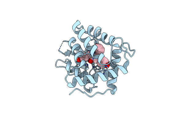 |
Crystal Structure Of F88Y Obelin Mutant From Obelia Longissima At 2.09 Angstrom Resolution
Organism: Obelia longissima
Method: X-RAY DIFFRACTION Resolution:2.09 Å Release Date: 2014-02-19 Classification: LUMINESCENT PROTEIN Ligands: CZH |
Organism: Obelia longissima
Method: X-RAY DIFFRACTION
Release Date: 2014-02-19
Ligands: CZH
 |
Crystal Structure Of Ca(2+)- Discharged F88Y Obelin Mutant From Obelia Longissima At 1.50 Angstrom Resolution
Organism: Obelia longissima
Method: X-RAY DIFFRACTION Resolution:1.50 Å Release Date: 2014-02-19 Classification: LUMINESCENT PROTEIN Ligands: CEI, CA |
Organism: Obelia longissima
Method: X-RAY DIFFRACTION
Release Date: 2014-02-19
Ligands: CEI, CA
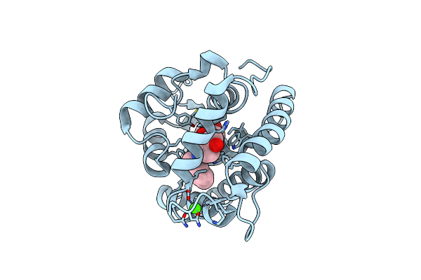 |
Crystal Structure Analysis Of Photoprotein Clytin
Organism: Clytia gregaria
Method: X-RAY DIFFRACTION Resolution:1.90 Å Release Date: 2010-10-06 Classification: FLUORESCENT PROTEIN, HYDROLASE Ligands: CZH, CA |
Organism: Clytia gregaria
Method: X-RAY DIFFRACTION
Release Date: 2010-10-06
Ligands: CZH, CA
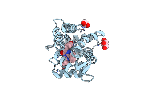 |
Crystal Structure Of Coelenterazine-Binding Protein From Renilla Muelleri
Organism: Renilla muelleri
Method: X-RAY DIFFRACTION Resolution:1.72 Å Release Date: 2007-01-09 Classification: LUMINESCENT PROTEIN Ligands: CTZ, GOL |
Organism: Renilla muelleri
Method: X-RAY DIFFRACTION
Release Date: 2007-01-09
Ligands: CTZ, GOL
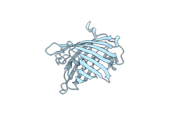 |
Green Fluorescent Protein From Clytia Gregaria
Organism: Clytia gregaria
Method: X-RAY DIFFRACTION Resolution:1.55 Å Release Date: 2006-09-12 Classification: LUMINESCENT PROTEIN |
Organism: Clytia gregaria
Method: X-RAY DIFFRACTION
Release Date: 2006-09-12
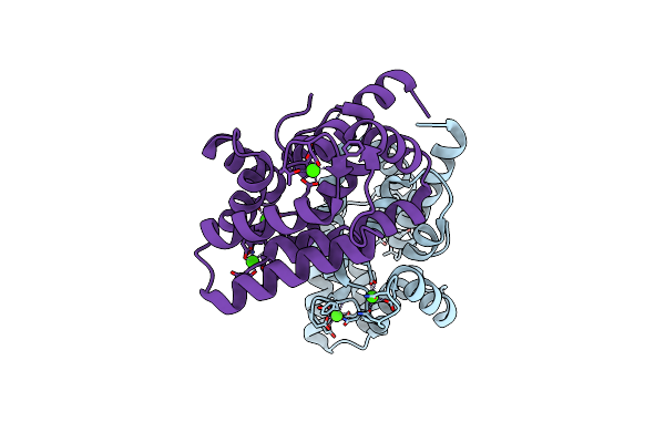 |
Crystal Structure Of Coelenterazine-Binding Protein From Renilla Muelleri In The Ca Loaded Apo Form
Organism: Renilla muelleri
Method: X-RAY DIFFRACTION Resolution:1.80 Å Release Date: 2006-09-12 Classification: STRUCTURAL GENOMICS, UNKNOWN FUNCTION Ligands: CA |
Organism: Renilla muelleri
Method: X-RAY DIFFRACTION
Release Date: 2006-09-12
Ligands: CA
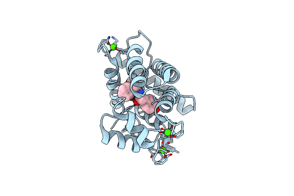 |
Crystal Structure Of Obelin Following Ca2+ Triggered Bioluminescence Suggests Neutral Coelenteramide As The Primary Excited State
Organism: Obelia longissima
Method: X-RAY DIFFRACTION Resolution:1.93 Å Release Date: 2006-02-14 Classification: LUMINESCENT PROTEIN Ligands: CA, CEI |
Organism: Obelia longissima
Method: X-RAY DIFFRACTION
Release Date: 2006-02-14
Ligands: CA, CEI
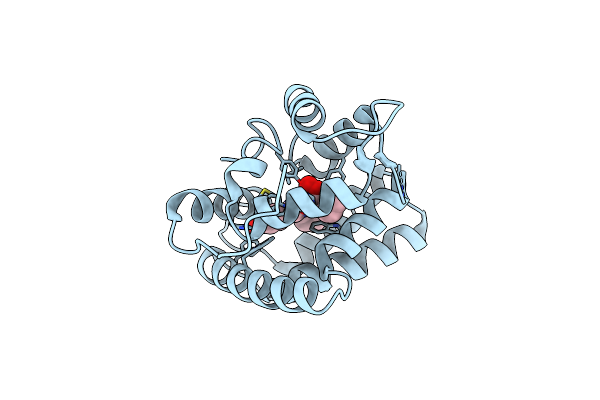 |
Obelin From Obelia Longissima
Organism: Obelia longissima
Method: X-RAY DIFFRACTION Resolution:1.17 Å Release Date: 2005-07-19 Classification: LUMINESCENT PROTEIN Ligands: CZH |
Organism: Obelia longissima
Method: X-RAY DIFFRACTION
Release Date: 2005-07-19
Ligands: CZH
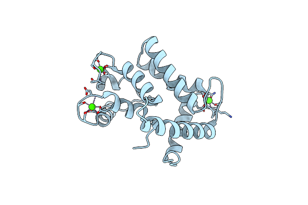 |
Crystal Structure Of Calcium-Loaded Apo-Obelin From Obelia Longissima
Organism: Obelia longissima
Method: X-RAY DIFFRACTION Resolution:2.20 Å Release Date: 2004-12-28 Classification: LUMINESCENT PROTEIN Ligands: CA |
Organism: Obelia longissima
Method: X-RAY DIFFRACTION
Release Date: 2004-12-28
Ligands: CA
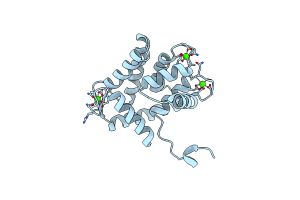 |
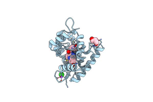
Calcium-Loaded Apo-Aequorin From Aequorea Victoria
Organism: Aequorea victoria
Method: X-RAY DIFFRACTION Resolution:1.70 Å Release Date: 2004-12-28 Classification: LUMINESCENT PROTEIN Ligands: CA |
Organism: Aequorea victoria
Method: X-RAY DIFFRACTION
Release Date: 2004-12-28
Ligands: CA
 |
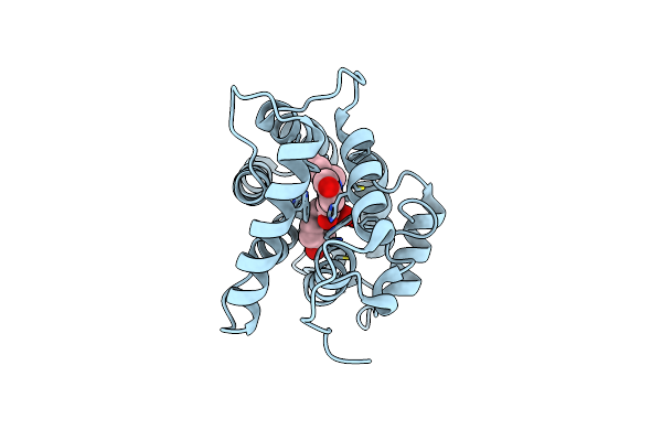
Crystal Structure Of A Ca2+-Discharged Photoprotein: Implications For The Mechanisms Of The Calcium Trigger And The Bioluminescence
Organism: Obelia longissima
Method: X-RAY DIFFRACTION Resolution:1.96 Å Release Date: 2004-10-05 Classification: LUMINESCENT PROTEIN |
Organism: Obelia longissima
Method: X-RAY DIFFRACTION
Release Date: 2004-10-05
 |
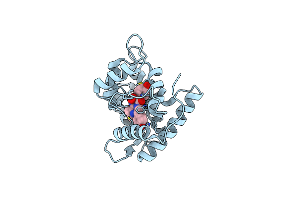
The Crystal Structure Of Obelin From Obelia Geniculata At 1.82 A Resolution
Organism: Obelia longissima
Method: X-RAY DIFFRACTION Resolution:1.82 Å Release Date: 2001-07-11 Classification: LUMINESCENT PROTEIN Ligands: CZH |
Organism: Obelia longissima
Method: X-RAY DIFFRACTION
Release Date: 2001-07-11
Ligands: CZH
 |
Crystal Structure Of W92F Obelin Mutant From Obelia Longissima At 1.72 Angstrom Resolution
Organism: Obelia longissima
Method: X-RAY DIFFRACTION Resolution:1.72 Å Release Date: 2001-07-11 Classification: LUMINESCENT PROTEIN Ligands: CZH |
Organism: Obelia longissima
Method: X-RAY DIFFRACTION
Release Date: 2001-07-11
Ligands: CZH