

Search Count: 86
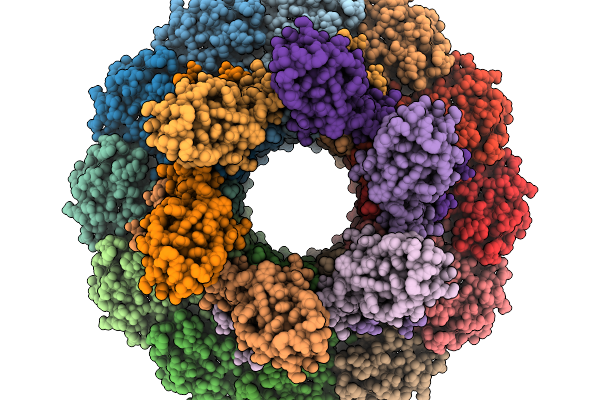 |
Structure Of Bacteriophage T4 Neck Protein Gp13 And Gp14 Assembled In Vitro In C6 Symmetry
Organism: Escherichia phage t4
Method: ELECTRON MICROSCOPY Release Date: 2026-02-18 Classification: VIRAL PROTEIN |
Organism: Escherichia phage t4
Method: ELECTRON MICROSCOPY
Release Date: 2026-02-18
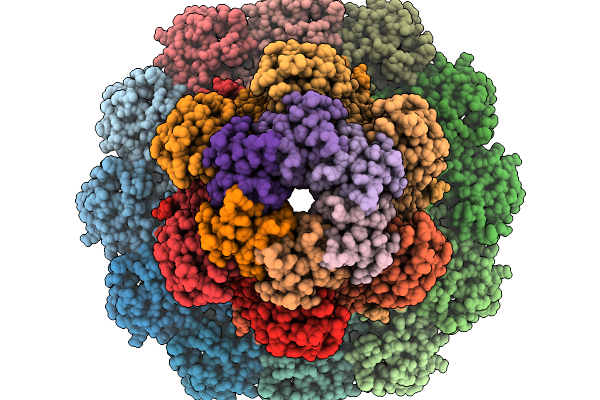 |
Structure Of Bacteriophage T4 Neck Protein Gp13 And Gp14 And Hfq Assembled In Vitro In C6 Symmetry
Organism: Escherichia phage t4
Method: ELECTRON MICROSCOPY Release Date: 2026-02-18 Classification: VIRAL PROTEIN |
Organism: Escherichia phage t4
Method: ELECTRON MICROSCOPY
Release Date: 2026-02-18
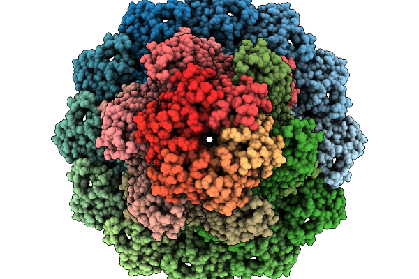 |
Structure Of Bacteriophage T4 Protal-Neck Protein Gp20-Gp13-Gp14-Hfq Assembled In Vitro In C6 Symmetry
Organism: Escherichia phage t4
Method: ELECTRON MICROSCOPY Resolution:2.91 Å Release Date: 2026-02-18 Classification: VIRAL PROTEIN |
Organism: Escherichia phage t4
Method: ELECTRON MICROSCOPY
Release Date: 2026-02-18
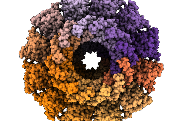 |
Structure Of Bacteriophage T4 Protal-Neck Mismatch Complex Gp20-Gp14-Gp13 Assembled In Vitro In C6 Symmetry
Organism: Escherichia phage t4
Method: ELECTRON MICROSCOPY Release Date: 2026-02-18 Classification: VIRAL PROTEIN |
Organism: Escherichia phage t4
Method: ELECTRON MICROSCOPY
Release Date: 2026-02-18
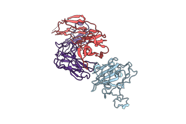 |
The Local Refined Map Of Sars-Cov-2 Eg.5.1 Variant Spike Protein Complexed With Antibody Xgi-171
Organism: Homo sapiens, Severe acute respiratory syndrome coronavirus 2
Method: ELECTRON MICROSCOPY Release Date: 2025-12-10 Classification: VIRAL PROTEIN/IMMUNE SYSTEM |
Organism: Homo sapiens, Severe acute respiratory syndrome coronavirus 2
Method: ELECTRON MICROSCOPY
Release Date: 2025-12-10
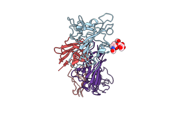 |
The Local Refined Map Of Sars-Cov-2 Eg.5.1 Variant Spike Protein Complexed With Antibody Xgi-183
Organism: Homo sapiens, Severe acute respiratory syndrome coronavirus 2
Method: ELECTRON MICROSCOPY Release Date: 2025-12-10 Classification: VIRAL PROTEIN/IMMUNE SYSTEM |
Organism: Homo sapiens, Severe acute respiratory syndrome coronavirus 2
Method: ELECTRON MICROSCOPY
Release Date: 2025-12-10
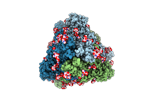 |
Structure Of Sars-Cov-2 Eg.5.1 Variant Spike Protein Complexed With Antibody Xgi-171
Organism: Homo sapiens, Severe acute respiratory syndrome coronavirus 2
Method: ELECTRON MICROSCOPY Release Date: 2025-12-10 Classification: VIRAL PROTEIN/IMMUNE SYSTEM Ligands: NAG |
Organism: Homo sapiens, Severe acute respiratory syndrome coronavirus 2
Method: ELECTRON MICROSCOPY
Release Date: 2025-12-10
Ligands: NAG
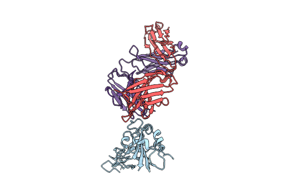 |
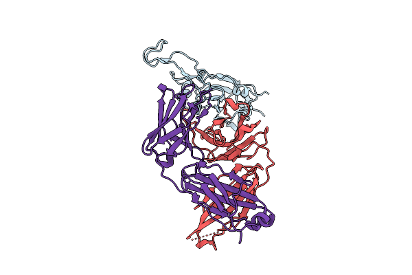
The Local Refined Map Of Sars-Cov-2 Eg.5.1 Variant Spike Protein Complexed With Antibody Xgi-198
Organism: Homo sapiens, Severe acute respiratory syndrome coronavirus 2
Method: ELECTRON MICROSCOPY Release Date: 2025-12-10 Classification: VIRAL PROTEIN/IMMUNE SYSTEM |
Organism: Homo sapiens, Severe acute respiratory syndrome coronavirus 2
Method: ELECTRON MICROSCOPY
Release Date: 2025-12-10
 |
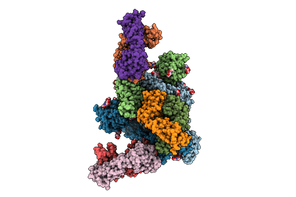
The Local Refined Map Of Sars-Cov-2 Eg.5.1 Variant Spike Protein Complexed With Antibody Xgi-203
Organism: Severe acute respiratory syndrome coronavirus 2, Homo sapiens
Method: ELECTRON MICROSCOPY Release Date: 2025-12-10 Classification: VIRAL PROTEIN/IMMUNE SYSTEM |
Organism: Severe acute respiratory syndrome coronavirus 2, Homo sapiens
Method: ELECTRON MICROSCOPY
Release Date: 2025-12-10
 |
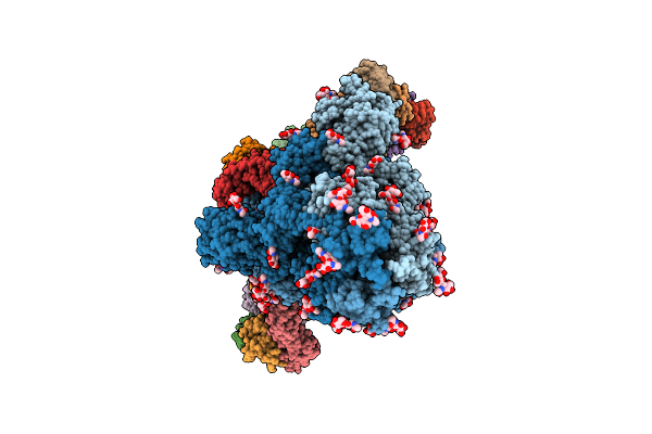
Structure Of Sars-Cov-2 Eg.5.1 Variant Spike Protein Complexed With Antibody Xgi-203
Organism: Homo sapiens, Severe acute respiratory syndrome coronavirus 2
Method: ELECTRON MICROSCOPY Release Date: 2025-12-10 Classification: VIRAL PROTEIN/IMMUNE SYSTEM Ligands: NAG |
Organism: Homo sapiens, Severe acute respiratory syndrome coronavirus 2
Method: ELECTRON MICROSCOPY
Release Date: 2025-12-10
Ligands: NAG
 |
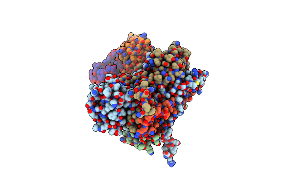
Structure Of Sars-Cov-2 Eg.5.1 Variant Spike Protein Complexed With Antibody Xgi-198
Organism: Severe acute respiratory syndrome coronavirus 2, Homo sapiens
Method: ELECTRON MICROSCOPY Release Date: 2025-12-10 Classification: VIRAL PROTEIN/IMMUNE SYSTEM Ligands: NAG |
Organism: Severe acute respiratory syndrome coronavirus 2, Homo sapiens
Method: ELECTRON MICROSCOPY
Release Date: 2025-12-10
Ligands: NAG
 |
Cryo-Em Structure Of Enterovirus A71 Mature Virion In Complex With Fab Ct11F9
Organism: Mus musculus, Enterovirus a71
Method: ELECTRON MICROSCOPY Resolution:3.04 Å Release Date: 2025-04-23 Classification: VIRUS Ligands: SPH |
Organism: Mus musculus, Enterovirus a71
Method: ELECTRON MICROSCOPY
Release Date: 2025-04-23
Ligands: SPH
 |
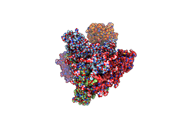
Cryo-Em Structure Of Sars-Cov-2 D614G S With One Ace2 Receptor Binding (Rb1) In Prefusion Conformation
Organism: Severe acute respiratory syndrome coronavirus 2, Homo sapiens
Method: ELECTRON MICROSCOPY Release Date: 2025-02-05 Classification: VIRAL PROTEIN/HYDROLASE Ligands: NAG |
Organism: Severe acute respiratory syndrome coronavirus 2, Homo sapiens
Method: ELECTRON MICROSCOPY
Release Date: 2025-02-05
Ligands: NAG
 |
Cryo-Em Structure Of Sars-Cov-2 D614G S With Two Ace2 Receptors Binding (Rb2) In Prefusion Conformation
Organism: Severe acute respiratory syndrome coronavirus 2, Homo sapiens
Method: ELECTRON MICROSCOPY Release Date: 2025-02-05 Classification: VIRAL PROTEIN/HYDROLASE Ligands: NAG |
Organism: Severe acute respiratory syndrome coronavirus 2, Homo sapiens
Method: ELECTRON MICROSCOPY
Release Date: 2025-02-05
Ligands: NAG
 |
Cryo-Em Structure Of Sars-Cov-2 D614G S With Three Ace2 Receptors Binding (Rb3) In Prefusion Conformation (Focused Refinement Of Ntd-Sd1-Rbd-Ace2)
Organism: Severe acute respiratory syndrome coronavirus 2, Homo sapiens
Method: ELECTRON MICROSCOPY Release Date: 2025-02-05 Classification: VIRAL PROTEIN/HYDROLASE Ligands: NAG |
Organism: Severe acute respiratory syndrome coronavirus 2, Homo sapiens
Method: ELECTRON MICROSCOPY
Release Date: 2025-02-05
Ligands: NAG
 |
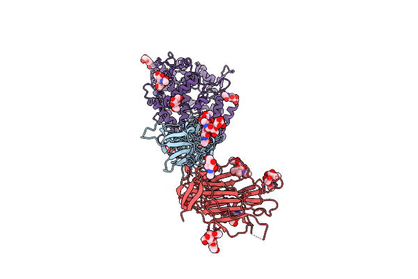
Cryo-Em Structure Of Sars-Cov-2 D614G S With Three Ace2 Receptors Binding (Rb3) In Prefusion Conformation
Organism: Severe acute respiratory syndrome coronavirus 2, Homo sapiens
Method: ELECTRON MICROSCOPY Release Date: 2025-02-05 Classification: VIRAL PROTEIN/HYDROLASE Ligands: NAG |
Organism: Severe acute respiratory syndrome coronavirus 2, Homo sapiens
Method: ELECTRON MICROSCOPY
Release Date: 2025-02-05
Ligands: NAG
 |
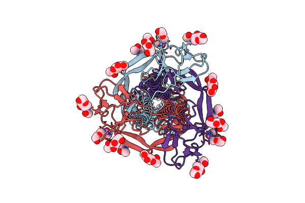
Cryo-Em Structure Of Sars-Cov-2 S Trimer In The Early Fusion Intermediate Conformation (E-Fic) (Focused Refinement Of Ntd-Sd1-Rbd-Ace2)
Organism: Homo sapiens, Severe acute respiratory syndrome coronavirus 2
Method: ELECTRON MICROSCOPY Release Date: 2025-02-05 Classification: VIRAL PROTEIN Ligands: NAG |
Organism: Homo sapiens, Severe acute respiratory syndrome coronavirus 2
Method: ELECTRON MICROSCOPY
Release Date: 2025-02-05
Ligands: NAG
 |
Cryo-Em Structure Of Sars-Cov-2 S Trimer In The Early Fusion Intermediate Conformation (E-Fic) (Focused Refinement Of Intact S2)
Organism: Severe acute respiratory syndrome coronavirus 2
Method: ELECTRON MICROSCOPY Release Date: 2025-02-05 Classification: VIRAL PROTEIN Ligands: NAG |
Organism: Severe acute respiratory syndrome coronavirus 2
Method: ELECTRON MICROSCOPY
Release Date: 2025-02-05
Ligands: NAG
 |
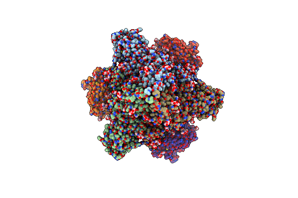
Cryo-Em Structure Of Sars-Cov-2 S Trimer In The Early Fusion Intermediate Conformation (E-Fic) (Focused Refinement Of S-Bottom)
Organism: Severe acute respiratory syndrome coronavirus 2
Method: ELECTRON MICROSCOPY Release Date: 2025-02-05 Classification: VIRAL PROTEIN Ligands: NAG |
Organism: Severe acute respiratory syndrome coronavirus 2
Method: ELECTRON MICROSCOPY
Release Date: 2025-02-05
Ligands: NAG
 |
Cryo-Em Structure Of Sars-Cov-2 S Trimer In The Early Fusion Intermediate Conformation (E-Fic)
Organism: Homo sapiens, Severe acute respiratory syndrome coronavirus 2
Method: ELECTRON MICROSCOPY Release Date: 2025-02-05 Classification: VIRAL PROTEIN/HTDROLASE Ligands: NAG |
Organism: Homo sapiens, Severe acute respiratory syndrome coronavirus 2
Method: ELECTRON MICROSCOPY
Release Date: 2025-02-05
Ligands: NAG