

Search Count: 37
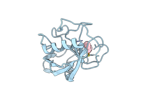 |
Crystal Structure Of Human Cyclophilin D (Cypd) With In Complex With The Covalently Bound Ebselen Inhibitor
Organism: Homo sapiens
Method: X-RAY DIFFRACTION Resolution:1.65 Å Release Date: 2023-10-18 Classification: ISOMERASE Ligands: 9JT |
Organism: Homo sapiens
Method: X-RAY DIFFRACTION
Release Date: 2023-10-18
Ligands: 9JT
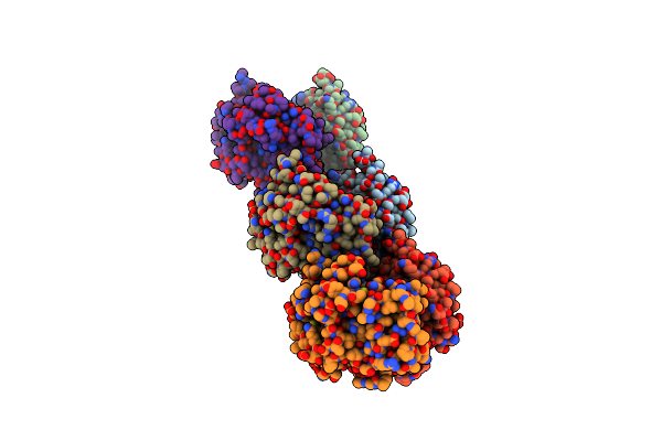 |
Glutathione S-Transferase Omega 1 Bound To Covalent Inhibitor 18
Organism: Homo sapiens
Method: X-RAY DIFFRACTION Resolution:2.75 Å Release Date: 2019-02-20 Classification: Transferase/Transferase Inhibitor Ligands: JR7, MES |
Organism: Homo sapiens
Method: X-RAY DIFFRACTION
Release Date: 2019-02-20
Ligands: JR7, MES
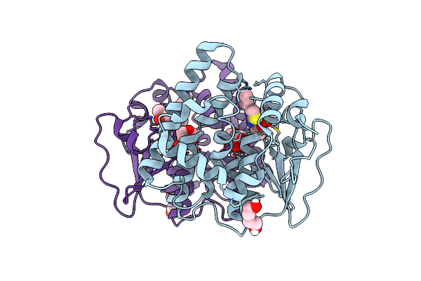 |
Glutathione S-Transferase Omega 1 Bound To Covalent Inhibitor 37
Organism: Homo sapiens
Method: X-RAY DIFFRACTION Resolution:2.00 Å Release Date: 2019-02-20 Classification: Transferase/Transferase Inhibitor Ligands: JRM, DMS, PEG, MES |
Organism: Homo sapiens
Method: X-RAY DIFFRACTION
Release Date: 2019-02-20
Ligands: JRM, DMS, PEG, MES
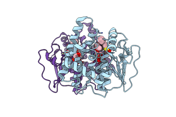 |
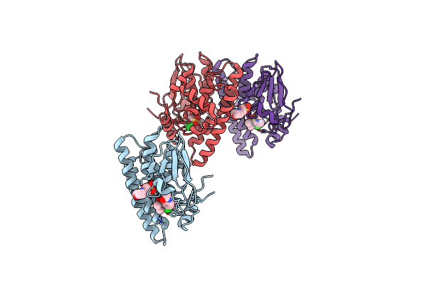
Glutathione S-Transferase Omega 1 Bound To Covalent Inhibitor 44
Organism: Homo sapiens
Method: X-RAY DIFFRACTION Resolution:2.16 Å Release Date: 2019-02-20 Classification: Transferase/Transferase Inhibitor Ligands: JRD, MES, ACN |
Organism: Homo sapiens
Method: X-RAY DIFFRACTION
Release Date: 2019-02-20
Ligands: JRD, MES, ACN
 |
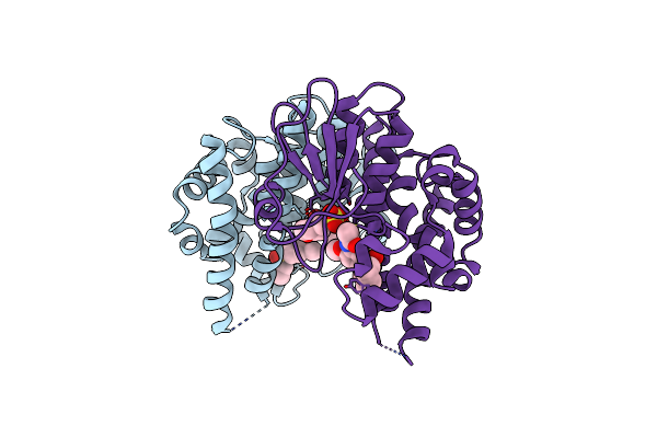
Glutathione S-Transferase Omega 1 Bound To Covalent Inhibitor C1-27
Organism: Homo sapiens
Method: X-RAY DIFFRACTION Resolution:2.38 Å Release Date: 2016-10-12 Classification: Transferase/Transferase Inhibitor Ligands: 4G9, MES |
Organism: Homo sapiens
Method: X-RAY DIFFRACTION
Release Date: 2016-10-12
Ligands: 4G9, MES
 |
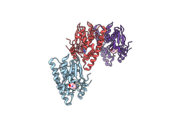
Glutathione S-Transferase Omega 1 Bound To Covalent Inhibitor C1-31
Organism: Homo sapiens
Method: X-RAY DIFFRACTION Resolution:1.94 Å Release Date: 2016-10-12 Classification: Transferase/Transferase Inhibitor Ligands: 4GB, MES |
Organism: Homo sapiens
Method: X-RAY DIFFRACTION
Release Date: 2016-10-12
Ligands: 4GB, MES
 |
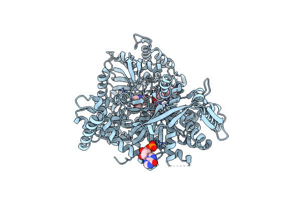
Glutathione S-Transferase Omega 1 Bound To Covalent Inhibitor C4-10
Organism: Homo sapiens
Method: X-RAY DIFFRACTION Resolution:2.06 Å Release Date: 2016-10-12 Classification: Transferase/Transferase Inhibitor Ligands: 4GG |
Organism: Homo sapiens
Method: X-RAY DIFFRACTION
Release Date: 2016-10-12
Ligands: 4GG
 |
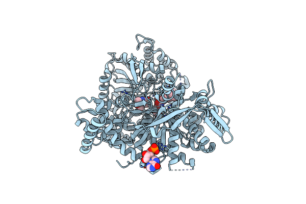
Crystal Structure Of Gpb In Complex With 4A
Organism: Oryctolagus cuniculus
Method: X-RAY DIFFRACTION Resolution:1.80 Å Release Date: 2015-11-18 Classification: TRANSFERASE Ligands: PLP, 4D0, IMP, DMS |
Organism: Oryctolagus cuniculus
Method: X-RAY DIFFRACTION
Release Date: 2015-11-18
Ligands: PLP, 4D0, IMP, DMS
 |
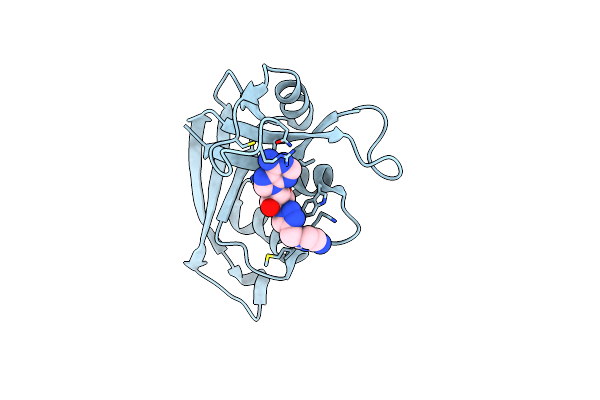
Crystal Structure Of Gpb In Complex With 4B
Organism: Oryctolagus cuniculus
Method: X-RAY DIFFRACTION Resolution:1.80 Å Release Date: 2015-11-18 Classification: TRANSFERASE Ligands: PLP, 4D1, IMP |
Organism: Oryctolagus cuniculus
Method: X-RAY DIFFRACTION
Release Date: 2015-11-18
Ligands: PLP, 4D1, IMP
 |
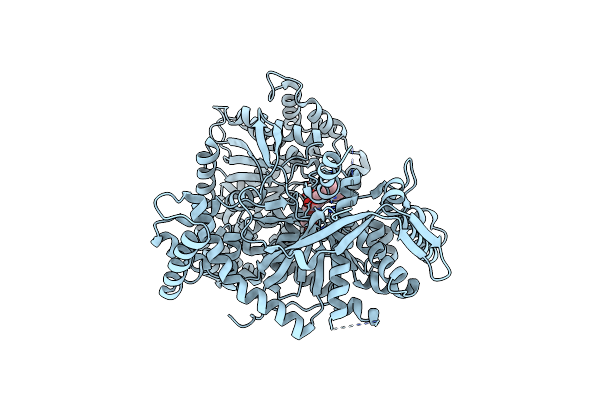
Crystal Structure Of Eosinophil-Derived Neurotoxin In Complex With The Triazole Double-Headed Ribonucleoside 11C
Organism: Homo sapiens
Method: X-RAY DIFFRACTION Resolution:1.34 Å Release Date: 2015-11-18 Classification: HYDROLASE Ligands: 5J9 |
Organism: Homo sapiens
Method: X-RAY DIFFRACTION
Release Date: 2015-11-18
Ligands: 5J9
 |
Crystal Structure Of Gpb In Complex With S3, Sugar (N-[(Biphenyl-4-Yloxy)Acetyl]-Beta-D-Glucopyranosylamine)
Organism: Oryctolagus cuniculus
Method: X-RAY DIFFRACTION Resolution:2.00 Å Release Date: 2014-07-23 Classification: transferase/transferase inhibitor Ligands: 26M |
Organism: Oryctolagus cuniculus
Method: X-RAY DIFFRACTION
Release Date: 2014-07-23
Ligands: 26M
 |
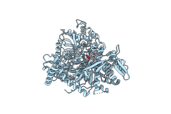
Crystal Structure Of Gpb In Complex With Sugar (N-[(2E)-3-(Biphenyl-4-Yl)Prop-2-Enoyl]-Beta-D-Glucopyranosylamine
Organism: Oryctolagus cuniculus
Method: X-RAY DIFFRACTION Resolution:2.00 Å Release Date: 2014-07-23 Classification: transferase/transferase inhibitor Ligands: 26Q, DMS |
Organism: Oryctolagus cuniculus
Method: X-RAY DIFFRACTION
Release Date: 2014-07-23
Ligands: 26Q, DMS
 |
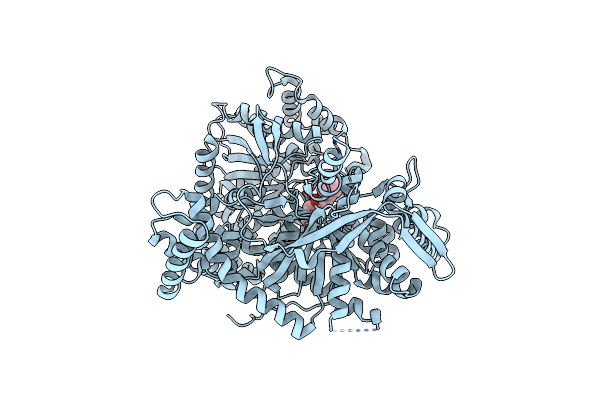
Crystal Structure Of Gpb In Complex With Sugar (N-{(2R)-2-Methyl-3-[4-(Propan-2-Yl)Phenyl]Propanoyl}-Beta-D-Glucopyranosylamine) (S21)
Organism: Oryctolagus cuniculus
Method: X-RAY DIFFRACTION Resolution:2.15 Å Release Date: 2014-07-23 Classification: transferase/transferase inhibitor Ligands: 26R |
Organism: Oryctolagus cuniculus
Method: X-RAY DIFFRACTION
Release Date: 2014-07-23
Ligands: 26R
 |
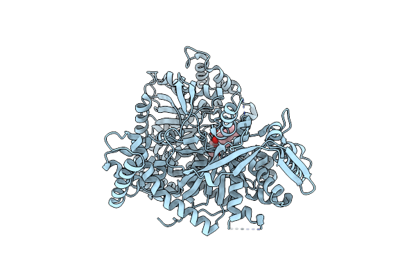
Crystal Structure Of Gpb In Complex With Sugar (N-[4-(5,6,7,8-Tetrahydronaphthalen-2-Yl)Butanoyl]-Beta-D-Glucopyranosylamine)
Organism: Oryctolagus cuniculus
Method: X-RAY DIFFRACTION Resolution:1.90 Å Release Date: 2014-07-23 Classification: transferase/transferase inhibitor Ligands: 26V |
Organism: Oryctolagus cuniculus
Method: X-RAY DIFFRACTION
Release Date: 2014-07-23
Ligands: 26V
 |
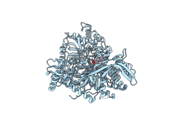
Crystal Structure Of Gpb In Complex With Sugar (N-[(3R)-3-(4-Ethylphenyl)Butanoyl]-Beta-D-Glucopyranosylamine) (S20)
Organism: Oryctolagus cuniculus
Method: X-RAY DIFFRACTION Resolution:1.85 Å Release Date: 2014-07-23 Classification: transferase/transferase inhibitor Ligands: 26W |
Organism: Oryctolagus cuniculus
Method: X-RAY DIFFRACTION
Release Date: 2014-07-23
Ligands: 26W
 |
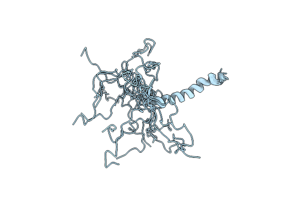
Crystal Structure Of Gpb In Complex With Sugar (N-{(2E)-3-[4-(Propan-2-Yl)Phenyl]Prop-2-Enoyl}-Beta-D-Glucopyranosylamine) (S6)
Organism: Oryctolagus cuniculus
Method: X-RAY DIFFRACTION Resolution:2.45 Å Release Date: 2014-07-23 Classification: transferase/transferase inhibitor Ligands: 26Y |
Organism: Oryctolagus cuniculus
Method: X-RAY DIFFRACTION
Release Date: 2014-07-23
Ligands: 26Y
 |
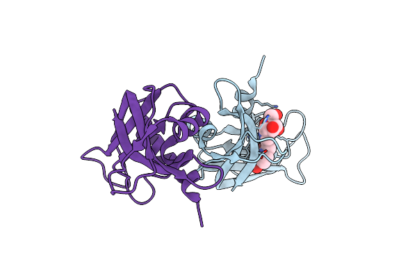
Structure Of The Regulatory Domain Of Human Brain Carnitine Palmitoyltransferase 1
Organism: Homo sapiens
Method: SOLUTION NMR Release Date: 2013-09-11 Classification: SIGNALING PROTEIN |
Organism: Homo sapiens
Method: SOLUTION NMR
Release Date: 2013-09-11
 |
Crystal Structure Of Ribonuclease A In Complex With 5A
Organism: Bos taurus
Method: X-RAY DIFFRACTION Resolution:1.70 Å Release Date: 2012-11-21 Classification: Hydrolase/Hydrolase Inhibitor Ligands: 0EY |
Organism: Bos taurus
Method: X-RAY DIFFRACTION
Release Date: 2012-11-21
Ligands: 0EY
 |
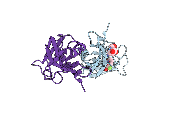
Crystal Structure Of Ribonuclease A In Complex With 5B
Organism: Bos taurus
Method: X-RAY DIFFRACTION Resolution:1.80 Å Release Date: 2012-11-21 Classification: Hydrolase/Hydrolase Inhibitor Ligands: 0FT |
Organism: Bos taurus
Method: X-RAY DIFFRACTION
Release Date: 2012-11-21
Ligands: 0FT
 |
Crystal Structure Of Ribonuclease A In Complex With 5E
Organism: Bos taurus
Method: X-RAY DIFFRACTION Resolution:1.90 Å Release Date: 2012-11-21 Classification: Hydrolase/Hydrolase Inhibitor Ligands: 0G0 |
Organism: Bos taurus
Method: X-RAY DIFFRACTION
Release Date: 2012-11-21
Ligands: 0G0