

Search Count: 15
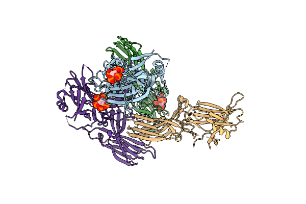 |
X-Ray Crystal Structure Of Visual Arrestin Complexed With Inositol Hexaphosphate
Organism: Bos taurus
Method: X-RAY DIFFRACTION Resolution:3.10 Å Release Date: 2021-10-27 Classification: SIGNALING PROTEIN Ligands: IHP |
Organism: Bos taurus
Method: X-RAY DIFFRACTION
Release Date: 2021-10-27
Ligands: IHP
 |
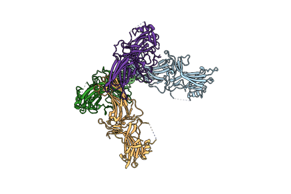
X-Ray Crystal Structure Of Visual Arrestin Complexed With Inositol 1,4,5-Triphosphate
Organism: Bos taurus
Method: X-RAY DIFFRACTION Resolution:3.00 Å Release Date: 2021-10-27 Classification: SIGNALING PROTEIN Ligands: I3P, PTD, EDO |
Organism: Bos taurus
Method: X-RAY DIFFRACTION
Release Date: 2021-10-27
Ligands: I3P, PTD, EDO
 |
Crystal Structure Of Native Bovine Arrestin 1
Organism: Bos taurus
Method: X-RAY DIFFRACTION Resolution:2.50 Å Release Date: 2021-10-13 Classification: PROTEIN BINDING |
Organism: Bos taurus
Method: X-RAY DIFFRACTION
Release Date: 2021-10-13
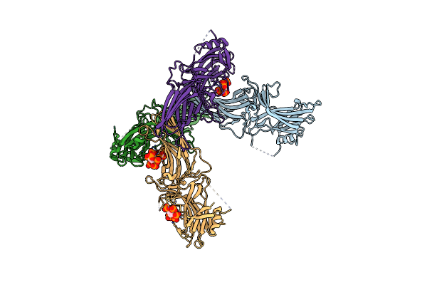 |
Crystal Structure Of Native Bovine Arrestin 1 In Complex With Inositol Hexakisphosphate
Organism: Bos taurus
Method: X-RAY DIFFRACTION Resolution:2.60 Å Release Date: 2021-10-13 Classification: PROTEIN BINDING Ligands: IHP |
Organism: Bos taurus
Method: X-RAY DIFFRACTION
Release Date: 2021-10-13
Ligands: IHP
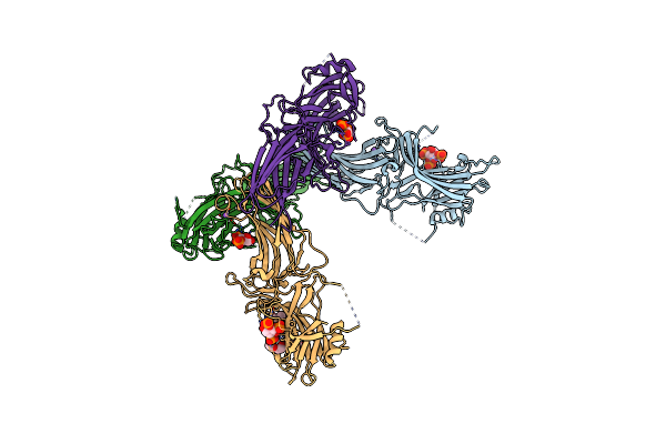 |
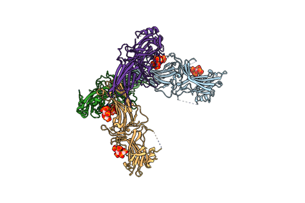
Crystal Structure Of Native Bovine Arrestin 1 In Complex With Inositol 1,4,5-Triphosphate
Organism: Bos taurus
Method: X-RAY DIFFRACTION Resolution:2.40 Å Release Date: 2021-10-13 Classification: PROTEIN BINDING Ligands: I3P, NA, ETX |
Organism: Bos taurus
Method: X-RAY DIFFRACTION
Release Date: 2021-10-13
Ligands: I3P, NA, ETX
 |
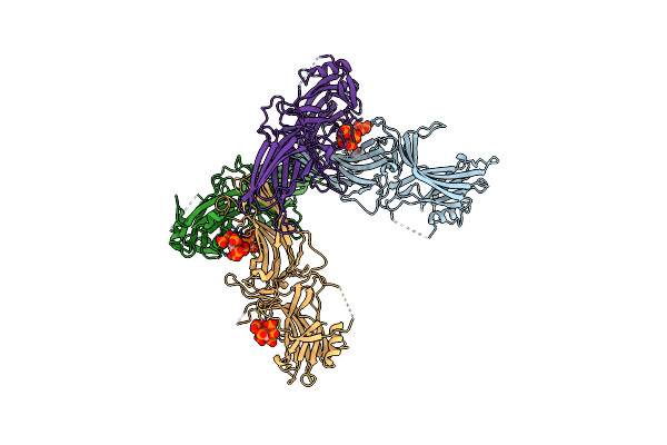
Crystal Structure Of Native Bovine Arrestin 1 In Complex With 5-Methylenebiphosphonate Inositol Pentakisphaophate (5-Pcp Ip5)
Organism: Bos taurus
Method: X-RAY DIFFRACTION Resolution:2.80 Å Release Date: 2021-10-13 Classification: PROTEIN BINDING Ligands: 5A3 |
Organism: Bos taurus
Method: X-RAY DIFFRACTION
Release Date: 2021-10-13
Ligands: 5A3
 |
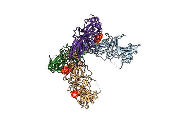
Crystal Structure Of Native Bovine Arrestin 1 In Complex With 1D-Myo-Inositol 5-Diphosphate Pentakisphosphate (5-Pp Ip5)
Organism: Bos taurus
Method: X-RAY DIFFRACTION Resolution:2.60 Å Release Date: 2021-10-13 Classification: PROTEIN BINDING Ligands: ETX, I7P |
Organism: Bos taurus
Method: X-RAY DIFFRACTION
Release Date: 2021-10-13
Ligands: ETX, I7P
 |
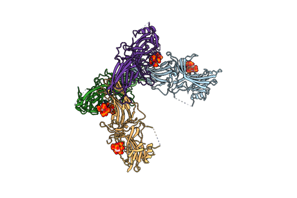
Crystal Structure Of Native Bovine Arrestin 1 In Complex With 1,5-Di-Methylenebisphosphonate Inositol Tetrakisphosphate (1,5-Pcp-Ip4)
Organism: Bos taurus
Method: X-RAY DIFFRACTION Resolution:2.66 Å Release Date: 2021-10-13 Classification: PROTEIN BINDING Ligands: 4WZ |
Organism: Bos taurus
Method: X-RAY DIFFRACTION
Release Date: 2021-10-13
Ligands: 4WZ
 |
Crystal Structure Of Native Bovine Arrestin 1 In Complex With 1D-Myo-Inositol 1,5-Bisdiphosphate Tetrakisphosphate (1,5-Pp Ip4)
Organism: Bos taurus
Method: X-RAY DIFFRACTION Resolution:3.00 Å Release Date: 2021-10-13 Classification: PROTEIN BINDING Ligands: I8P |
Organism: Bos taurus
Method: X-RAY DIFFRACTION
Release Date: 2021-10-13
Ligands: I8P
 |
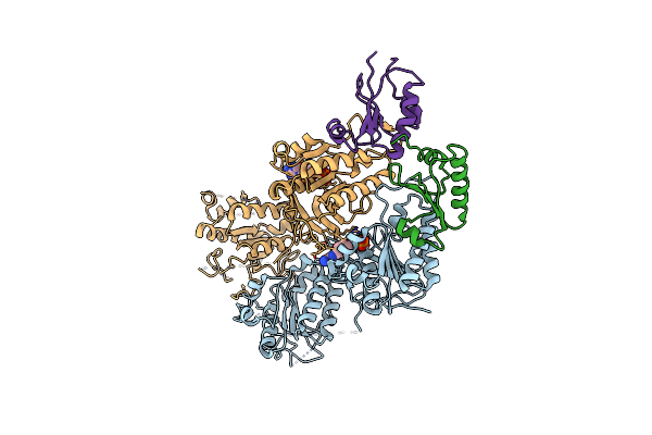
Crystal Structure Of Foldswitch-Stabilized Kaib In Complex With The N-Terminal Ci Domain Of Kaic From Thermosynechococcus Elongatus
Organism: Thermosynechococcus elongatus, Thermosynechococcus elongatus (strain bp-1)
Method: X-RAY DIFFRACTION Resolution:1.80 Å Release Date: 2017-03-29 Classification: Transcription Regulator Ligands: ADP |
Organism: Thermosynechococcus elongatus, Thermosynechococcus elongatus (strain bp-1)
Method: X-RAY DIFFRACTION
Release Date: 2017-03-29
Ligands: ADP
 |
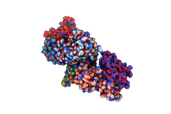
Crystal Structure Of Kaic S431E In Complex With Foldswitch-Stabilized Kaib From Thermosynechococcus Elongatus
Organism: Thermosynechococcus elongatus (strain bp-1)
Method: X-RAY DIFFRACTION Resolution:3.87 Å Release Date: 2017-03-29 Classification: Transcription Regulator Ligands: ADP |
Organism: Thermosynechococcus elongatus (strain bp-1)
Method: X-RAY DIFFRACTION
Release Date: 2017-03-29
Ligands: ADP
 |
Crystal Structure Of Foldswitch-Stabilized Kaib In Complex With The N-Terminal Ci Domain Of Kaic And A Dimer Of Kaia C-Terminal Domains From Thermosynechococcus Elongatus
Organism: Thermosynechococcus elongatus, Thermosynechococcus elongatus (strain bp-1)
Method: X-RAY DIFFRACTION Resolution:2.61 Å Release Date: 2017-03-29 Classification: Transcription Regulator Ligands: PO4 |
Organism: Thermosynechococcus elongatus, Thermosynechococcus elongatus (strain bp-1)
Method: X-RAY DIFFRACTION
Release Date: 2017-03-29
Ligands: PO4
 |
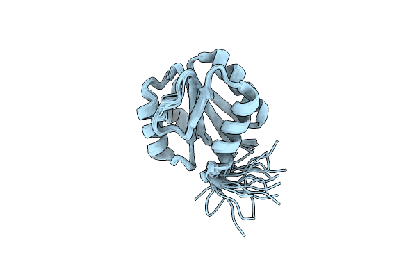
Nmr Structure Of Foldswitch-Stablized Kaib From Thermosynechococcus Elongatus
Organism: Thermosynechococcus elongatus
Method: SOLUTION NMR Release Date: 2017-03-29 Classification: Sigaling protein |
Organism: Thermosynechococcus elongatus
Method: SOLUTION NMR
Release Date: 2017-03-29
 |
Nmr Structure Of Pseudo Receiver Domain Of Cika From Thermosynechococcus Elongatus
Organism: Thermosynechococcus elongatus (strain bp-1)
Method: SOLUTION NMR Release Date: 2017-03-29 Classification: Sigaling protein |
Organism: Thermosynechococcus elongatus (strain bp-1)
Method: SOLUTION NMR
Release Date: 2017-03-29
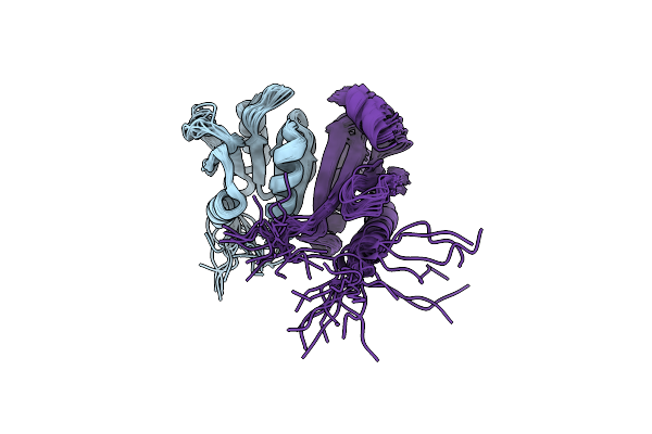 |
Nmr Structure Of Foldswitch-Stablized Kaib In Complex With Pseudo Receiver Domain Of Cika From Thermosynechococcus Elongatus
Organism: Thermosynechococcus elongatus (strain bp-1), Thermosynechococcus elongatus
Method: SOLUTION NMR Release Date: 2017-03-29 Classification: Sigaling protein |
Organism: Thermosynechococcus elongatus (strain bp-1), Thermosynechococcus elongatus
Method: SOLUTION NMR
Release Date: 2017-03-29