
Search Count: 202
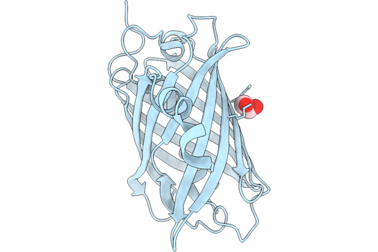 |
Organism: Discosoma sp.
Method: X-RAY DIFFRACTION Resolution:1.90 Å Release Date: 2026-04-22 Classification: FLUORESCENT PROTEIN Ligands: EDO |
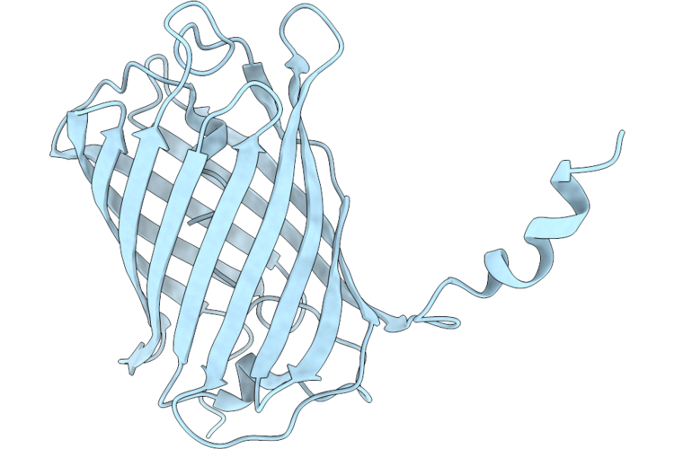 |
Organism: Discosoma sp.
Method: X-RAY DIFFRACTION Resolution:1.70 Å Release Date: 2026-04-22 Classification: FLUORESCENT PROTEIN |
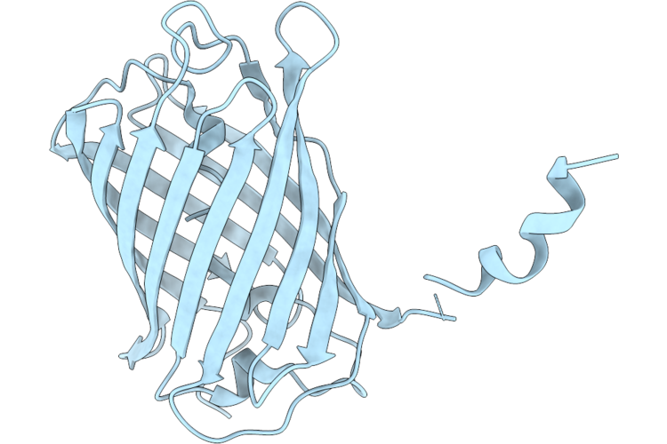 |
Crystal Structure Of Lssmorange Under Room Temperature At Ph 8.0, 250Ps After Pump Laser, Refined Against 8.33 Times Extrapolated Structure Factor
Organism: Discosoma sp.
Method: X-RAY DIFFRACTION Resolution:2.00 Å Release Date: 2026-04-22 Classification: FLUORESCENT PROTEIN |
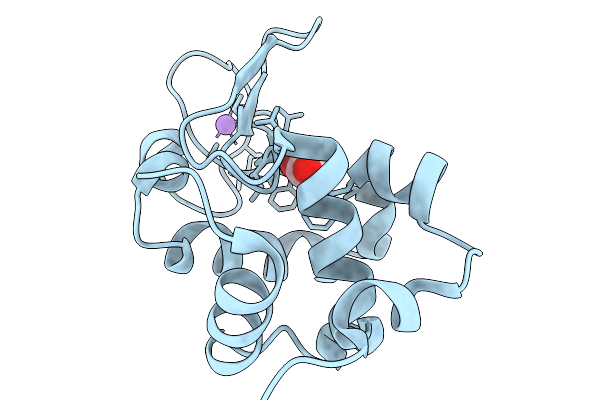 |
Organism: Gallus gallus
Method: X-RAY DIFFRACTION Resolution:1.68 Å Release Date: 2026-03-25 Classification: HYDROLASE |
 |
Organism: Gallus gallus
Method: X-RAY DIFFRACTION Resolution:1.74 Å Release Date: 2026-03-25 Classification: HYDROLASE |
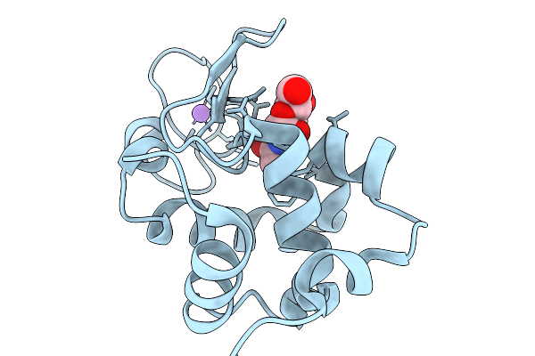 |
Time-Resolved Mixing Crystallography Structure Of 3-5 Micrometers Lysozyme Microcrystals With 226 Mm N-Acetyl-D-Glucosamine At 2 Seconds Delay
Organism: Gallus gallus
Method: X-RAY DIFFRACTION Resolution:1.68 Å Release Date: 2026-03-25 Classification: HYDROLASE Ligands: CL, NA, NDG |
 |
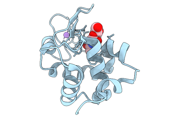
Time-Resolved Mixing Crystallography Structure Of 3-5 Micrometers Lysozyme Microcrystals With 226 Mm N-Acetyl-D-Glucosamine At 5 Seconds Delay
Organism: Gallus gallus
Method: X-RAY DIFFRACTION Resolution:1.68 Å Release Date: 2026-03-25 Classification: HYDROLASE Ligands: CL, NA, NDG |
 |
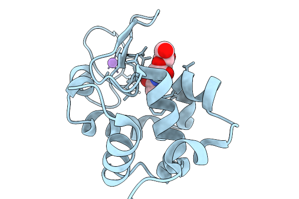
Time-Resolved Mixing Crystallography Structure Of 3-5 Micrometers Lysozyme Microcrystals With 226 Mm N-Acetyl-D-Glucosamine At 9.7 Seconds Delay
Organism: Gallus gallus
Method: X-RAY DIFFRACTION Resolution:1.68 Å Release Date: 2026-03-25 Classification: HYDROLASE Ligands: CL, NA, NDG |
 |
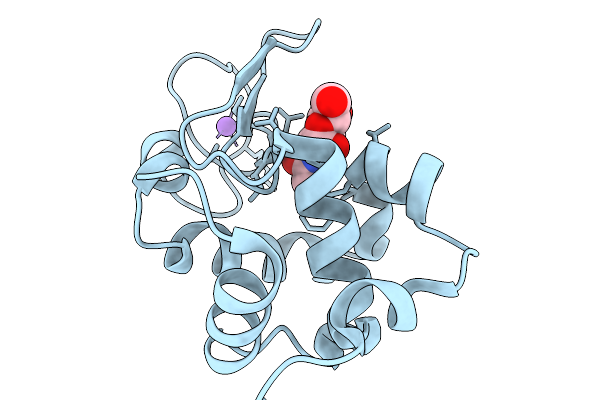
Time-Resolved Mixing Crystallography Structure Of 1 Micrometer Lysozyme Microcrystals With 226 Mm N-Acetyl-D-Glucosamine At 1.3 Seconds Delay
Organism: Gallus gallus
Method: X-RAY DIFFRACTION Resolution:1.72 Å Release Date: 2026-03-25 Classification: HYDROLASE Ligands: CL, NA, NDG |
 |
Time-Resolved Mixing Crystallography Structure Of 1 Micrometer Lysozyme Microcrystals With 226 Mm N-Acetyl-D-Glucosamine At 5.0 Seconds Delay
Organism: Gallus gallus
Method: X-RAY DIFFRACTION Resolution:1.75 Å Release Date: 2026-03-25 Classification: HYDROLASE Ligands: CL, NA, NDG |
 |
Time-Resolved Mixing Crystallography Structure Of 1 Micrometer Lysozyme Microcrystals With 226 Mm N-Acetyl-D-Glucosamine At 7.5 Seconds Delay
Organism: Gallus gallus
Method: X-RAY DIFFRACTION Resolution:1.77 Å Release Date: 2026-03-25 Classification: HYDROLASE Ligands: CL, NA, NDG |
 |
Time-Resolved Mixing Crystallography Structure Of 1 Micrometer Lysozyme Microcrystals With 226 Mm N-Acetyl-D-Glucosamine At 9.7 Seconds Delay
Organism: Gallus gallus
Method: X-RAY DIFFRACTION Resolution:1.78 Å Release Date: 2026-03-25 Classification: HYDROLASE Ligands: CL, NA, NDG |
 |
Time-Resolved Mixing Crystallography Structure Of 1 Micrometer Lysozyme Microcrystals With 452 Mm N-Acetyl-D-Glucosamine At 1.3 Seconds Delay
Organism: Gallus gallus
Method: X-RAY DIFFRACTION Resolution:1.80 Å Release Date: 2026-03-25 Classification: HYDROLASE Ligands: CL, NA, NDG |
 |
Time-Resolved Mixing Crystallography Structure Of 1 Micrometer Lysozyme Microcrystals With 452 Mm N-Acetyl-D-Glucosamine At 2.5 Seconds Delay
Organism: Gallus gallus
Method: X-RAY DIFFRACTION Resolution:1.81 Å Release Date: 2026-03-25 Classification: HYDROLASE Ligands: CL, NA, NDG |
 |
Time-Resolved Mixing Crystallography Structure Of 1 Micrometer Lysozyme Microcrystals With 452 Mm N-Acetyl-D-Glucosamine At 4 Seconds Delay
Organism: Gallus gallus
Method: X-RAY DIFFRACTION Resolution:1.80 Å Release Date: 2026-03-25 Classification: HYDROLASE Ligands: CL, NA, NDG |
 |
Time-Resolved Mixing Crystallography Structure Of 1 Micrometer Lysozyme Microcrystals With 452 Mm N-Acetyl-D-Glucosamine At 5.0 Seconds Delay
Organism: Gallus gallus
Method: X-RAY DIFFRACTION Resolution:1.82 Å Release Date: 2026-03-25 Classification: HYDROLASE Ligands: CL, NA, NDG |
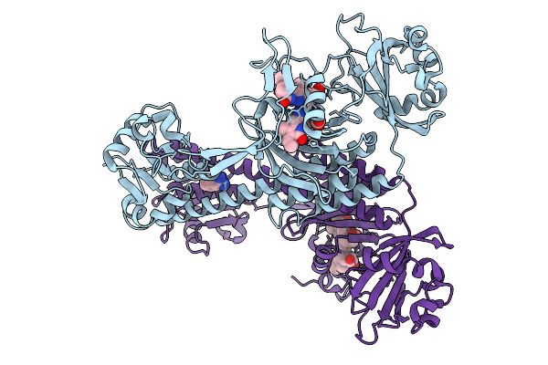 |
Photoactivation In Bacteriophytochromes, Reference (Dark) Structure For The 3 Ps Time Point
Organism: Stigmatella aurantiaca
Method: X-RAY DIFFRACTION Resolution:2.30 Å Release Date: 2025-10-08 Classification: SIGNALING PROTEIN Ligands: 3Q8, BEN |
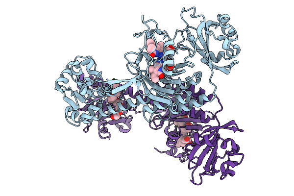 |
Photoactivation In Bacteriophytochrome, High Resolution Cryo Structure In The Dark.
Organism: Stigmatella aurantiaca
Method: X-RAY DIFFRACTION Resolution:1.40 Å Release Date: 2025-10-08 Classification: SIGNALING PROTEIN Ligands: EL5, P33 |
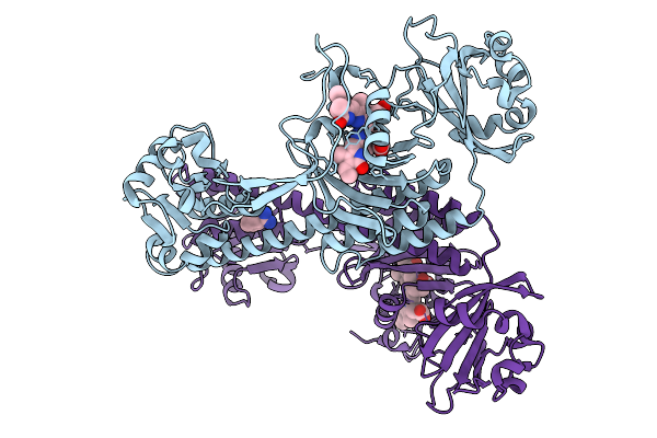 |
Photoactivation In Bacteriophytochromes, Reference (Dark) Structure For The 100 Ps Time Point
Organism: Stigmatella aurantiaca
Method: X-RAY DIFFRACTION Resolution:1.93 Å Release Date: 2025-10-08 Classification: SIGNALING PROTEIN Ligands: EL5, BEN |
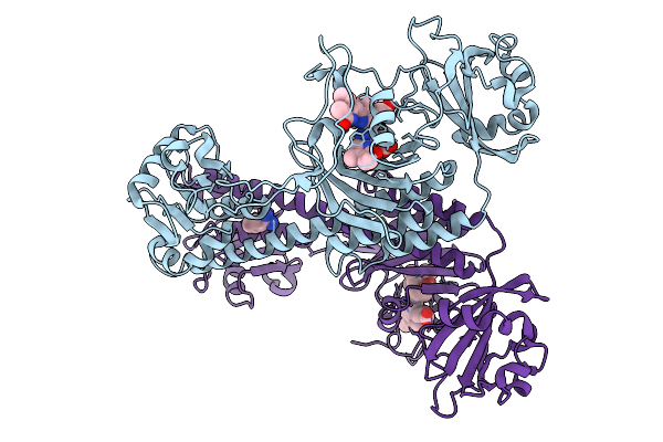 |
Organism: Stigmatella aurantiaca
Method: X-RAY DIFFRACTION Resolution:2.30 Å Release Date: 2025-10-08 Classification: SIGNALING PROTEIN Ligands: BLA, BEN |