

Search Count: 34
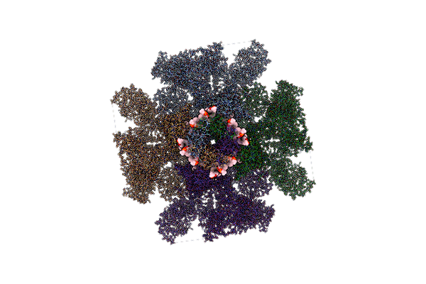 |
Structure Of The Full-Length Ip3R1 Channel Determined At High Ca2+
Organism: Rattus norvegicus
Method: ELECTRON MICROSCOPY Release Date: 2022-11-23 Classification: MEMBRANE PROTEIN Ligands: ZN, CA, PLX |
Organism: Rattus norvegicus
Method: ELECTRON MICROSCOPY
Release Date: 2022-11-23
Ligands: ZN, CA, PLX
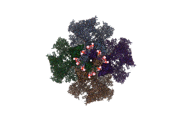 |
Structure Of The Full-Length Ip3R1 Channel Determined In The Presence Of Calcium/Ip3/Atp
Organism: Rattus norvegicus
Method: ELECTRON MICROSCOPY Release Date: 2022-11-23 Classification: MEMBRANE PROTEIN Ligands: ZN, ATP, I3P, CA, PLX |
Organism: Rattus norvegicus
Method: ELECTRON MICROSCOPY
Release Date: 2022-11-23
Ligands: ZN, ATP, I3P, CA, PLX
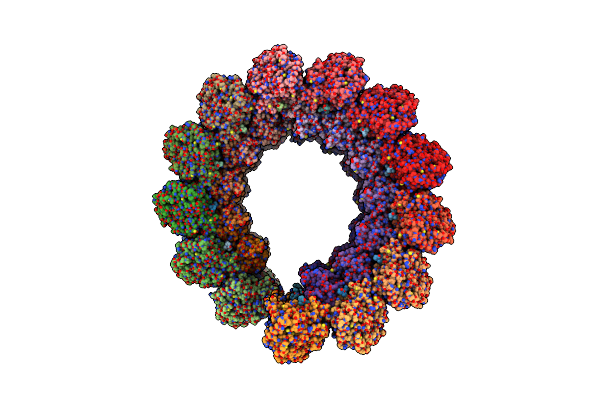 |
The Symmetry-Released Subpellicular Microtubule Map From Detergent-Extracted Toxoplasma Cells
Organism: Toxoplasma gondii
Method: ELECTRON MICROSCOPY Release Date: 2022-06-22 Classification: CELL INVASION |
Organism: Toxoplasma gondii
Method: ELECTRON MICROSCOPY
Release Date: 2022-06-22
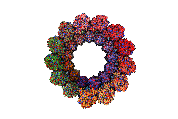 |
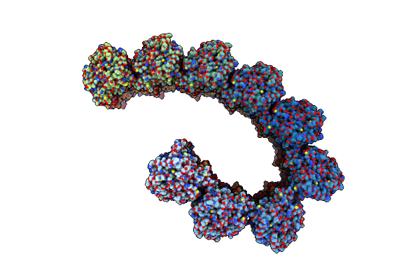
Subpellicular Microtubule From Detergent-Extract Toxoplasma Gondii Cells
Organism: Toxoplasma gondii
Method: ELECTRON MICROSCOPY Release Date: 2022-06-22 Classification: CELL INVASION |
Organism: Toxoplasma gondii
Method: ELECTRON MICROSCOPY
Release Date: 2022-06-22
 |
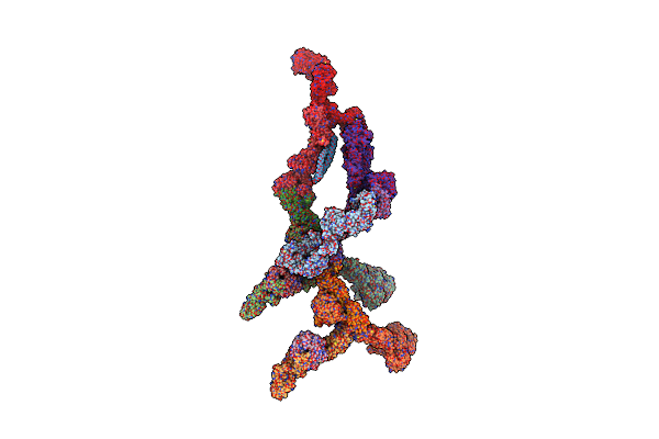
The Tubulin-Based Conoid From Detergent-Extract Toxoplasma Gondii Cells
Organism: Toxoplasma gondii
Method: ELECTRON MICROSCOPY Release Date: 2022-06-22 Classification: CELL INVASION |
Organism: Toxoplasma gondii
Method: ELECTRON MICROSCOPY
Release Date: 2022-06-22
 |
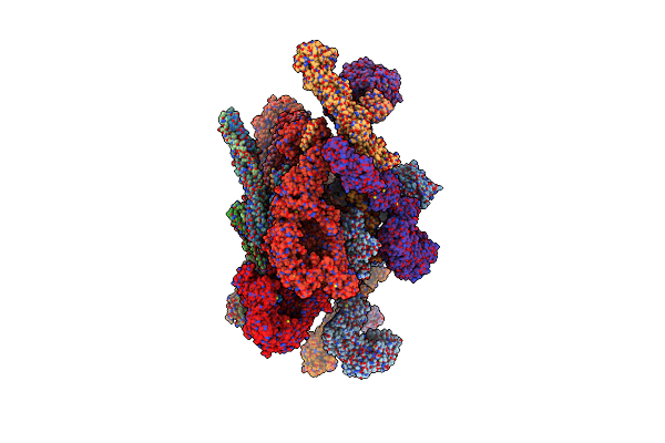
Double Nuclear Outer Ring From The Isolated Yeast Npc
Organism: Saccharomyces cerevisiae
Method: ELECTRON MICROSCOPY Release Date: 2022-01-26 Classification: TRANSLOCASE |
Organism: Saccharomyces cerevisiae
Method: ELECTRON MICROSCOPY
Release Date: 2022-01-26
 |
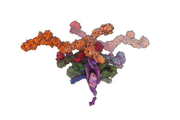
Inner Ring Spoke From The Isolated Yeast Npc
Organism: Saccharomyces cerevisiae
Method: ELECTRON MICROSCOPY Release Date: 2022-01-26 Classification: TRANSLOCASE |
Organism: Saccharomyces cerevisiae
Method: ELECTRON MICROSCOPY
Release Date: 2022-01-26
 |
Structure Of The In Situ Yeast Npc
Organism: Saccharomyces cerevisiae
Method: ELECTRON MICROSCOPY Release Date: 2022-01-26 Classification: TRANSLOCASE |
Organism: Saccharomyces cerevisiae
Method: ELECTRON MICROSCOPY
Release Date: 2022-01-26
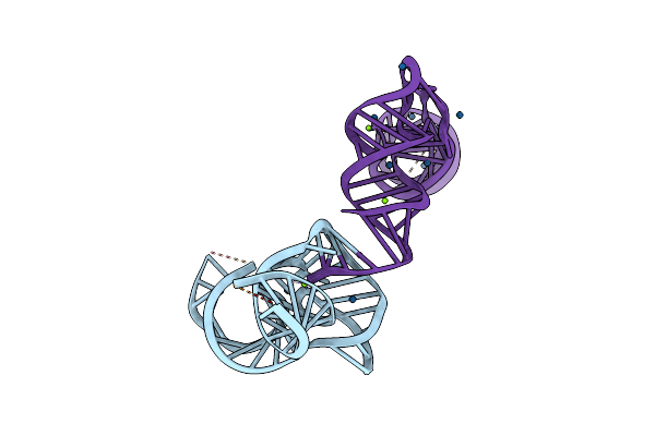 |
Co-Crystal Structure Of The Geobacillus Kaustophilus Glyq T-Box Riboswitch Discriminator Domain In Complex With Trna-Gly
Organism: Geobacillus kaustophilus
Method: X-RAY DIFFRACTION Resolution:2.66 Å Release Date: 2019-11-20 Classification: RNA Ligands: IR, MG |
Organism: Geobacillus kaustophilus
Method: X-RAY DIFFRACTION
Release Date: 2019-11-20
Ligands: IR, MG
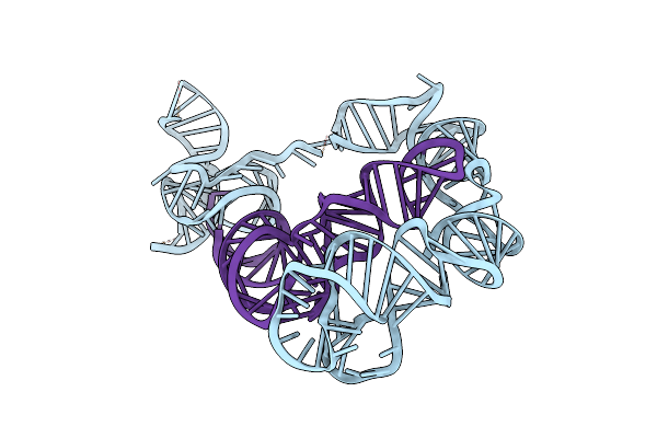 |
Cryo-Em Structure Of The Full-Length Bacillus Subtilis Glyqs T-Box Riboswitch In Complex With Trna-Gly
Organism: Bacillus subtilis
Method: ELECTRON MICROSCOPY Release Date: 2019-11-20 Classification: RNA |
Organism: Bacillus subtilis
Method: ELECTRON MICROSCOPY
Release Date: 2019-11-20
 |
Structure Of Full-Length Ip3R1 Channel Bound With Adenophostin A
Organism: Rattus norvegicus
Method: ELECTRON MICROSCOPY Release Date: 2018-12-05 Classification: MEMBRANE PROTEIN Ligands: JYP |
Organism: Rattus norvegicus
Method: ELECTRON MICROSCOPY
Release Date: 2018-12-05
Ligands: JYP
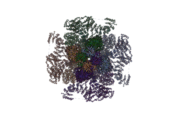 |
Structure Of Full-Length Ip3R1 Channel In The Apo-State
Organism: Rattus norvegicus
Method: ELECTRON MICROSCOPY Release Date: 2018-12-05 Classification: MEMBRANE PROTEIN |
Organism: Rattus norvegicus
Method: ELECTRON MICROSCOPY
Release Date: 2018-12-05
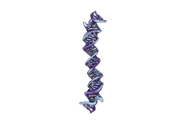 |
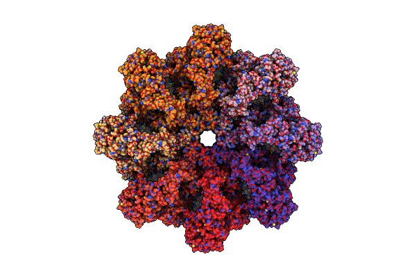
Hybrid Nmr/Cryo-Em Structure Of The Hiv-1 Rna Dimerization Signal
Organism: Homo sapiens
Method: ELECTRON MICROSCOPY, SOLUTION NMR Release Date: 2018-02-21 Classification: RNA |
Organism: Homo sapiens
Method: ELECTRON MICROSCOPY, SOLUTION NMR
Release Date: 2018-02-21
 |
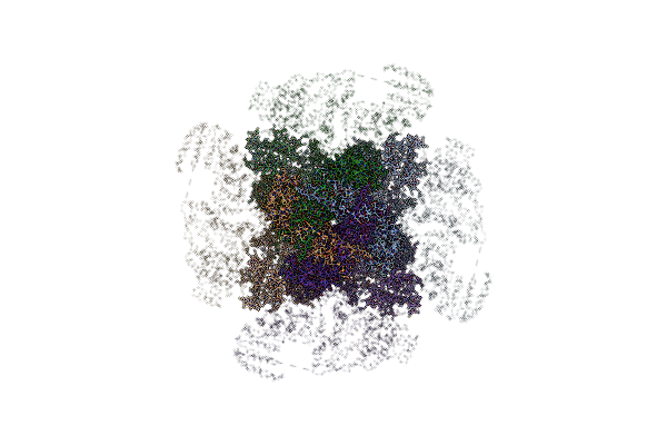
Near Atomic Structure Of The Dark Apoptosome
Organism: Drosophila melanogaster
Method: ELECTRON MICROSCOPY Release Date: 2017-02-22 Classification: APOPTOSIS Ligands: DTP |
Organism: Drosophila melanogaster
Method: ELECTRON MICROSCOPY
Release Date: 2017-02-22
Ligands: DTP
 |
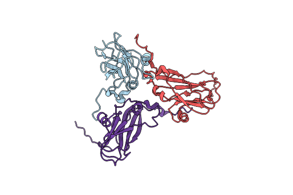
Structure Of Full-Length Ip3R1 Channel In The Apo-State Determined By Single Particle Cryo-Em
Organism: Rattus norvegicus
Method: ELECTRON MICROSCOPY Release Date: 2015-10-07 Classification: TRANSPORT PROTEIN |
Organism: Rattus norvegicus
Method: ELECTRON MICROSCOPY
Release Date: 2015-10-07
 |
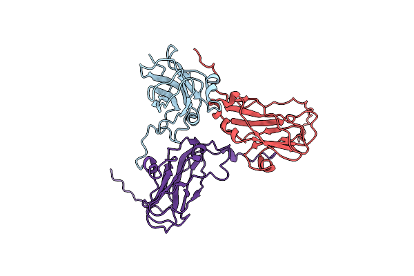
Full Virus Map Of Brome Mosaic Virus
Organism: Brome mosaic virus
Method: ELECTRON MICROSCOPY Release Date: 2014-09-10 Classification: VIRUS |
Organism: Brome mosaic virus
Method: ELECTRON MICROSCOPY
Release Date: 2014-09-10
 |
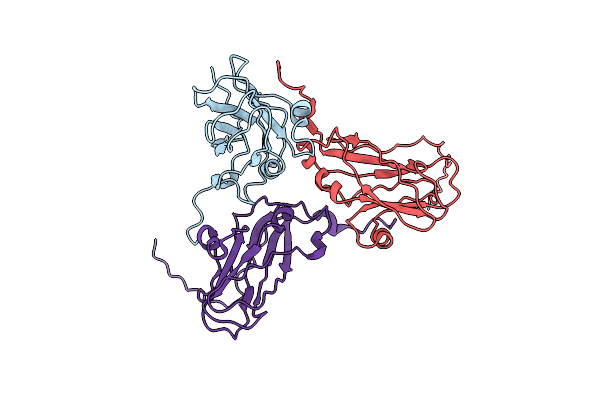
Virus Model Of Brome Mosaic Virus (First Half Data Set)
Organism: Brome mosaic virus
Method: ELECTRON MICROSCOPY Release Date: 2014-09-10 Classification: VIRUS |
Organism: Brome mosaic virus
Method: ELECTRON MICROSCOPY
Release Date: 2014-09-10
 |
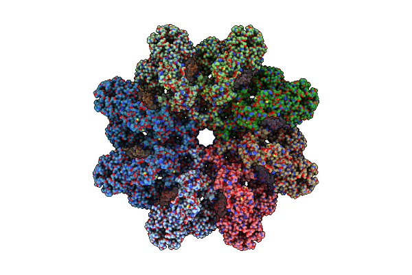
Virus Model Of Brome Mosaic Virus (Second Half Data Set)
Organism: Brome mosaic virus
Method: ELECTRON MICROSCOPY Release Date: 2014-09-10 Classification: VIRUS |
Organism: Brome mosaic virus
Method: ELECTRON MICROSCOPY
Release Date: 2014-09-10
 |
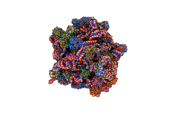
Structure Of The Drosophila Apoptosome
Organism: Drosophila melanogaster
Method: ELECTRON MICROSCOPY Release Date: 2014-07-09 Classification: APOPTOSIS Ligands: MG, DTP |
Organism: Drosophila melanogaster
Method: ELECTRON MICROSCOPY
Release Date: 2014-07-09
Ligands: MG, DTP
 |
Structure Of The Methanococcus Jannaschii Ribosome-Secyebeta Channel Complex
Organism: Methanococcus jannaschii, Methanococcus jannaschii
Method: ELECTRON MICROSCOPY Release Date: 2014-07-09 Classification: RIBOSOME/PROTEIN TRANSPORT |
Organism: Methanococcus jannaschii, Methanococcus jannaschii
Method: ELECTRON MICROSCOPY
Release Date: 2014-07-09