

Search Count: 13
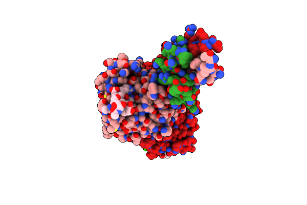 |
Crystal Structure Of Recombinant Gamma N308K Fibrinogen Fragment D With The Peptide Ligand Gly-Pro-Arg-Pro-Amide
Organism: Homo sapiens
Method: X-RAY DIFFRACTION Resolution:3.04 Å Release Date: 2009-08-18 Classification: BLOOD CLOTTING Ligands: CA |
Organism: Homo sapiens
Method: X-RAY DIFFRACTION
Release Date: 2009-08-18
Ligands: CA
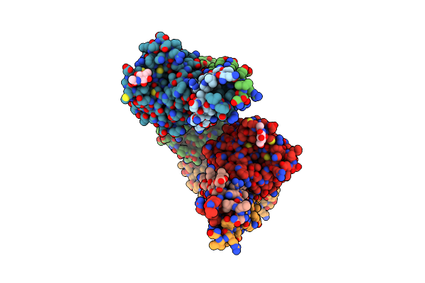 |
Crystal Structure Of Bbetad432A Variant Fibrinogen Fragment D With The Peptide Ligand Gly-His-Arg-Pro-Amide
Organism: Homo sapiens, Synthetic construct
Method: X-RAY DIFFRACTION Resolution:2.30 Å Release Date: 2009-01-13 Classification: BLOOD CLOTTING Ligands: NAG, CA |
Organism: Homo sapiens, Synthetic construct
Method: X-RAY DIFFRACTION
Release Date: 2009-01-13
Ligands: NAG, CA
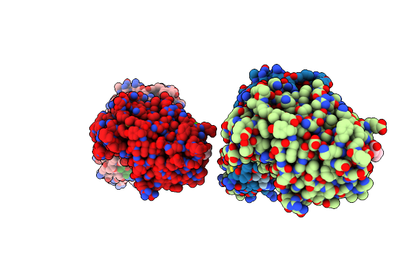 |
Crystal Structure Of Recombinant Gammad364A Fibrinogen Fragment D With The Peptide Ligand Gly-Pro-Arg-Pro-Amide
Organism: Homo sapiens, Synthetic construct
Method: X-RAY DIFFRACTION Resolution:2.60 Å Release Date: 2008-09-02 Classification: BLOOD CLOTTING Ligands: CA |
Organism: Homo sapiens, Synthetic construct
Method: X-RAY DIFFRACTION
Release Date: 2008-09-02
Ligands: CA
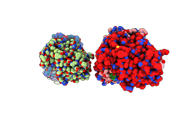 |
Crystal Structure Of Fragment D Of Gammad298,301A Fibrinogen With The Peptide Ligand Gly-His-Arg-Pro-Amide
Organism: Homo sapiens
Method: X-RAY DIFFRACTION Resolution:2.40 Å Release Date: 2007-05-15 Classification: BLOOD CLOTTING Ligands: CA |
Organism: Homo sapiens
Method: X-RAY DIFFRACTION
Release Date: 2007-05-15
Ligands: CA
 |
Crystal Structure Of Fragment D Of Gammad298,301A Fibrinogen With The Peptide Ligand Gly-Pro-Arg-Pro-Amide
Organism: Homo sapiens
Method: X-RAY DIFFRACTION Resolution:2.70 Å Release Date: 2007-05-15 Classification: BLOOD CLOTTING Ligands: CA |
Organism: Homo sapiens
Method: X-RAY DIFFRACTION
Release Date: 2007-05-15
Ligands: CA
 |
Fibrinogen Fragment D With "A" Knob Peptide Mimic Gprvve
Organism: Homo sapiens
Method: X-RAY DIFFRACTION Resolution:2.89 Å Release Date: 2006-07-04 Classification: BLOOD CLOTTING Ligands: CA |
Organism: Homo sapiens
Method: X-RAY DIFFRACTION
Release Date: 2006-07-04
Ligands: CA
 |
Crystal Structure Of Fragment D Of Bbetad398A Fibrinogen With The Peptide Ligand Gly-His-Arg-Pro-Amide
Organism: Homo sapiens
Method: X-RAY DIFFRACTION Resolution:2.45 Å Release Date: 2004-05-25 Classification: BLOOD CLOTTING Ligands: CA |
Organism: Homo sapiens
Method: X-RAY DIFFRACTION
Release Date: 2004-05-25
Ligands: CA
 |
Crystal Structure Of Fragment D Of Bbetad398A Fibrinogen
Organism: Homo sapiens
Method: X-RAY DIFFRACTION Resolution:2.70 Å Release Date: 2004-05-25 Classification: BLOOD CLOTTING Ligands: NAG, CA |
Organism: Homo sapiens
Method: X-RAY DIFFRACTION
Release Date: 2004-05-25
Ligands: NAG, CA
 |
Crystal Structure Of Fragment D Of Gammae132A Fibrinogen
Organism: Homo sapiens
Method: X-RAY DIFFRACTION Resolution:2.81 Å Release Date: 2004-03-16 Classification: BLOOD CLOTTING Ligands: NAG, CA |
Organism: Homo sapiens
Method: X-RAY DIFFRACTION
Release Date: 2004-03-16
Ligands: NAG, CA
 |
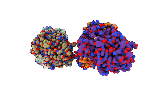
Crystal Structure Of Fragment D Of Gammae132A Fibrinogen With The Peptide Ligand Gly-His-Arg-Pro-Amide
Organism: Homo sapiens
Method: X-RAY DIFFRACTION Resolution:2.53 Å Release Date: 2004-03-16 Classification: BLOOD CLOTTING Ligands: CA |
Organism: Homo sapiens
Method: X-RAY DIFFRACTION
Release Date: 2004-03-16
Ligands: CA
 |
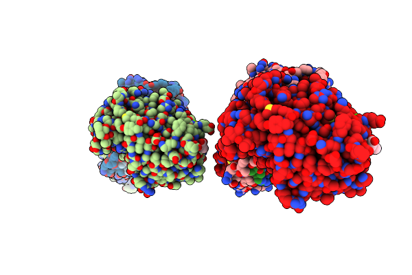
Crystal Structure Of Recombinant Human Fibrinogen Fragment D
Organism: Homo sapiens
Method: X-RAY DIFFRACTION Resolution:2.80 Å Release Date: 2002-11-06 Classification: BLOOD CLOTTING Ligands: NAG, CA |
Organism: Homo sapiens
Method: X-RAY DIFFRACTION
Release Date: 2002-11-06
Ligands: NAG, CA
 |
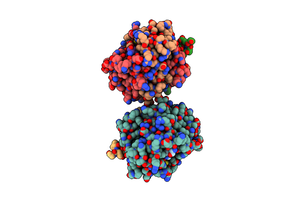
Crystal Structure Of Recombinant Human Fibrinogen Fragment D With The Peptide Ligands Gly-Pro-Arg-Pro-Amide And Gly-His-Arg-Pro-Amide
Organism: Homo sapiens
Method: X-RAY DIFFRACTION Resolution:2.80 Å Release Date: 2002-11-06 Classification: BLOOD CLOTTING Ligands: CA |
Organism: Homo sapiens
Method: X-RAY DIFFRACTION
Release Date: 2002-11-06
Ligands: CA
 |
The Crystal Structure Of Fibrinogen-Aa Peptide 1-23 (F8Y) Bound To Bovine Thrombin Explains Why The Mutation Of Phe-8 To Tyrosine Strongly Inhibits Normal Cleavage At Arginine-16
Organism: Bos taurus
Method: X-RAY DIFFRACTION Resolution:2.50 Å Release Date: 1998-05-06 Classification: HYDROLASE/HYDROLASE SUBSTRATE |
Organism: Bos taurus
Method: X-RAY DIFFRACTION
Release Date: 1998-05-06