

Search Count: 25
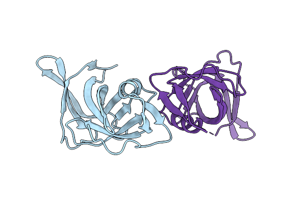 |
Pseudomonas Fluorescens Periplasmic Aspartic Peptidase Pfl01_1762 (Rloa)
Organism: Pseudomonas fluorescens pf0-1
Method: X-RAY DIFFRACTION Resolution:2.40 Å Release Date: 2025-04-09 Classification: HYDROLASE |
Organism: Pseudomonas fluorescens pf0-1
Method: X-RAY DIFFRACTION
Release Date: 2025-04-09
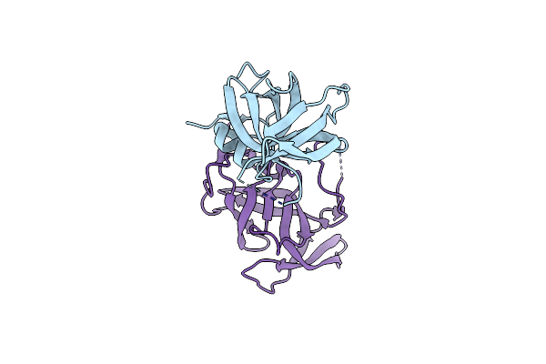 |
Pseudomonas Aeruginosa Periplasmic Aspartic Peptidase Pa0462 (Rloa2)
Organism: Pseudomonas aeruginosa pao1
Method: X-RAY DIFFRACTION Resolution:2.40 Å Release Date: 2025-04-09 Classification: HYDROLASE |
Organism: Pseudomonas aeruginosa pao1
Method: X-RAY DIFFRACTION
Release Date: 2025-04-09
 |
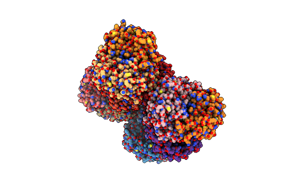
Crystal Structure Of Rimk From Pseudomonas Aeruginosa Pao1
Organism: Pseudomonas aeruginosa pao1, Synthetic construct
Method: X-RAY DIFFRACTION Resolution:2.40 Å Release Date: 2022-10-05 Classification: LIGASE Ligands: ADP |
Organism: Pseudomonas aeruginosa pao1, Synthetic construct
Method: X-RAY DIFFRACTION
Release Date: 2022-10-05
Ligands: ADP
 |
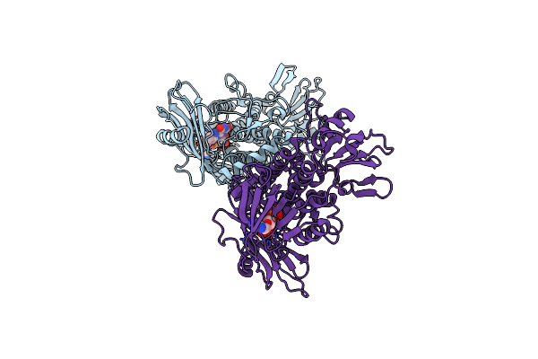
Crystal Structure Of Rimk From Pseudomonas Syringae Dc3000
Organism: Pseudomonas syringae pv. tomato str. dc3000
Method: X-RAY DIFFRACTION Resolution:2.90 Å Release Date: 2022-10-05 Classification: LIGASE Ligands: ADP |
Organism: Pseudomonas syringae pv. tomato str. dc3000
Method: X-RAY DIFFRACTION
Release Date: 2022-10-05
Ligands: ADP
 |
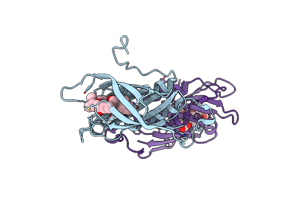
Tmn9 In Complex With Cofactor Fad
Organism: Streptomyces sp. nrrl 11266
Method: X-RAY DIFFRACTION Resolution:2.40 Å Release Date: 2020-10-07 Classification: OXIDOREDUCTASE Ligands: FAD |
Organism: Streptomyces sp. nrrl 11266
Method: X-RAY DIFFRACTION
Release Date: 2020-10-07
Ligands: FAD
 |
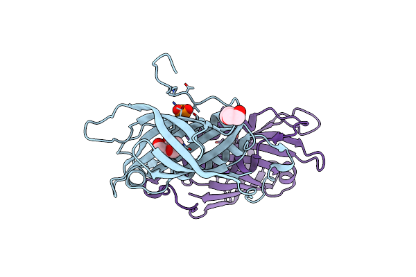
Tsn15 In Complex With Substrate Intermediate
Organism: Streptomyces longisporoflavus
Method: X-RAY DIFFRACTION Resolution:1.70 Å Release Date: 2019-10-23 Classification: UNKNOWN FUNCTION Ligands: KUJ, PO4, PPI |
Organism: Streptomyces longisporoflavus
Method: X-RAY DIFFRACTION
Release Date: 2019-10-23
Ligands: KUJ, PO4, PPI
 |
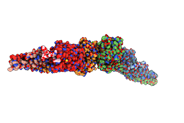
Crystal Structure Of Tsn15 In Apo Form
Organism: Streptomyces longisporoflavus
Method: X-RAY DIFFRACTION Resolution:1.80 Å Release Date: 2019-10-23 Classification: LYASE Ligands: PO4, PGE, PEG |
Organism: Streptomyces longisporoflavus
Method: X-RAY DIFFRACTION
Release Date: 2019-10-23
Ligands: PO4, PGE, PEG
 |
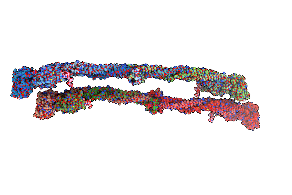
Crystal Structure Of D-Dimer From Human Fibrin Complexed With Gly-His-Arg-Pro-Tyr-Amide
Organism: Homo sapiens
Method: X-RAY DIFFRACTION Resolution:3.60 Å Release Date: 2009-07-28 Classification: BLOOD CLOTTING Ligands: CA |
Organism: Homo sapiens
Method: X-RAY DIFFRACTION
Release Date: 2009-07-28
Ligands: CA
 |
Crystal Structure Of Human Fibrinogen
Organism: Homo sapiens
Method: X-RAY DIFFRACTION Resolution:2.90 Å Release Date: 2009-05-19 Classification: BLOOD CLOTTING Ligands: CA |
Organism: Homo sapiens
Method: X-RAY DIFFRACTION
Release Date: 2009-05-19
Ligands: CA
 |
Crystal Structure Of D-Dimer From Human Fibrin Complexed With Met-His-Arg-Pro-Tyr-Amide.
Organism: Homo sapiens
Method: X-RAY DIFFRACTION Resolution:2.80 Å Release Date: 2007-12-11 Classification: BLOOD CLOTTING Ligands: CA |
Organism: Homo sapiens
Method: X-RAY DIFFRACTION
Release Date: 2007-12-11
Ligands: CA
 |
Crystal Structure Of D-Dimer From Human Fibrin Complexed With Gly-His-Arg-Pro-Tyr-Amide
Organism: Homo sapiens
Method: X-RAY DIFFRACTION Resolution:2.70 Å Release Date: 2007-10-23 Classification: BLOOD CLOTTING Ligands: CA, NAG |
Organism: Homo sapiens
Method: X-RAY DIFFRACTION
Release Date: 2007-10-23
Ligands: CA, NAG
 |
Crystal Structure Of Human Fragment D Complexed With Ala-His-Arg-Pro-Amide
Organism: Homo sapiens
Method: X-RAY DIFFRACTION Resolution:2.70 Å Release Date: 2006-12-05 Classification: BLOOD CLOTTING Ligands: CA |
Organism: Homo sapiens
Method: X-RAY DIFFRACTION
Release Date: 2006-12-05
Ligands: CA
 |
Fibrin D-Dimer, Lamprey Complexed With The Peptide Ligand: Gly-His-Arg-Pro-Amide
Organism: Petromyzon marinus
Method: X-RAY DIFFRACTION Resolution:2.90 Å Release Date: 2003-01-07 Classification: BLOOD CLOTTING Ligands: NAG, CA |
Organism: Petromyzon marinus
Method: X-RAY DIFFRACTION
Release Date: 2003-01-07
Ligands: NAG, CA
 |
Crystal Structure Of Human D-Dimer From Cross-Linked Fibrin Complexed With Gpr And Ghrpldk Peptide Ligands.
Organism: Homo sapiens
Method: X-RAY DIFFRACTION Resolution:3.20 Å Release Date: 2003-01-07 Classification: BLOOD CLOTTING Ligands: NDG, MAN, CA |
Organism: Homo sapiens
Method: X-RAY DIFFRACTION
Release Date: 2003-01-07
Ligands: NDG, MAN, CA
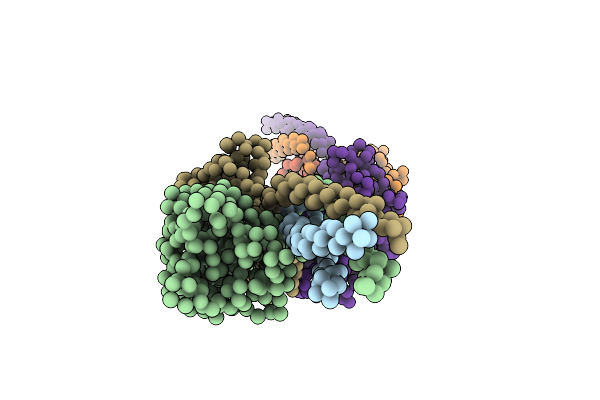 |
Fragment Double-D From Human Fibrin
Organism: Homo sapiens
Method: X-RAY DIFFRACTION Resolution:4.50 Å Release Date: 2003-01-07 Classification: BLOOD CLOTTING |
Organism: Homo sapiens
Method: X-RAY DIFFRACTION
Release Date: 2003-01-07
 |
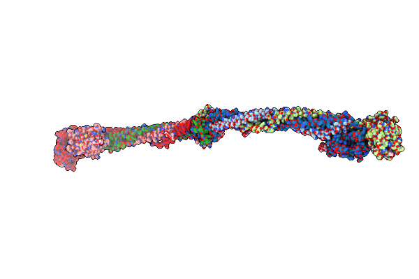
Crystal Structure Of Fragment D From Lamprey Fibrinogen Complexed With The Peptide Gly-His-Arg-Pro-Amide
Organism: Petromyzon marinus
Method: X-RAY DIFFRACTION Resolution:2.80 Å Release Date: 2002-08-23 Classification: BLOOD CLOTTING Ligands: NDG, MAN, CA, NAG, BMA, GAL |
Organism: Petromyzon marinus
Method: X-RAY DIFFRACTION
Release Date: 2002-08-23
Ligands: NDG, MAN, CA, NAG, BMA, GAL
 |
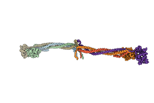
Crystal Structure Of Native Chicken Fibrinogen With Two Different Bound Ligands
Organism: Gallus gallus
Method: X-RAY DIFFRACTION Resolution:2.70 Å Release Date: 2002-06-26 Classification: BLOOD CLOTTING Ligands: NDG, CA, NAG |
Organism: Gallus gallus
Method: X-RAY DIFFRACTION
Release Date: 2002-06-26
Ligands: NDG, CA, NAG
 |
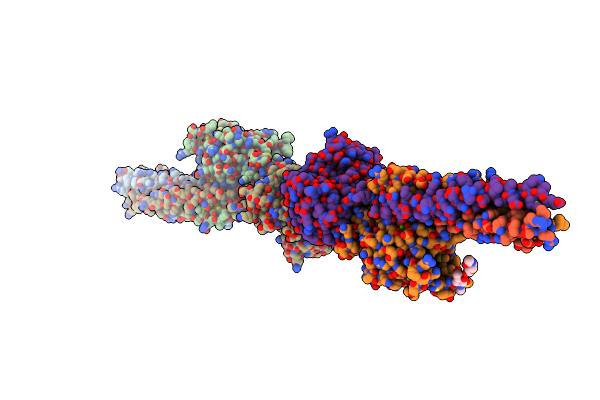
Crystal Structure Of Native Chicken Fibrinogen
Organism: Gallus gallus
Method: X-RAY DIFFRACTION Resolution:5.50 Å Release Date: 2000-05-10 Classification: BLOOD CLOTTING |
Organism: Gallus gallus
Method: X-RAY DIFFRACTION
Release Date: 2000-05-10
 |
Crystal Structure Of Fragment Double-D From Human Fibrin
Organism: Homo sapiens
Method: X-RAY DIFFRACTION Resolution:3.00 Å Release Date: 1999-06-08 Classification: BLOOD COAGULATION Ligands: NAG, CA |
Organism: Homo sapiens
Method: X-RAY DIFFRACTION
Release Date: 1999-06-08
Ligands: NAG, CA
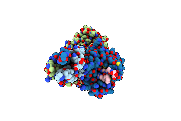 |
Crystal Structure Of Fragment Double-D From Human Fibrin With The Peptide Ligand Gly-His-Arg-Pro-Amide
Organism: Homo sapiens
Method: X-RAY DIFFRACTION Resolution:2.70 Å Release Date: 1999-06-08 Classification: BLOOD COAGULATION Ligands: NAG, CA |
Organism: Homo sapiens
Method: X-RAY DIFFRACTION
Release Date: 1999-06-08
Ligands: NAG, CA