

Search Count: 20
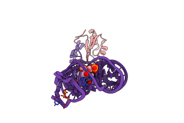 |
The C-Di-Gmp-I Riboswitch Bound To Pgpg
Organism: Homo sapiens
Method: X-RAY DIFFRACTION Resolution:2.80 Å Release Date: 2012-01-04 Classification: SIGNALING PROTEIN/RNA Ligands: MG |
Organism: Homo sapiens
Method: X-RAY DIFFRACTION
Release Date: 2012-01-04
Ligands: MG
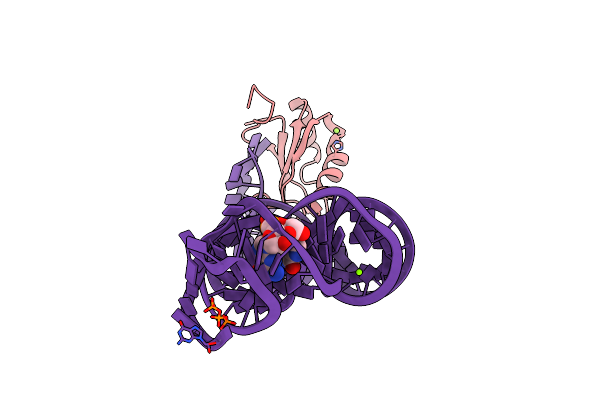 |
The C-Di-Gmp-I Riboswitch Bound To Gpg
Organism: Homo sapiens
Method: X-RAY DIFFRACTION Resolution:2.80 Å Release Date: 2012-01-04 Classification: SIGNALING PROTEIN/RNA Ligands: MG |
Organism: Homo sapiens
Method: X-RAY DIFFRACTION
Release Date: 2012-01-04
Ligands: MG
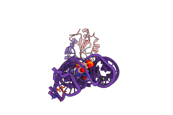 |
The C92U Mutant C-Di-Gmp-I Riboswitch Bound To Pgpa
Organism: Homo sapiens
Method: X-RAY DIFFRACTION Resolution:3.10 Å Release Date: 2012-01-04 Classification: SIGNALING PROTEIN/RNA Ligands: MG |
Organism: Homo sapiens
Method: X-RAY DIFFRACTION
Release Date: 2012-01-04
Ligands: MG
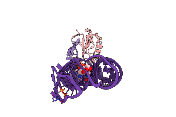 |
The C92U Mutant C-Di-Gmp-I Riboswitch Bound To Gpa
Organism: Homo sapiens
Method: X-RAY DIFFRACTION Resolution:2.70 Å Release Date: 2012-01-04 Classification: SIGNALING PROTEIN/RNA Ligands: MG |
Organism: Homo sapiens
Method: X-RAY DIFFRACTION
Release Date: 2012-01-04
Ligands: MG
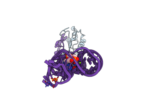 |
Crystal Structure Of The G20A Mutant C-Di-Gmp Riboswith Bound To C-Di-Gmp
Organism: Homo sapiens
Method: X-RAY DIFFRACTION Resolution:2.90 Å Release Date: 2010-08-25 Classification: RNA binding PROTEIN/RNA Ligands: C2E, MG |
Organism: Homo sapiens
Method: X-RAY DIFFRACTION
Release Date: 2010-08-25
Ligands: C2E, MG
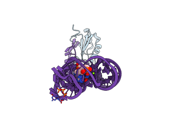 |
Crystal Structure Of The C92U Mutant C-Di-Gmp Riboswith Bound To C-Di-Gmp
Organism: Homo sapiens
Method: X-RAY DIFFRACTION Resolution:3.00 Å Release Date: 2010-08-25 Classification: RNA binding PROTEIN/RNA Ligands: C2E, MG |
Organism: Homo sapiens
Method: X-RAY DIFFRACTION
Release Date: 2010-08-25
Ligands: C2E, MG
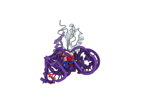 |
Crystal Structure Of The G20A/C92U Mutant C-Di-Gmp Riboswith Bound To C-Di-Gmp
Organism: Homo sapiens
Method: X-RAY DIFFRACTION Resolution:3.00 Å Release Date: 2010-08-25 Classification: RNA BINDING PROTEIN/RNA Ligands: C2E, MG |
Organism: Homo sapiens
Method: X-RAY DIFFRACTION
Release Date: 2010-08-25
Ligands: C2E, MG
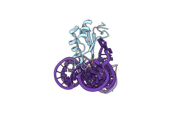 |
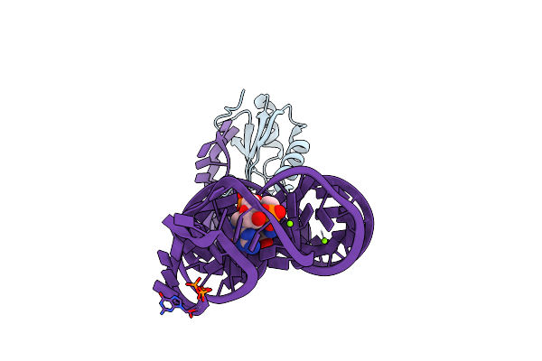
Crystal Structure Of The G20A/C92U Mutant C-Di-Gmp Riboswith Bound To C-Di-Amp
Organism: Homo sapiens
Method: X-RAY DIFFRACTION Resolution:3.20 Å Release Date: 2010-08-25 Classification: RNA binding PROTEIN/rna Ligands: 2BA, MG |
Organism: Homo sapiens
Method: X-RAY DIFFRACTION
Release Date: 2010-08-25
Ligands: 2BA, MG
 |
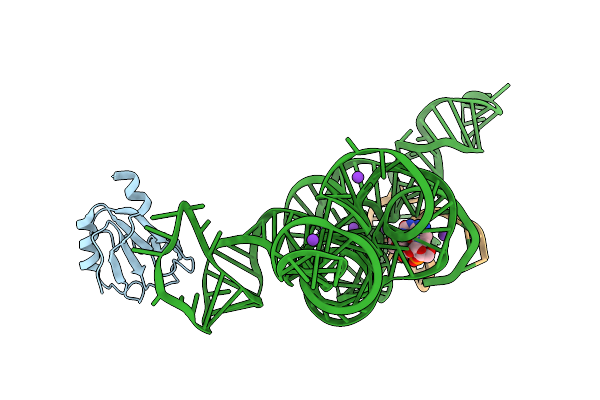
Native Structure Of A C-Di-Gmp Riboswitch From V. Cholerae
Organism: Homo sapiens
Method: X-RAY DIFFRACTION Resolution:2.30 Å Release Date: 2010-08-25 Classification: RNA BINDING PROTEIN/RNA Ligands: MG, C2E |
Organism: Homo sapiens
Method: X-RAY DIFFRACTION
Release Date: 2010-08-25
Ligands: MG, C2E
 |
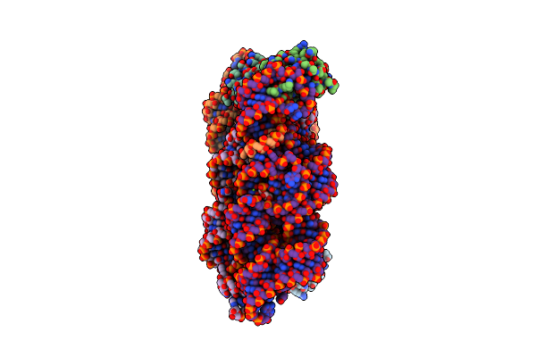
Plasticity Of The Kink Turn Structural Motif
Organism: Homo sapiens
Method: X-RAY DIFFRACTION Resolution:4.18 Å Release Date: 2010-03-09 Classification: RNA binding protein/DNA/RNA Ligands: K, MG |
Organism: Homo sapiens
Method: X-RAY DIFFRACTION
Release Date: 2010-03-09
Ligands: K, MG
 |
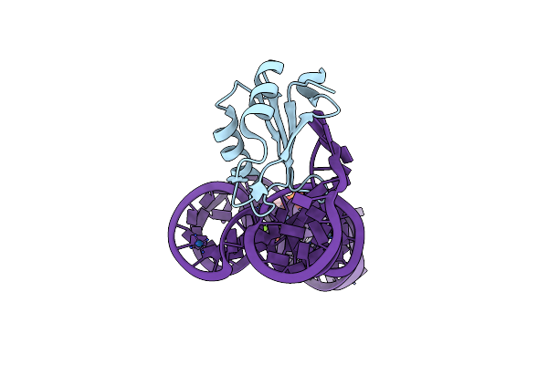
Crystal Structure Of The Bacillus Anthracis Glms Ribozyme Bound To Glc6P
Organism: Homo sapiens
Method: X-RAY DIFFRACTION Resolution:2.85 Å Release Date: 2009-12-29 Classification: RNA binding protein/RNA Ligands: MG, G6P |
Organism: Homo sapiens
Method: X-RAY DIFFRACTION
Release Date: 2009-12-29
Ligands: MG, G6P
 |
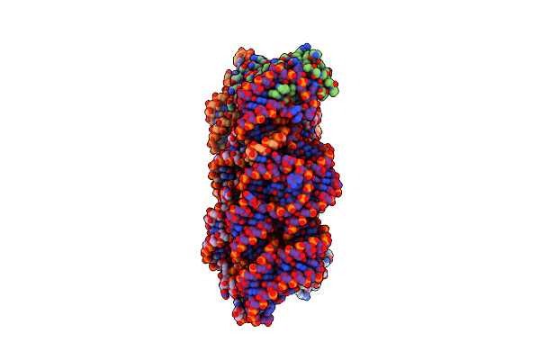
Structure Of A C-Di-Gmp Riboswitch From V. Cholerae
Organism: Homo sapiens, Vibrio cholerae
Method: X-RAY DIFFRACTION Resolution:2.70 Å Release Date: 2009-11-10 Classification: RNA binding protein/RNA Ligands: C2E, IRI, MG |
Organism: Homo sapiens, Vibrio cholerae
Method: X-RAY DIFFRACTION
Release Date: 2009-11-10
Ligands: C2E, IRI, MG
 |
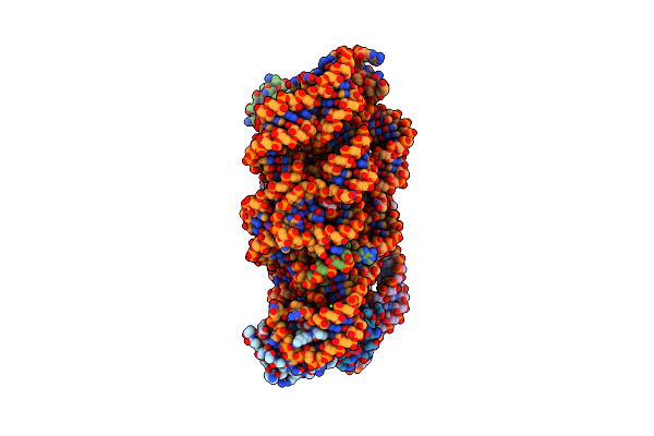
Crystal Structure Of The Pre-Cleaved Bacillus Anthracis Glms Ribozyme
Organism: Homo sapiens
Method: X-RAY DIFFRACTION Resolution:3.10 Å Release Date: 2009-11-03 Classification: RNA binding protein/RNA Ligands: MG |
Organism: Homo sapiens
Method: X-RAY DIFFRACTION
Release Date: 2009-11-03
Ligands: MG
 |
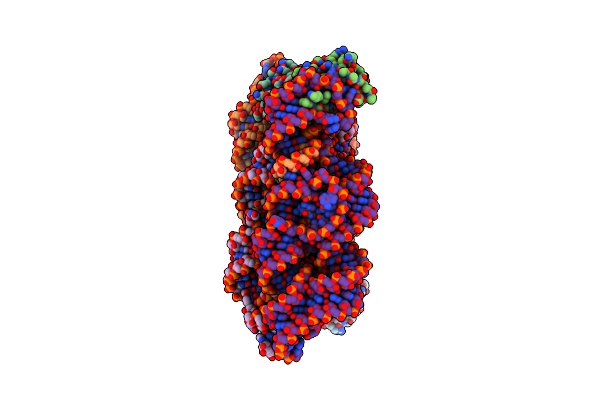
Crystal Structure Of The G33A Mutant Bacillus Anthracis Glms Ribozyme Bound To Glcn6P
Organism: Homo sapiens, Syntetic construct
Method: X-RAY DIFFRACTION Resolution:3.00 Å Release Date: 2009-11-03 Classification: RNA binding protein/RNA Ligands: GLP, MG |
Organism: Homo sapiens, Syntetic construct
Method: X-RAY DIFFRACTION
Release Date: 2009-11-03
Ligands: GLP, MG
 |
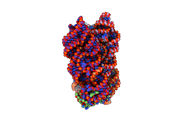
Crystal Structure Of The Bacillus Anthracis Glms Ribozyme Bound To Man6P
Organism: Homo sapiens, Syntetic construct
Method: X-RAY DIFFRACTION Resolution:3.01 Å Release Date: 2009-11-03 Classification: RNA binding protein/RNA Ligands: 6MN, MG |
Organism: Homo sapiens, Syntetic construct
Method: X-RAY DIFFRACTION
Release Date: 2009-11-03
Ligands: 6MN, MG
 |
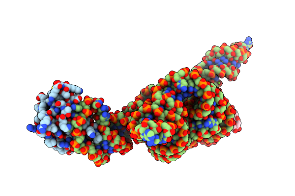
Crystal Structure Of The Product Bacillus Anthracis Glms Ribozyme
Organism: Homo sapiens, Syntetic construct
Method: X-RAY DIFFRACTION Resolution:2.90 Å Release Date: 2009-11-03 Classification: RNA binding protein/RNA Ligands: GLP, MG |
Organism: Homo sapiens, Syntetic construct
Method: X-RAY DIFFRACTION
Release Date: 2009-11-03
Ligands: GLP, MG
 |
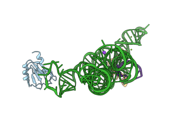
A Relaxed Active Site Following Exon Ligation By A Group I Intron
Organism: Homo sapiens, Synthetic construct
Method: X-RAY DIFFRACTION Resolution:3.31 Å Release Date: 2008-04-01 Classification: NUCLEAR PROTEIN/RNA Ligands: MG |
Organism: Homo sapiens, Synthetic construct
Method: X-RAY DIFFRACTION
Release Date: 2008-04-01
Ligands: MG
 |
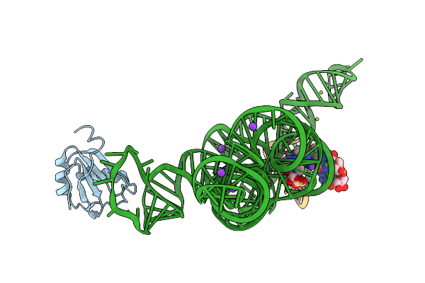
A Relaxed Active Site Following Exon Ligation By A Group I Intron
Organism: Homo sapiens, Synthetic construct
Method: X-RAY DIFFRACTION Resolution:3.40 Å Release Date: 2008-04-01 Classification: NUCLEAR PROTEIN/RNA Ligands: MG, K |
Organism: Homo sapiens, Synthetic construct
Method: X-RAY DIFFRACTION
Release Date: 2008-04-01
Ligands: MG, K
 |
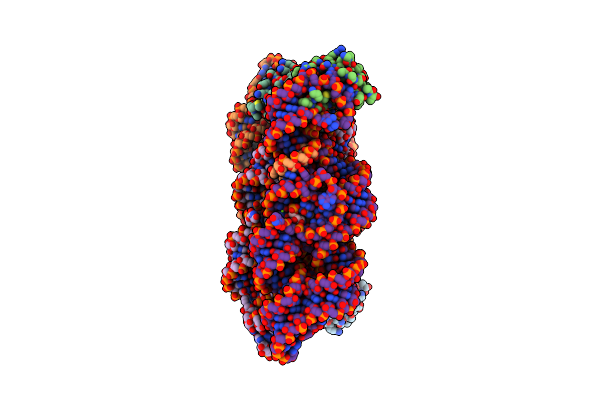
A Relaxed Active Site Following Exon Ligation By A Group I Intron
Organism: Homo sapiens, Synthetic construct
Method: X-RAY DIFFRACTION Resolution:3.33 Å Release Date: 2008-04-01 Classification: NUCLEAR PROTEIN/RNA Ligands: MG, K |
Organism: Homo sapiens, Synthetic construct
Method: X-RAY DIFFRACTION
Release Date: 2008-04-01
Ligands: MG, K
 |
Structural Investigation Of The Glms Ribozyme Bound To Its Catalytic Cofactor
Organism: Homo sapiens
Method: X-RAY DIFFRACTION Resolution:2.50 Å Release Date: 2007-01-16 Classification: STRUCTURAL PROTEIN/RNA Ligands: GLP, MG |
Organism: Homo sapiens
Method: X-RAY DIFFRACTION
Release Date: 2007-01-16
Ligands: GLP, MG