

Search Count: 65
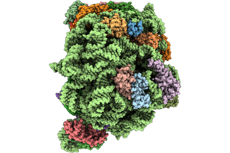 |
Pseudomonas Aeruginosa 50S Ribosome Bound To Rsfs (L1 Stalk In 'Mid' Conformation)
Organism: Pseudomonas aeruginosa pao1
Method: ELECTRON MICROSCOPY Resolution:2.29 Å Release Date: 2026-08-26 Classification: RIBOSOME Ligands: MG, ZN |
Organism: Pseudomonas aeruginosa pao1
Method: ELECTRON MICROSCOPY
Release Date: 2026-08-26
Ligands: MG, ZN
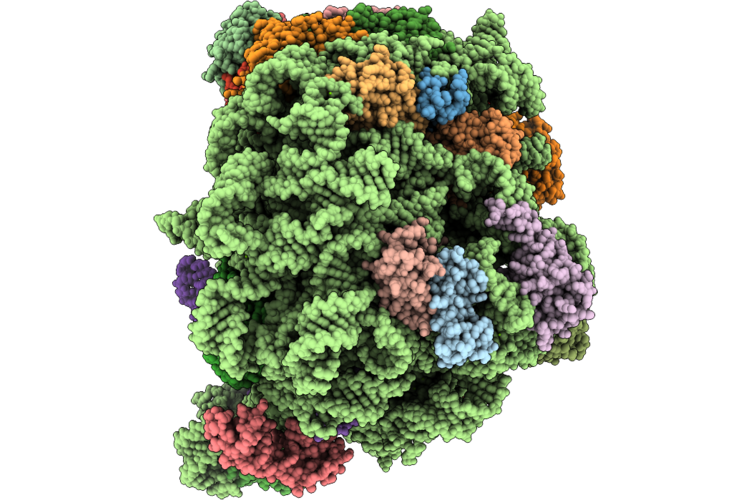 |
Pseudomonas Aeruginosa 50S Ribosome Bound To Rsfs (L1 Stalk 'In' Conformation)
Organism: Pseudomonas aeruginosa pao1
Method: ELECTRON MICROSCOPY Resolution:2.29 Å Release Date: 2026-08-26 Classification: RIBOSOME Ligands: MG, ZN |
Organism: Pseudomonas aeruginosa pao1
Method: ELECTRON MICROSCOPY
Release Date: 2026-08-26
Ligands: MG, ZN
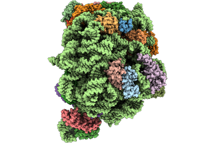 |
Pseudomonas Aeruginosa 50S Ribosome Bound To Rsfs (L1 Stalk In 'Out' Conformation)
Organism: Pseudomonas aeruginosa pao1
Method: ELECTRON MICROSCOPY Resolution:2.29 Å Release Date: 2026-08-26 Classification: RIBOSOME Ligands: MG, ZN |
Organism: Pseudomonas aeruginosa pao1
Method: ELECTRON MICROSCOPY
Release Date: 2026-08-26
Ligands: MG, ZN
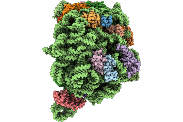 |
Pseudomonas Aeruginosa 50S Ribosome Bound To Rsfs (L1 Stalk In 'Out' Conformation And Missing Ul1 And Trna)
Organism: Pseudomonas aeruginosa pao1
Method: ELECTRON MICROSCOPY Resolution:2.29 Å Release Date: 2026-08-26 Classification: RIBOSOME Ligands: MG, ZN |
Organism: Pseudomonas aeruginosa pao1
Method: ELECTRON MICROSCOPY
Release Date: 2026-08-26
Ligands: MG, ZN
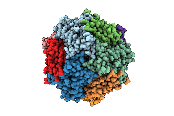 |
Structure Of Thioferritin (Pfdpsl) With Ferrihydrite Growth At A Single Three-Fold Pore.
Organism: Pyrococcus furiosus
Method: ELECTRON MICROSCOPY Release Date: 2025-10-29 Classification: METAL BINDING PROTEIN Ligands: FE, OXY, O |
Organism: Pyrococcus furiosus
Method: ELECTRON MICROSCOPY
Release Date: 2025-10-29
Ligands: FE, OXY, O
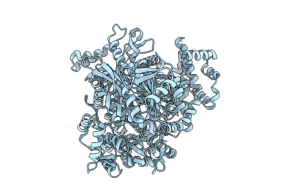 |
Hmus Heme Dechelatase: Disordered Domain 1, Heme Free.
Organism: Bacteroides thetaiotaomicron
Method: ELECTRON MICROSCOPY Release Date: 2025-09-24 Classification: METAL TRANSPORT |
Organism: Bacteroides thetaiotaomicron
Method: ELECTRON MICROSCOPY
Release Date: 2025-09-24
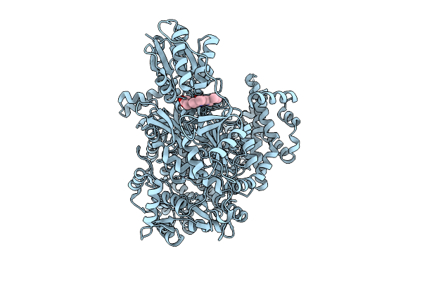 |
A Widespread Heme Dechelatase In Healthy And Pathogenic Human Microbiomes.
Organism: Bacteroides
Method: ELECTRON MICROSCOPY Release Date: 2025-08-20 Classification: METAL TRANSPORT Ligands: HEM, NA |
Organism: Bacteroides
Method: ELECTRON MICROSCOPY
Release Date: 2025-08-20
Ligands: HEM, NA
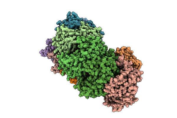 |
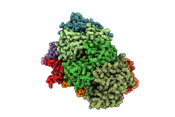
Post-Targeting Acascade Type Ia Crispr-Cas Surveillance Complexes
Organism: Saccharolobus solfataricus p2, Saccharolobus solfataricus
Method: ELECTRON MICROSCOPY Release Date: 2025-08-13 Classification: RNA BINDING PROTEIN/RNA Ligands: TRS |
Organism: Saccharolobus solfataricus p2, Saccharolobus solfataricus
Method: ELECTRON MICROSCOPY
Release Date: 2025-08-13
Ligands: TRS
 |
Post-Targeting Acascade Type Ia Crispr-Cas Surveillance Complexes
Organism: Saccharolobus solfataricus, Saccharolobus solfataricus p2
Method: ELECTRON MICROSCOPY Release Date: 2025-08-13 Classification: RNA BINDING PROTEIN/RNA/DNA Ligands: TRS |
Organism: Saccharolobus solfataricus, Saccharolobus solfataricus p2
Method: ELECTRON MICROSCOPY
Release Date: 2025-08-13
Ligands: TRS
 |
Post-Targeting Acascade Type Ia Crispr-Cas Surveillance Complexes
Organism: Saccharolobus solfataricus, Saccharolobus solfataricus p2
Method: ELECTRON MICROSCOPY Release Date: 2025-08-13 Classification: RNA BINDING PROTEIN/RNA Ligands: TRS |
Organism: Saccharolobus solfataricus, Saccharolobus solfataricus p2
Method: ELECTRON MICROSCOPY
Release Date: 2025-08-13
Ligands: TRS
 |
Structure Of Thioferritin From Pyrococcus Furiosis
Organism: Pyrococcus furiosus
Method: ELECTRON MICROSCOPY Release Date: 2025-08-06 Classification: METAL BINDING PROTEIN Ligands: FE |
Organism: Pyrococcus furiosus
Method: ELECTRON MICROSCOPY
Release Date: 2025-08-06
Ligands: FE
 |
Structure Of Thioferritin Exhibiting Iron Mineral Nucleation, From Pyrococcus Furiosis
Organism: Pyrococcus furiosus
Method: ELECTRON MICROSCOPY Release Date: 2025-08-06 Classification: METAL BINDING PROTEIN Ligands: FE, O |
Organism: Pyrococcus furiosus
Method: ELECTRON MICROSCOPY
Release Date: 2025-08-06
Ligands: FE, O
 |
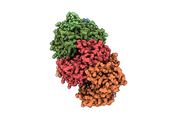
Structure Of Thioferritin With Averaged Iron Mineral Core, From Pyrococcus Furiosis
Organism: Pyrococcus furiosus
Method: ELECTRON MICROSCOPY Release Date: 2025-08-06 Classification: METAL BINDING PROTEIN Ligands: FE |
Organism: Pyrococcus furiosus
Method: ELECTRON MICROSCOPY
Release Date: 2025-08-06
Ligands: FE
 |
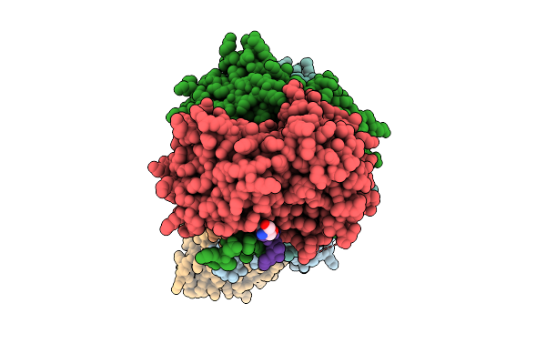
Post-Targeting Acascade Type I-A Crispr-Cas Surveillance Complexes
Organism: Saccharolobus solfataricus p2, Saccharolobus solfataricus
Method: ELECTRON MICROSCOPY Release Date: 2025-07-23 Classification: RNA BINDING PROTEIN/RNA/DNA Ligands: TRS |
Organism: Saccharolobus solfataricus p2, Saccharolobus solfataricus
Method: ELECTRON MICROSCOPY
Release Date: 2025-07-23
Ligands: TRS
 |
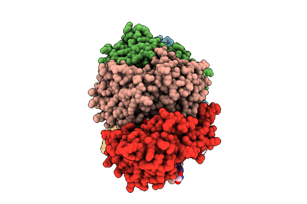
Post-Targeting Acascade Type Ia Crispr_Cas Surveillance Complexes
Organism: Saccharolobus solfataricus p2, Saccharolobus solfataricus
Method: ELECTRON MICROSCOPY Release Date: 2025-07-23 Classification: RNA BINDING PROTEIN/RNA/DNA Ligands: TRS |
Organism: Saccharolobus solfataricus p2, Saccharolobus solfataricus
Method: ELECTRON MICROSCOPY
Release Date: 2025-07-23
Ligands: TRS
 |
Post-Targeting Acascade Type Ia Crispr-Cas Surveillance Complexes
Organism: Saccharolobus solfataricus p2, Saccharolobus solfataricus
Method: ELECTRON MICROSCOPY Release Date: 2025-07-23 Classification: RNA BINDING PROTEIN/RNA/DNA Ligands: TRS |
Organism: Saccharolobus solfataricus p2, Saccharolobus solfataricus
Method: ELECTRON MICROSCOPY
Release Date: 2025-07-23
Ligands: TRS
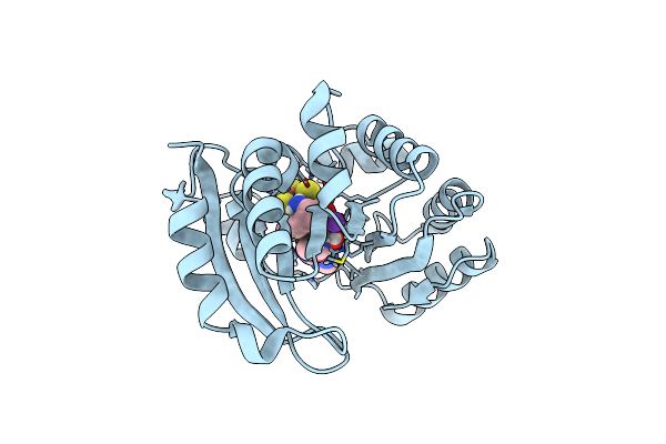 |
The Structure Of A Crystallizable Variant Of E. Coli Pyruvate Formate-Lyase Activating Enzyme Bound To A Partially Cleaved Sam Molecule
Organism: Escherichia coli k-12
Method: X-RAY DIFFRACTION Resolution:1.69 Å Release Date: 2023-05-24 Classification: OXIDOREDUCTASE Ligands: SF4, SAM, K |
Organism: Escherichia coli k-12
Method: X-RAY DIFFRACTION
Release Date: 2023-05-24
Ligands: SF4, SAM, K
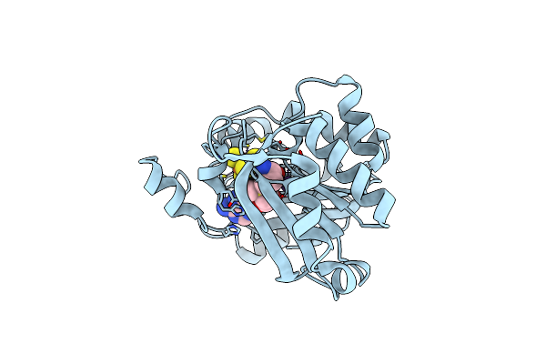 |
The Structure Of A Crystallizable Variant Of E. Coli Pyruvate Formate-Lyase Activating Enzyme Bound To Sam, Alternate Crystal Form
Organism: Escherichia coli k-12
Method: X-RAY DIFFRACTION Resolution:2.65 Å Release Date: 2023-05-24 Classification: OXIDOREDUCTASE Ligands: SF4, SAM, K, CL |
Organism: Escherichia coli k-12
Method: X-RAY DIFFRACTION
Release Date: 2023-05-24
Ligands: SF4, SAM, K, CL
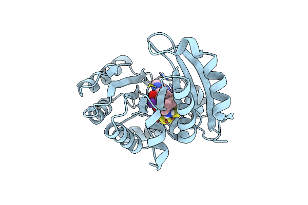 |
The Structure Of A Crystallizable Variant Of E. Coli Pyruvate Formate-Lyase Activating Enzyme Bound To Sam
Organism: Escherichia coli k-12
Method: X-RAY DIFFRACTION Resolution:1.46 Å Release Date: 2023-05-24 Classification: OXIDOREDUCTASE Ligands: SF4, SAM, K, CL |
Organism: Escherichia coli k-12
Method: X-RAY DIFFRACTION
Release Date: 2023-05-24
Ligands: SF4, SAM, K, CL
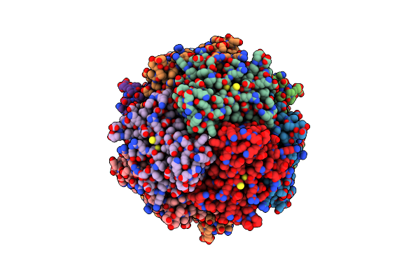 |
Structure Of Dpsl (Dna Protection In Starved Cells - Like) From Pyrococcus Furiosus
Organism: Pyrococcus furiosus
Method: ELECTRON MICROSCOPY Release Date: 2021-12-01 Classification: METAL BINDING PROTEIN Ligands: ZN, FE |
Organism: Pyrococcus furiosus
Method: ELECTRON MICROSCOPY
Release Date: 2021-12-01
Ligands: ZN, FE