

Search Count: 74
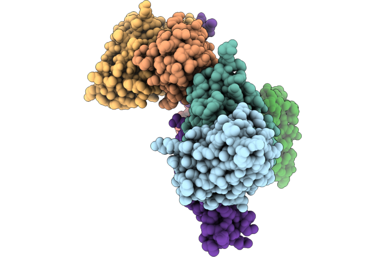 |
Cryo-Electron Microscopy Structure Of Pfripr Bound To Monoclonal Antibodies Rp.047, Rp.057 And Rp.035
Organism: Mus musculus, Plasmodium falciparum 3d7
Method: ELECTRON MICROSCOPY Resolution:3.35 Å Release Date: 2026-06-24 Classification: IMMUNE SYSTEM |
Organism: Mus musculus, Plasmodium falciparum 3d7
Method: ELECTRON MICROSCOPY
Release Date: 2026-06-24
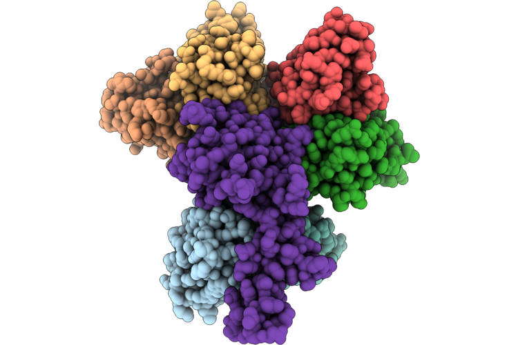 |
Cryo-Electron Microscopy Structure Of Pfripr Bound To Monoclonal Antibodies Rp.093, Rp.073 And Rp.063
Organism: Mus musculus, Plasmodium falciparum 3d7
Method: ELECTRON MICROSCOPY Resolution:2.97 Å Release Date: 2026-06-24 Classification: IMMUNE SYSTEM |
Organism: Mus musculus, Plasmodium falciparum 3d7
Method: ELECTRON MICROSCOPY
Release Date: 2026-06-24
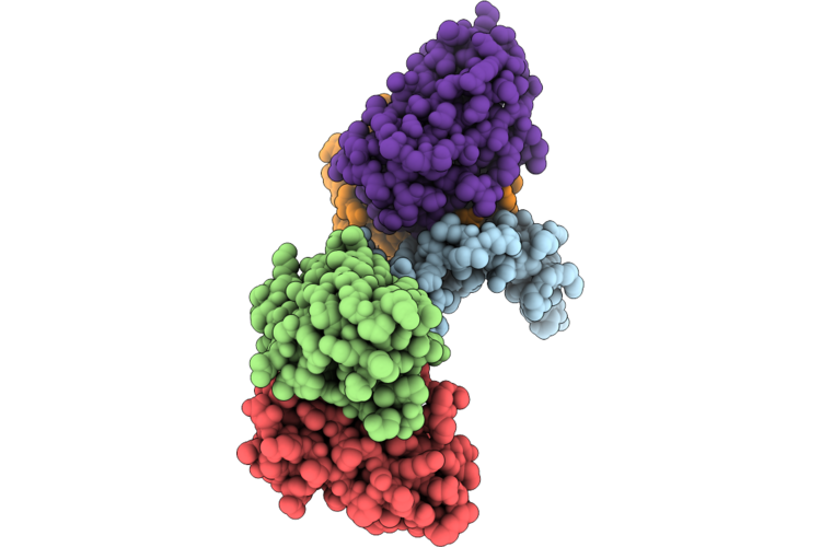 |
Cryo-Electron Microscopy Structure Of Pfripr Bound To Monoclonal Antibodies Rp.092 And Rp.052
Organism: Plasmodium falciparum 3d7, Mus musculus
Method: ELECTRON MICROSCOPY Resolution:4.01 Å Release Date: 2026-06-24 Classification: IMMUNE SYSTEM |
Organism: Plasmodium falciparum 3d7, Mus musculus
Method: ELECTRON MICROSCOPY
Release Date: 2026-06-24
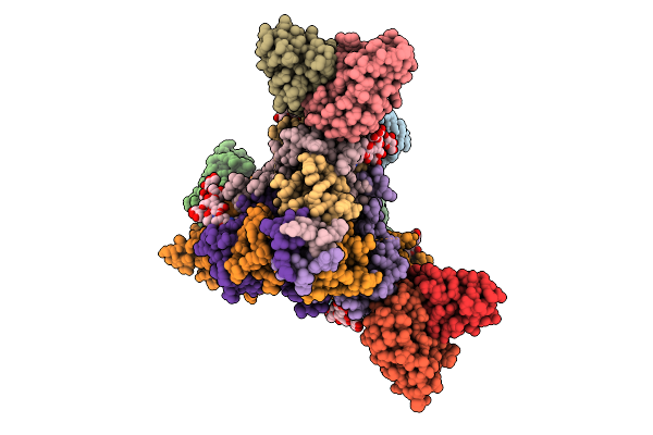 |
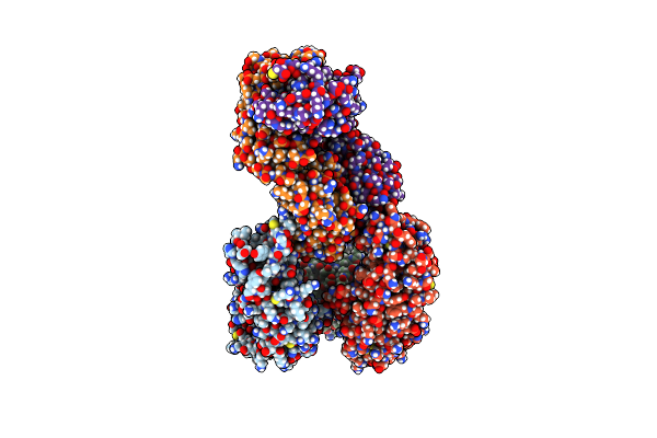
A Broadly-Neutralizing Antibody Against Ebolavirus Glycoprotein That Can Potentiate The Breadth And Neutralization Potency Of Other Anti-Glycoprotein Antibodies
Organism: Oryctolagus cuniculus, Ebolavirus
Method: ELECTRON MICROSCOPY Release Date: 2025-11-05 Classification: VIRAL PROTEIN/IMMUNE SYSTEM Ligands: NAG |
Organism: Oryctolagus cuniculus, Ebolavirus
Method: ELECTRON MICROSCOPY
Release Date: 2025-11-05
Ligands: NAG
 |
Plasmodium Falciparum Reticulocyte-Binding Protein Homologue 5 (Pfrh5) Bound To R5.251
Organism: Plasmodium falciparum, Homo sapiens
Method: X-RAY DIFFRACTION Resolution:3.23 Å Release Date: 2024-07-31 Classification: IMMUNE SYSTEM |
Organism: Plasmodium falciparum, Homo sapiens
Method: X-RAY DIFFRACTION
Release Date: 2024-07-31
 |
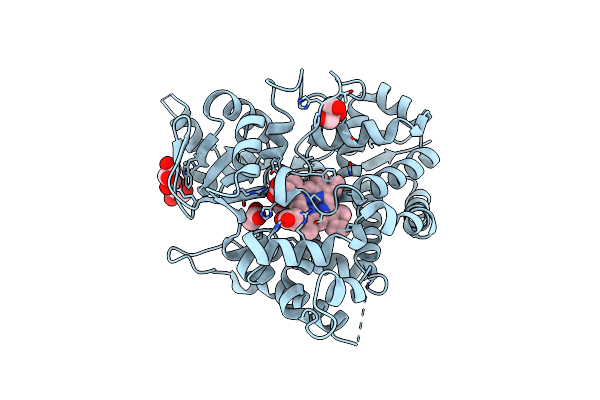
Plasmodium Falciparum Reticulocyte-Binding Protein Homologue 5 (Pfrh5) Bound To R5.034
Organism: Plasmodium falciparum, Homo sapiens
Method: X-RAY DIFFRACTION Resolution:3.99 Å Release Date: 2024-07-31 Classification: IMMUNE SYSTEM |
Organism: Plasmodium falciparum, Homo sapiens
Method: X-RAY DIFFRACTION
Release Date: 2024-07-31
 |
Crystal Structure Of Substrate-Free Syoa
Organism: Amycolatopsis thermoflava n1165
Method: X-RAY DIFFRACTION Resolution:1.98 Å Release Date: 2024-07-24 Classification: OXIDOREDUCTASE Ligands: HEM, GOL, ACT |
Organism: Amycolatopsis thermoflava n1165
Method: X-RAY DIFFRACTION
Release Date: 2024-07-24
Ligands: HEM, GOL, ACT
 |
Crystal Structure Of Syoa Bound To Syringol
Organism: Amycolatopsis thermoflava n1165
Method: X-RAY DIFFRACTION Resolution:1.26 Å Release Date: 2024-07-24 Classification: OXIDOREDUCTASE Ligands: HEM, 3DM, NO3, MG |
Organism: Amycolatopsis thermoflava n1165
Method: X-RAY DIFFRACTION
Release Date: 2024-07-24
Ligands: HEM, 3DM, NO3, MG
 |
Crystal Structure Of Syoa Bound To 4-Methylsyringol
Organism: Amycolatopsis thermoflava n1165
Method: X-RAY DIFFRACTION Resolution:1.12 Å Release Date: 2024-07-24 Classification: OXIDOREDUCTASE Ligands: HEM, UB9, NO3 |
Organism: Amycolatopsis thermoflava n1165
Method: X-RAY DIFFRACTION
Release Date: 2024-07-24
Ligands: HEM, UB9, NO3
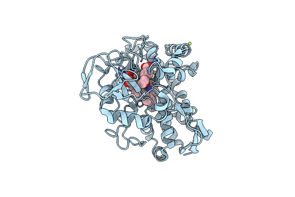 |
The Crystal Structure Of Wild-Type Cyp199A4 Bound To 4-Propionylbenzoic Acid
Organism: Rhodopseudomonas palustris haa2
Method: X-RAY DIFFRACTION Resolution:1.70 Å Release Date: 2024-05-08 Classification: OXIDOREDUCTASE Ligands: HEM, LVK, CL, MG |
Organism: Rhodopseudomonas palustris haa2
Method: X-RAY DIFFRACTION
Release Date: 2024-05-08
Ligands: HEM, LVK, CL, MG
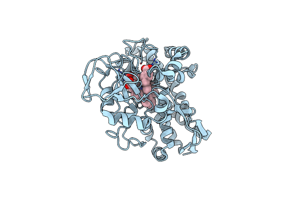 |
The Crystal Structure Of Wild-Type Cyp199A4 Bound To 4-(2-Oxopropyl)Benzoic Acid
Organism: Rhodopseudomonas palustris haa2
Method: X-RAY DIFFRACTION Resolution:1.84 Å Release Date: 2024-05-08 Classification: OXIDOREDUCTASE Ligands: LVC, HEM, CL |
Organism: Rhodopseudomonas palustris haa2
Method: X-RAY DIFFRACTION
Release Date: 2024-05-08
Ligands: LVC, HEM, CL
 |
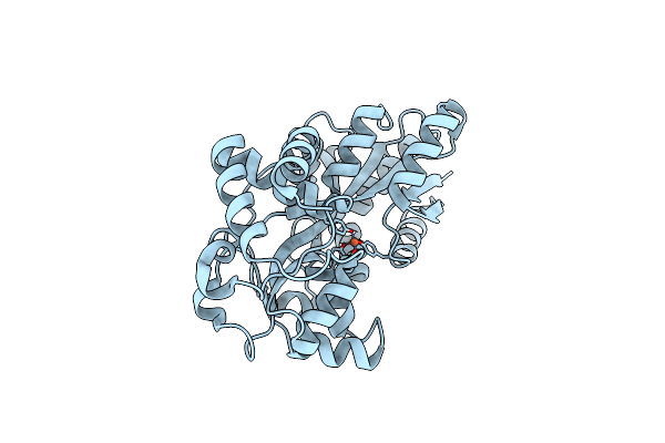
Crystal Structure Of Futa Bound To Fe(Iii) Solved By Neutron Diffraction
Organism: Prochlorococcus marinus subsp. pastoris str. ccmp1986
Method: NEUTRON DIFFRACTION Resolution:2.10 Å Release Date: 2024-01-17 Classification: METAL BINDING PROTEIN Ligands: FE |
Organism: Prochlorococcus marinus subsp. pastoris str. ccmp1986
Method: NEUTRON DIFFRACTION
Release Date: 2024-01-17
Ligands: FE
 |
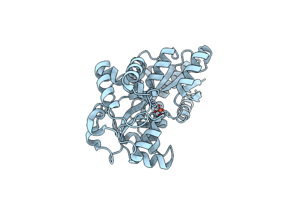
Sfx Structure Of Futa Bound To Fe(Iii)
Organism: Prochlorococcus marinus subsp. pastoris str. ccmp1986
Method: X-RAY DIFFRACTION Resolution:1.60 Å Release Date: 2023-08-30 Classification: METAL BINDING PROTEIN Ligands: FE |
Organism: Prochlorococcus marinus subsp. pastoris str. ccmp1986
Method: X-RAY DIFFRACTION
Release Date: 2023-08-30
Ligands: FE
 |
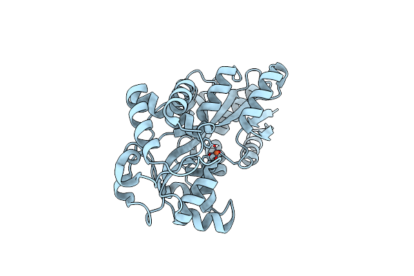
Sfx Structure Of Futa After An Accumulated Dose Of 350 Kgy
Organism: Prochlorococcus marinus subsp. pastoris str. ccmp1986
Method: X-RAY DIFFRACTION Resolution:1.65 Å Release Date: 2023-08-30 Classification: METAL BINDING PROTEIN Ligands: FE |
Organism: Prochlorococcus marinus subsp. pastoris str. ccmp1986
Method: X-RAY DIFFRACTION
Release Date: 2023-08-30
Ligands: FE
 |
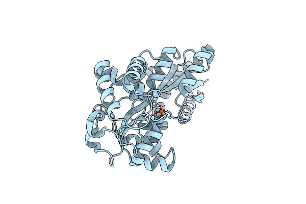
Crystal Structure Of Futa Bound To Fe(Ii)
Organism: Prochlorococcus marinus subsp. pastoris str. ccmp1986
Method: X-RAY DIFFRACTION Resolution:1.70 Å Release Date: 2023-08-30 Classification: METAL BINDING PROTEIN Ligands: FE2 |
Organism: Prochlorococcus marinus subsp. pastoris str. ccmp1986
Method: X-RAY DIFFRACTION
Release Date: 2023-08-30
Ligands: FE2
 |
Crystal Structure Of Futa After An Accumulated Dose Of 5 Kgy
Organism: Prochlorococcus marinus subsp. pastoris str. ccmp1986
Method: X-RAY DIFFRACTION Resolution:1.76 Å Release Date: 2023-08-30 Classification: METAL BINDING PROTEIN Ligands: FE |
Organism: Prochlorococcus marinus subsp. pastoris str. ccmp1986
Method: X-RAY DIFFRACTION
Release Date: 2023-08-30
Ligands: FE
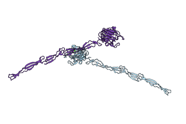 |
Structure Of Aap A Domain And B-Repeats (Residues 351-813) From Staphylococcus Epidermidis
Organism: Staphylococcus epidermidis rp62a
Method: X-RAY DIFFRACTION Resolution:2.30 Å Release Date: 2023-05-03 Classification: CELL ADHESION Ligands: CA, CL |
Organism: Staphylococcus epidermidis rp62a
Method: X-RAY DIFFRACTION
Release Date: 2023-05-03
Ligands: CA, CL
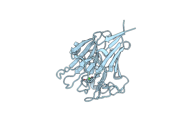 |
Structure Of Pls A-Domain (Residues 391-656; 513-518 Deletion Mutant) From Staphylococcus Aureus
Organism: Staphylococcus aureus
Method: X-RAY DIFFRACTION Resolution:2.75 Å Release Date: 2022-11-09 Classification: CELL ADHESION Ligands: CA |
Organism: Staphylococcus aureus
Method: X-RAY DIFFRACTION
Release Date: 2022-11-09
Ligands: CA
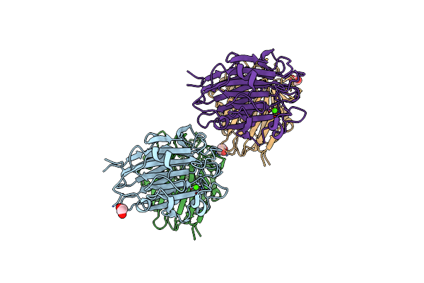 |
Structure Of Sasg A-Domain (Residues 163-419) From Staphylococcus Aureus
Organism: Staphylococcus aureus (strain nctc 8325 / ps 47)
Method: X-RAY DIFFRACTION Resolution:1.65 Å Release Date: 2022-11-02 Classification: CELL ADHESION Ligands: CA, EDO |
Organism: Staphylococcus aureus (strain nctc 8325 / ps 47)
Method: X-RAY DIFFRACTION
Release Date: 2022-11-02
Ligands: CA, EDO
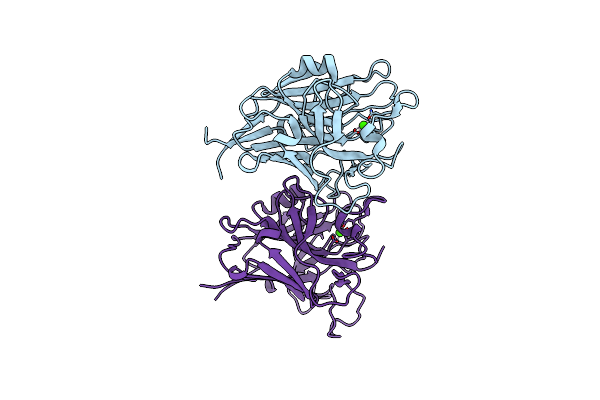 |
Structure Of Pls A-Domain (Residues 391-656) From Staphylococcus Aureus
Organism: Staphylococcus aureus subsp. aureus nctc 8325
Method: X-RAY DIFFRACTION Resolution:1.21 Å Release Date: 2022-10-26 Classification: CELL ADHESION Ligands: CA |
Organism: Staphylococcus aureus subsp. aureus nctc 8325
Method: X-RAY DIFFRACTION
Release Date: 2022-10-26
Ligands: CA