

Search Count: 17
 |
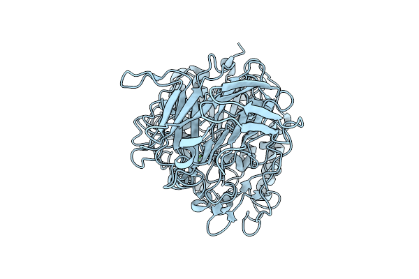
Non-Hemolytic Phospholipase C (Plc N)
Organism: Pseudomonas aeruginosa ucbpp-pa14
Method: X-RAY DIFFRACTION Resolution:1.57 Å Release Date: 2025-10-15 Classification: HYDROLASE Ligands: CA |
Organism: Pseudomonas aeruginosa ucbpp-pa14
Method: X-RAY DIFFRACTION
Release Date: 2025-10-15
Ligands: CA
 |
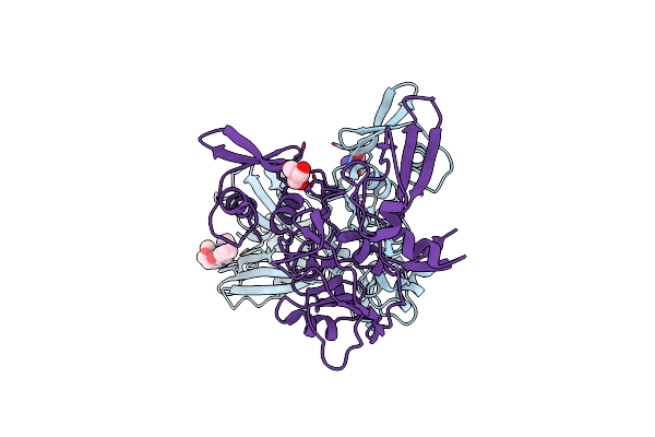
Hemolytic Phospholipase C (Plc H)
Organism: Pseudomonas aeruginosa pa14
Method: ELECTRON MICROSCOPY Release Date: 2025-10-15 Classification: HYDROLASE Ligands: CA |
Organism: Pseudomonas aeruginosa pa14
Method: ELECTRON MICROSCOPY
Release Date: 2025-10-15
Ligands: CA
 |
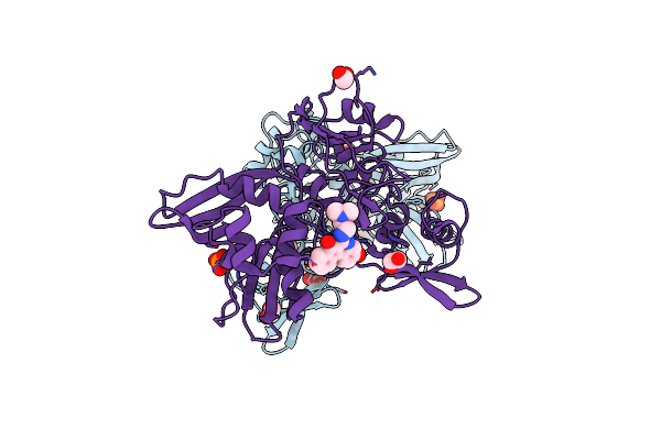
Crystal Structure Of Nsp15 Endoribonuclease From Sars Cov-2 In Complex With Sepantronium (Ym-155)
Organism: Severe acute respiratory syndrome coronavirus 2
Method: X-RAY DIFFRACTION Resolution:2.08 Å Release Date: 2025-05-28 Classification: VIRAL PROTEIN Ligands: TRS, GXU |
Organism: Severe acute respiratory syndrome coronavirus 2
Method: X-RAY DIFFRACTION
Release Date: 2025-05-28
Ligands: TRS, GXU
 |
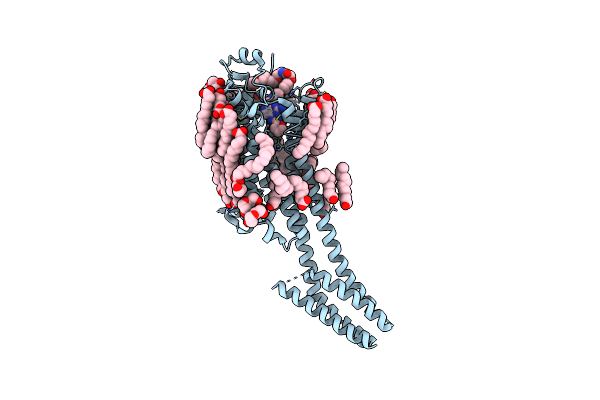
Crystal Structure Of Nsp15 Endoribonuclease From Sars Cov-2 In Complex With Tas-103
Organism: Severe acute respiratory syndrome coronavirus 2
Method: X-RAY DIFFRACTION, SOLUTION SCATTERING Resolution:2.00 Å Release Date: 2025-05-28 Classification: VIRAL PROTEIN Ligands: A1IUV, PO4, EDO |
Organism: Severe acute respiratory syndrome coronavirus 2
Method: X-RAY DIFFRACTION, SOLUTION SCATTERING
Release Date: 2025-05-28
Ligands: A1IUV, PO4, EDO
 |
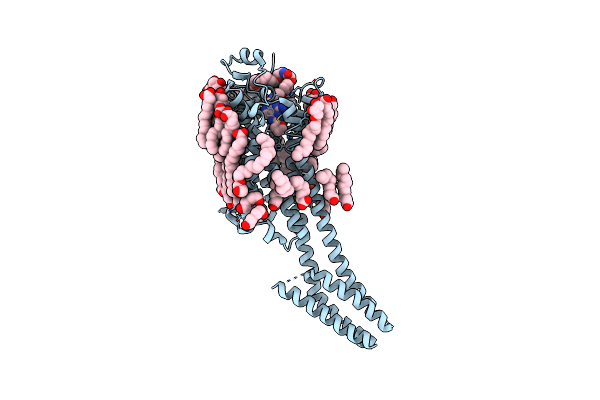
Crystal Structure Of The Adenosine A2A Receptor (A2A-Psb1-Bril) In Complex With Preladenant Conjugate Psb-2113
Organism: Homo sapiens, Escherichia coli
Method: X-RAY DIFFRACTION Resolution:2.25 Å Release Date: 2022-03-02 Classification: MEMBRANE PROTEIN Ligands: CLR, 8E2, OLA, OLC, PEG |
Organism: Homo sapiens, Escherichia coli
Method: X-RAY DIFFRACTION
Release Date: 2022-03-02
Ligands: CLR, 8E2, OLA, OLC, PEG
 |
Crystal Structure Of The Adenosine A2A Receptor (A2A-Psb1-Bril) In Complex With Preladenant Conjugate Psb-2115
Organism: Homo sapiens, Escherichia coli
Method: X-RAY DIFFRACTION Resolution:2.60 Å Release Date: 2022-03-02 Classification: MEMBRANE PROTEIN Ligands: CLR, 8IM, OLA, OLC, PEG |
Organism: Homo sapiens, Escherichia coli
Method: X-RAY DIFFRACTION
Release Date: 2022-03-02
Ligands: CLR, 8IM, OLA, OLC, PEG
 |
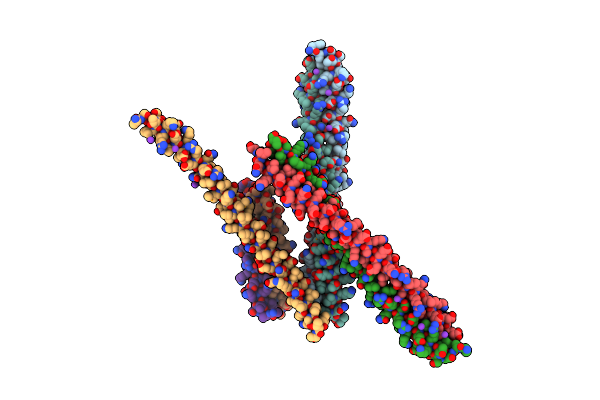
Near-Atomic Resolution Fibril Structure Of Complete Amyloid-Beta(1-42) By Cryo-Em
Organism: Homo sapiens
Method: ELECTRON MICROSCOPY Release Date: 2017-09-13 Classification: PROTEIN FIBRIL |
Organism: Homo sapiens
Method: ELECTRON MICROSCOPY
Release Date: 2017-09-13
 |
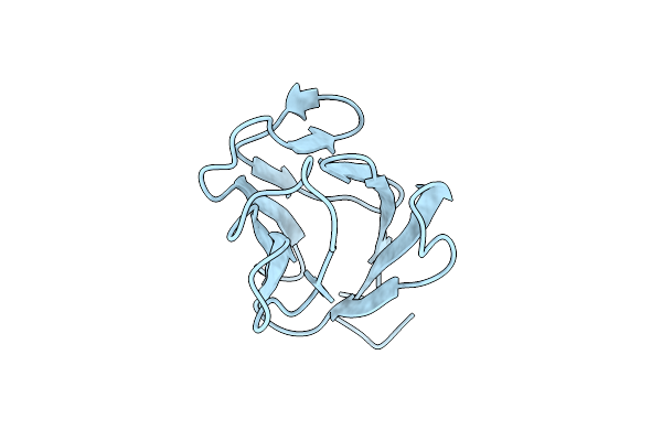
Crystal Structure Of Coiled Coil Domain Of Pawr
Organism: Rattus norvegicus
Method: X-RAY DIFFRACTION Resolution:3.00 Å Release Date: 2016-11-16 Classification: APOPTOSIS Ligands: K |
Organism: Rattus norvegicus
Method: X-RAY DIFFRACTION
Release Date: 2016-11-16
Ligands: K
 |
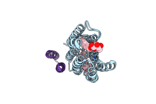
Crystal Structure Of Odhi From Corynebacterium Glutamicum
Organism: Corynebacterium glutamicum
Method: X-RAY DIFFRACTION Resolution:2.00 Å Release Date: 2014-07-02 Classification: PROTEIN BINDING |
Organism: Corynebacterium glutamicum
Method: X-RAY DIFFRACTION
Release Date: 2014-07-02
 |
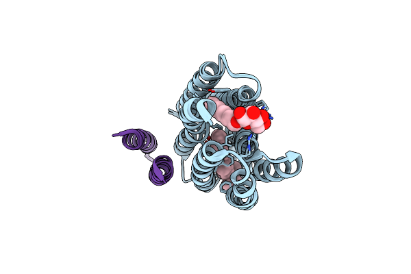
K Intermediate Structure Of Sensory Rhodopsin Ii/Transducer Complex In Combination With The Ground State Structure
Organism: Natronomonas pharaonis
Method: X-RAY DIFFRACTION Resolution:2.00 Å Release Date: 2006-03-07 Classification: MEMBRANE PROTEIN Ligands: BOG, RET |
Organism: Natronomonas pharaonis
Method: X-RAY DIFFRACTION
Release Date: 2006-03-07
Ligands: BOG, RET
 |
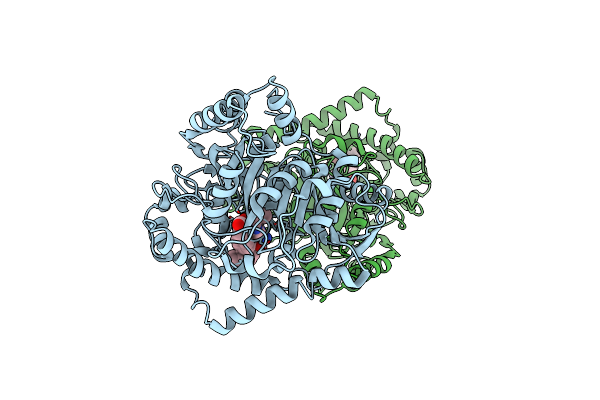
M Intermediate Structure Of Sensory Rhodopsin Ii/Transducer Complex In Combination With The Ground State Structure
Organism: Natronomonas pharaonis
Method: X-RAY DIFFRACTION Resolution:2.20 Å Release Date: 2006-03-07 Classification: MEMBRANE PROTEIN Ligands: BOG, RET |
Organism: Natronomonas pharaonis
Method: X-RAY DIFFRACTION
Release Date: 2006-03-07
Ligands: BOG, RET
 |
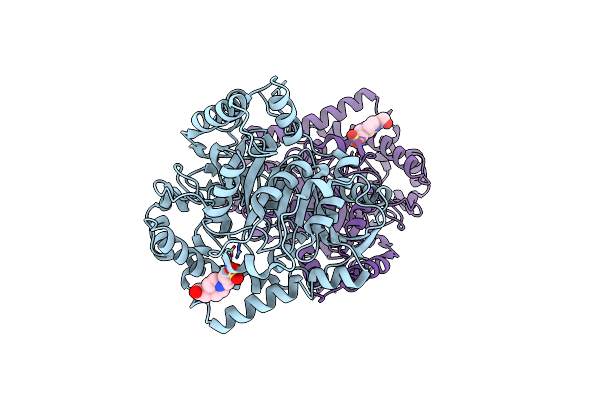
Crystal Structure Analysis Of The Peptide Amidase Pam In Complex With The Competitive Inhibitor Chymostatin
Organism: Stenotrophomonas maltophilia, Streptomyces hygroscopicus, mc521-c8
Method: X-RAY DIFFRACTION Resolution:1.80 Å Release Date: 2002-10-16 Classification: HYDROLASE/HYDROLASE INHIBITOR |
Organism: Stenotrophomonas maltophilia, Streptomyces hygroscopicus, mc521-c8
Method: X-RAY DIFFRACTION
Release Date: 2002-10-16
 |
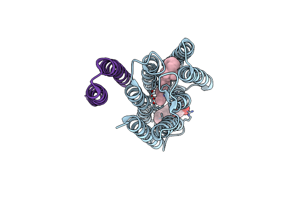
X-Ray Structure Of Native Peptide Amidase From Stenotrophomonas Maltophilia At 1.4 A
Organism: Stenotrophomonas maltophilia
Method: X-RAY DIFFRACTION Resolution:1.40 Å Release Date: 2002-10-16 Classification: HYDROLASE Ligands: EPE |
Organism: Stenotrophomonas maltophilia
Method: X-RAY DIFFRACTION
Release Date: 2002-10-16
Ligands: EPE
 |
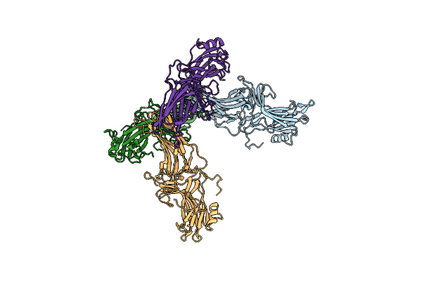
Molecular Basis Of Transmenbrane Signalling By Sensory Rhodopsin Ii-Transducer Complex
Organism: Natronomonas pharaonis
Method: X-RAY DIFFRACTION Resolution:1.93 Å Release Date: 2002-10-10 Classification: MEMBRANE PROTEIN Ligands: BOG, RET |
Organism: Natronomonas pharaonis
Method: X-RAY DIFFRACTION
Release Date: 2002-10-10
Ligands: BOG, RET
 |
Arrestin From Bovine Rod Outer Segments
Organism: Bos taurus
Method: X-RAY DIFFRACTION Resolution:3.30 Å Release Date: 1998-11-25 Classification: SENSORY TRANSDUCTION |
Organism: Bos taurus
Method: X-RAY DIFFRACTION
Release Date: 1998-11-25
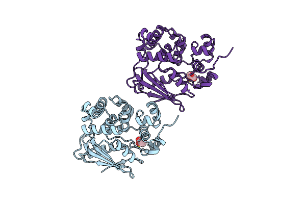 |
3-Methyladenine Dna Glycosylase Ii From Escherichia Coli
Organism: Escherichia coli
Method: X-RAY DIFFRACTION Resolution:1.80 Å Release Date: 1998-01-28 Classification: HYDROLASE Ligands: GOL |
Organism: Escherichia coli
Method: X-RAY DIFFRACTION
Release Date: 1998-01-28
Ligands: GOL
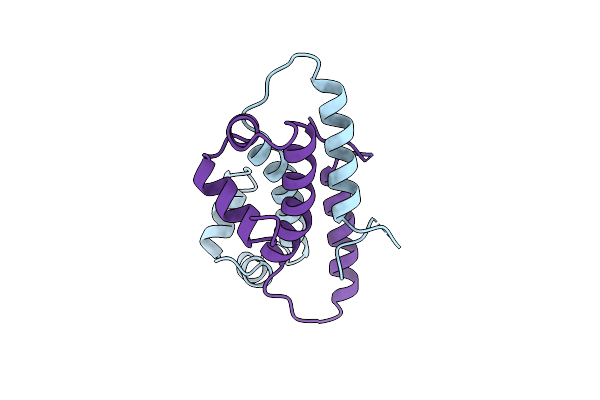 |
Crystal Structure Of The Factor For Inversion Stimulation Fis At 2.0 Angstroms Resolution
Organism: Escherichia coli
Method: X-RAY DIFFRACTION Resolution:2.00 Å Release Date: 1993-10-31 Classification: DNA BINDING PROTEIN |
Organism: Escherichia coli
Method: X-RAY DIFFRACTION
Release Date: 1993-10-31