

Search Count: 33
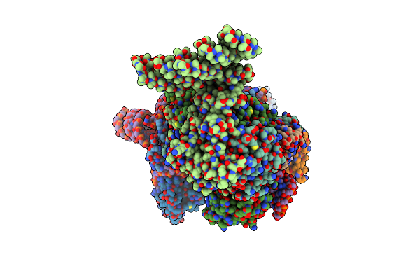 |
Yeast U1 Snrnp With Humanized U1C Zinc-Finger Domain
Organism: Saccharomyces cerevisiae s288c
Method: ELECTRON MICROSCOPY Release Date: 2024-05-15 Classification: SPLICING |
Organism: Saccharomyces cerevisiae s288c
Method: ELECTRON MICROSCOPY
Release Date: 2024-05-15
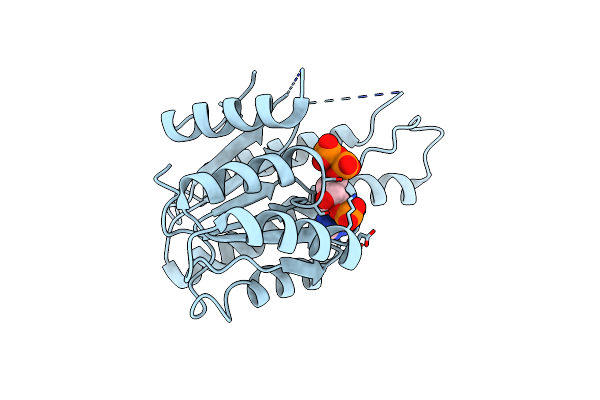 |
Crystal Structure Of The 3-Ketodihydrosphingosine Reductase Tsc10 From Cryptococcus Neoformans
Organism: Cryptococcus neoformans var. neoformans jec21
Method: X-RAY DIFFRACTION Resolution:3.20 Å Release Date: 2023-06-21 Classification: OXIDOREDUCTASE Ligands: NDP |
Organism: Cryptococcus neoformans var. neoformans jec21
Method: X-RAY DIFFRACTION
Release Date: 2023-06-21
Ligands: NDP
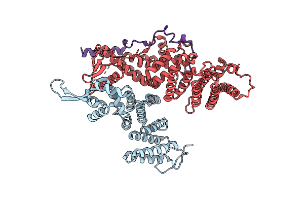 |
Selenomethionine-Substituted Structure Of S. Cerevisiae Csn12 In Complex With Thp3 And Sem1
Organism: Saccharomyces cerevisiae s288c
Method: X-RAY DIFFRACTION Resolution:2.85 Å Release Date: 2022-09-07 Classification: TRANSCRIPTION |
Organism: Saccharomyces cerevisiae s288c
Method: X-RAY DIFFRACTION
Release Date: 2022-09-07
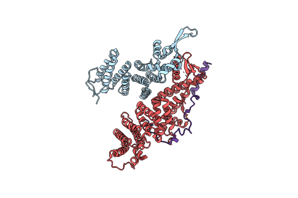 |
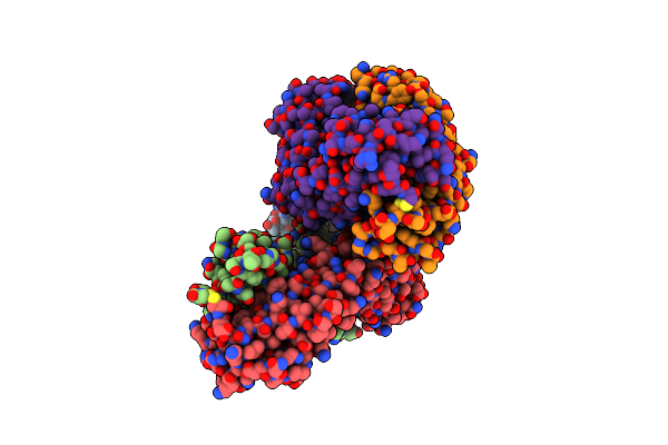
Native Crystal Structure Of S. Cerevisiae Csn12 In Complex With Thp3 And Sem1
Organism: Saccharomyces cerevisiae s288c
Method: X-RAY DIFFRACTION Resolution:2.90 Å Release Date: 2022-09-07 Classification: TRANSCRIPTION |
Organism: Saccharomyces cerevisiae s288c
Method: X-RAY DIFFRACTION
Release Date: 2022-09-07
 |
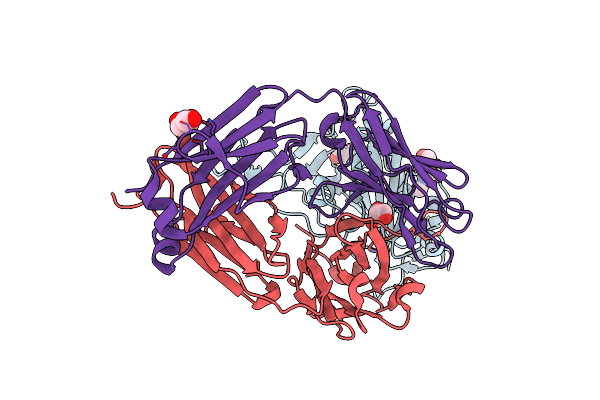
Crystallographic Structure Of Two Neutralizing Antibodies In Complex With Sars-Cov-2 Spike Receptor-Binding Domain (Rbd)
Organism: Severe acute respiratory syndrome coronavirus 2, Homo sapiens
Method: X-RAY DIFFRACTION Resolution:2.40 Å Release Date: 2022-06-15 Classification: ANTIVIRAL PROTEIN Ligands: TRS, NAG, GOL |
Organism: Severe acute respiratory syndrome coronavirus 2, Homo sapiens
Method: X-RAY DIFFRACTION
Release Date: 2022-06-15
Ligands: TRS, NAG, GOL
 |
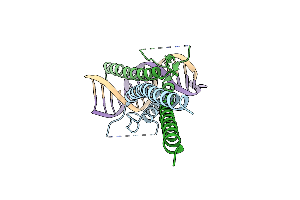
Crystallographic Structure Of Neutralizing Antibody P14-44 In Complex With Sars-Cov-2 Spike Receptor-Binding Domain (Rbd)
Organism: Severe acute respiratory syndrome coronavirus 2, Homo sapiens
Method: X-RAY DIFFRACTION Resolution:1.89 Å Release Date: 2022-06-15 Classification: ANTIVIRAL PROTEIN Ligands: NAG, GOL |
Organism: Severe acute respiratory syndrome coronavirus 2, Homo sapiens
Method: X-RAY DIFFRACTION
Release Date: 2022-06-15
Ligands: NAG, GOL
 |
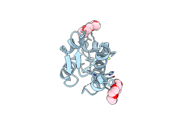
The Complex Of Dna With The C-Terminal Domain Of Tye7 From Saccharomyces Cerevisiae.
Organism: Saccharomyces cerevisiae (strain atcc 204508 / s288c), Synthetic construct
Method: X-RAY DIFFRACTION Resolution:2.55 Å Release Date: 2021-10-13 Classification: TRANSCRIPTION |
 |
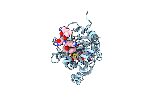
The Crystal Structure Of P53P Peptide Fragment In Complex With The N-Terminal Domain Of Mdmx
Organism: Homo sapiens
Method: X-RAY DIFFRACTION Resolution:2.11 Å Release Date: 2021-06-09 Classification: ONCOPROTEIN Ligands: O4B, MG |
Organism: Homo sapiens
Method: X-RAY DIFFRACTION
Release Date: 2021-06-09
Ligands: O4B, MG
 |
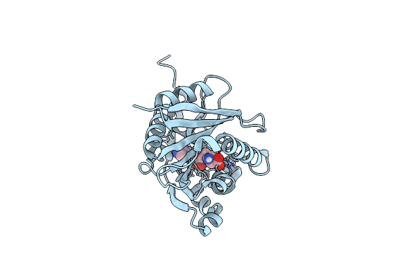
The Crystal Structure Of Scntm1 In Complex With Sah And Rps25A Hexapeptide
Organism: Saccharomyces cerevisiae
Method: X-RAY DIFFRACTION Resolution:1.05 Å Release Date: 2021-05-26 Classification: TRANSFERASE Ligands: SAH |
Organism: Saccharomyces cerevisiae
Method: X-RAY DIFFRACTION
Release Date: 2021-05-26
Ligands: SAH
 |
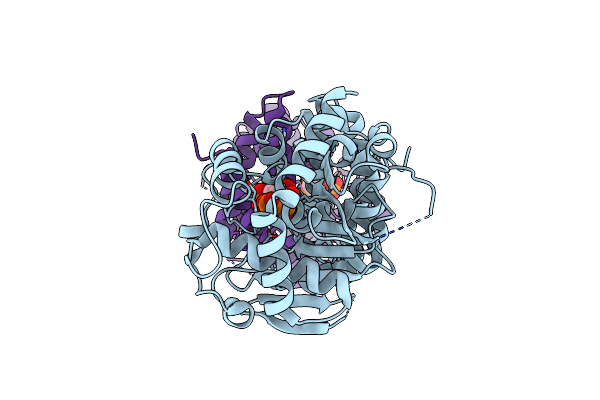
The Crystal Structure Of Scntm1 In Complex With Sah
Organism: Saccharomyces cerevisiae
Method: X-RAY DIFFRACTION Resolution:1.15 Å Release Date: 2021-05-26 Classification: TRANSFERASE Ligands: SAH |
Organism: Saccharomyces cerevisiae
Method: X-RAY DIFFRACTION
Release Date: 2021-05-26
Ligands: SAH
 |
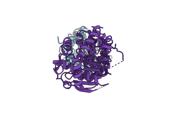
Anthranilate Phosphoribosyltransferase From Saccharomyces Cerevisiae In Complex With Prpp And Mg
Organism: Saccharomyces cerevisiae s288c
Method: X-RAY DIFFRACTION Resolution:2.44 Å Release Date: 2021-03-17 Classification: TRANSFERASE Ligands: PRP, MG |
Organism: Saccharomyces cerevisiae s288c
Method: X-RAY DIFFRACTION
Release Date: 2021-03-17
Ligands: PRP, MG
 |
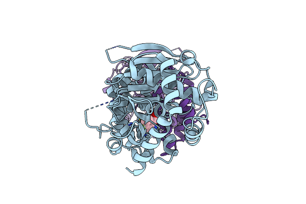
Anthranilate Phosphoribosyltransferase From Saccharomyces Cerevisiae
Organism: Saccharomyces cerevisiae s288c
Method: X-RAY DIFFRACTION Resolution:2.39 Å Release Date: 2021-03-17 Classification: TRANSFERASE |
Organism: Saccharomyces cerevisiae s288c
Method: X-RAY DIFFRACTION
Release Date: 2021-03-17
 |
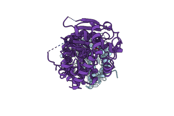
Anthranilate Phosphoribosyltransferase From Saccharomyces Cerevisiae In Complex With 4-Fluoroanthranilate
Organism: Saccharomyces cerevisiae s288c
Method: X-RAY DIFFRACTION Resolution:2.34 Å Release Date: 2021-03-17 Classification: TRANSFERASE Ligands: FA0 |
Organism: Saccharomyces cerevisiae s288c
Method: X-RAY DIFFRACTION
Release Date: 2021-03-17
Ligands: FA0
 |
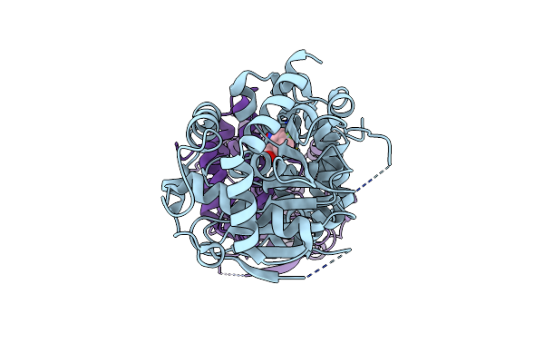
Anthranilate Phosphoribosyltransferase Variant Ser121Ala From Saccharomyces Cerevisiae With Mg Bound
Organism: Saccharomyces cerevisiae s288c
Method: X-RAY DIFFRACTION Resolution:1.95 Å Release Date: 2021-03-17 Classification: TRANSFERASE Ligands: MG |
Organism: Saccharomyces cerevisiae s288c
Method: X-RAY DIFFRACTION
Release Date: 2021-03-17
Ligands: MG
 |
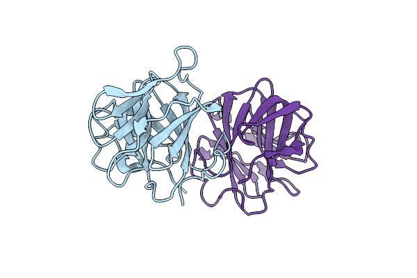
Anthranilate Phosphoribosyltransferase Variant Gly141Asn From Saccharomyces Cerevisiae In Complex With 4-Fluoroanthranilate
Organism: Saccharomyces cerevisiae s288c
Method: X-RAY DIFFRACTION Resolution:2.50 Å Release Date: 2021-03-17 Classification: TRANSFERASE Ligands: FA0 |
Organism: Saccharomyces cerevisiae s288c
Method: X-RAY DIFFRACTION
Release Date: 2021-03-17
Ligands: FA0
 |
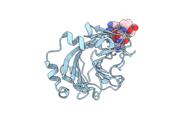
Crystal Structure Of The Spry Domain-Containing Protein 7 (Spry7)
Organism: Homo sapiens
Method: X-RAY DIFFRACTION Resolution:1.62 Å Release Date: 2020-10-14 Classification: UNKNOWN FUNCTION |
Organism: Homo sapiens
Method: X-RAY DIFFRACTION
Release Date: 2020-10-14
 |
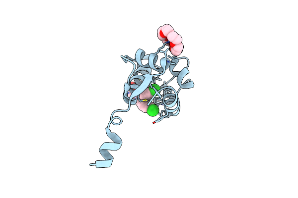
Structural Basis For The Regulation Of Inducible Nitric Oxide Synthase (Inos) By The Spry Domain-Containing Socs Box Protein 2 (Spsb2)
Organism: Homo sapiens
Method: X-RAY DIFFRACTION Resolution:1.24 Å Release Date: 2020-07-01 Classification: PROTEIN BINDING/INHIBITOR |
Organism: Homo sapiens
Method: X-RAY DIFFRACTION
Release Date: 2020-07-01
 |
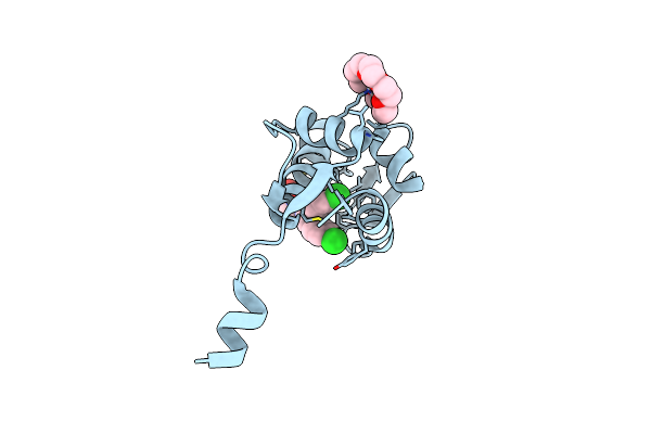
Crystal Structure Of The N-Terminal Domain Of Human Mdmx Protein In Complex With Nutlin3A
Organism: Homo sapiens
Method: X-RAY DIFFRACTION Resolution:1.63 Å Release Date: 2020-06-17 Classification: ONCOPROTEIN Ligands: O4B, NUT |
Organism: Homo sapiens
Method: X-RAY DIFFRACTION
Release Date: 2020-06-17
Ligands: O4B, NUT
 |
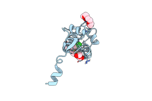
Crystal Structure Of The P53-Binding Domain Of Human Mdmx Protein In Complex With Nutlin3A
Organism: Homo sapiens
Method: X-RAY DIFFRACTION Resolution:1.65 Å Release Date: 2020-06-17 Classification: ONCOPROTEIN Ligands: NUT, O4B |
Organism: Homo sapiens
Method: X-RAY DIFFRACTION
Release Date: 2020-06-17
Ligands: NUT, O4B
 |
Human Mdmx Protein In Complex With Nutlin3A
Organism: Homo sapiens
Method: X-RAY DIFFRACTION Resolution:1.80 Å Release Date: 2020-06-10 Classification: ONCOPROTEIN Ligands: O4B, NUT, PEG |
Organism: Homo sapiens
Method: X-RAY DIFFRACTION
Release Date: 2020-06-10
Ligands: O4B, NUT, PEG