

Search Count: 19
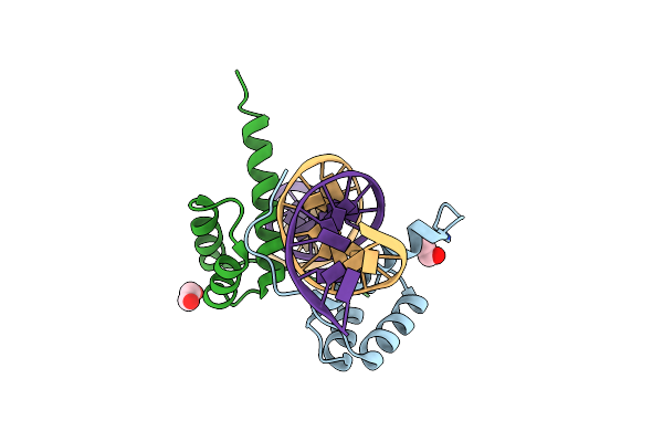 |
X-Ray Structure Of Alx4 Homeodomain Dimer Bound To Dna
Organism: Homo sapiens
Method: X-RAY DIFFRACTION Resolution:2.39 Å Release Date: 2025-04-09 Classification: TRANSCRIPTION/DNA Ligands: EDO |
Organism: Homo sapiens
Method: X-RAY DIFFRACTION
Release Date: 2025-04-09
Ligands: EDO
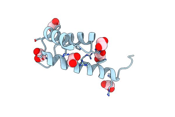 |
X-Ray Structure Of Alx4 Homeodomain
Organism: Homo sapiens
Method: X-RAY DIFFRACTION Resolution:2.39 Å Release Date: 2025-04-09 Classification: TRANSCRIPTION Ligands: EDO |
Organism: Homo sapiens
Method: X-RAY DIFFRACTION
Release Date: 2025-04-09
Ligands: EDO
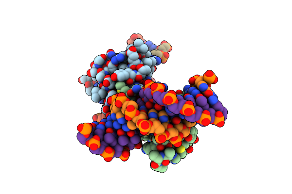 |
Crystal Structure Of Gsx2 Homeodomain In Complex With Dna
Organism: Mus musculus
Method: X-RAY DIFFRACTION Resolution:2.21 Å Release Date: 2023-10-18 Classification: TRANSCRIPTION/DNA Ligands: PGE |
Organism: Mus musculus
Method: X-RAY DIFFRACTION
Release Date: 2023-10-18
Ligands: PGE
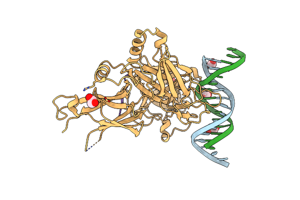 |
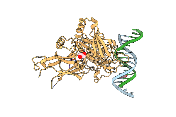
X-Ray Structure Of Wild Type Rbpj-L3Mbtl3-Dna Complex
Organism: Mus musculus, Synthetic dna, Homo sapiens
Method: X-RAY DIFFRACTION Resolution:2.06 Å Release Date: 2022-08-24 Classification: DNA BINDING PROTEIN/DNA Ligands: EDO, XYZ |
Organism: Mus musculus, Synthetic dna, Homo sapiens
Method: X-RAY DIFFRACTION
Release Date: 2022-08-24
Ligands: EDO, XYZ
 |
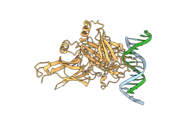
X-Ray Structure Of Rbpj-L3Mbtl3(Dt62)-Dna Complex
Organism: Mus musculus, Synthetic dna, Homo sapiens
Method: X-RAY DIFFRACTION Resolution:2.05 Å Release Date: 2022-08-24 Classification: DNA BINDING PROTEIN/DNA Ligands: EDO |
Organism: Mus musculus, Synthetic dna, Homo sapiens
Method: X-RAY DIFFRACTION
Release Date: 2022-08-24
Ligands: EDO
 |
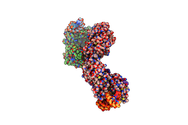
Csl (Rbpj) Bound To Notch3 Ram And Dna
Organism: Mus musculus, Homo sapiens
Method: X-RAY DIFFRACTION Resolution:2.41 Å Release Date: 2021-03-31 Classification: TRANSCRIPTION/SIGNALING PROTEIN/DNA |
Organism: Mus musculus, Homo sapiens
Method: X-RAY DIFFRACTION
Release Date: 2021-03-31
 |
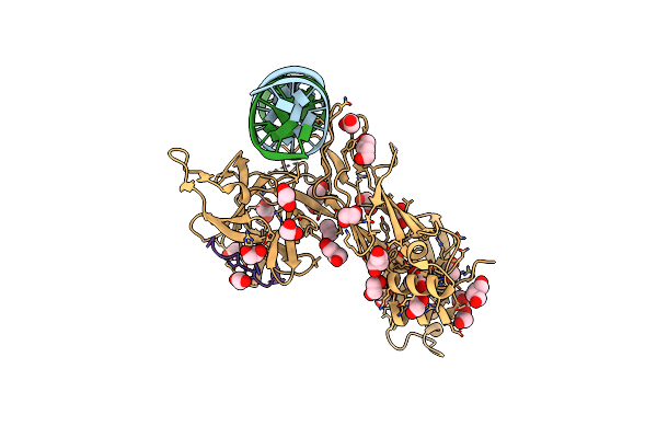
Structure Of The Rbpj-Sharp-Dna Repressor Complex
Organism: Mus musculus, Escherichia coli o157:h7
Method: X-RAY DIFFRACTION Resolution:2.78 Å Release Date: 2019-01-02 Classification: TRANSCRIPTION/DNA |
Organism: Mus musculus, Escherichia coli o157:h7
Method: X-RAY DIFFRACTION
Release Date: 2019-01-02
 |
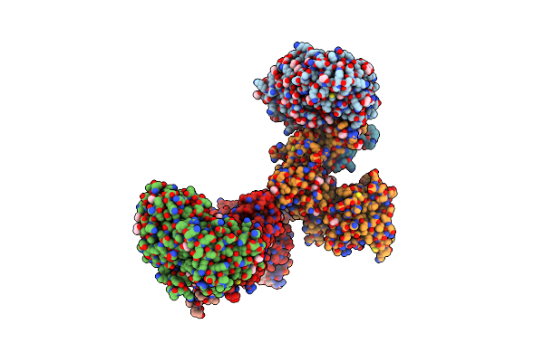
Csl-Rita Complex Bound To Dna
Organism: Mus musculus, Synthetic construct
Method: X-RAY DIFFRACTION Resolution:2.09 Å Release Date: 2017-04-26 Classification: TRANSCRIPTION/DNA BINDING PROTEIN/DNA Ligands: EDO, BU1 |
Organism: Mus musculus, Synthetic construct
Method: X-RAY DIFFRACTION
Release Date: 2017-04-26
Ligands: EDO, BU1
 |
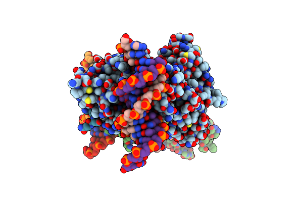
Structure Of The Su(H)-Hairless-Dna Repressor Complex
Organism: Escherichia coli o157:h7, Drosophila melanogaster, Mus musculus
Method: X-RAY DIFFRACTION Resolution:2.14 Å Release Date: 2016-06-15 Classification: TRANSPORT/DNA Binding/DNA Ligands: EDO |
Organism: Escherichia coli o157:h7, Drosophila melanogaster, Mus musculus
Method: X-RAY DIFFRACTION
Release Date: 2016-06-15
Ligands: EDO
 |
Csl (Rbp-Jk) With Corepressor Kyot2 Bound To Dna
Organism: Mus musculus, Homo sapiens
Method: X-RAY DIFFRACTION Resolution:2.85 Å Release Date: 2013-11-06 Classification: TRANSCRIPTION/DNA BINDING PROTEIN/DNA Ligands: EDO |
Organism: Mus musculus, Homo sapiens
Method: X-RAY DIFFRACTION
Release Date: 2013-11-06
Ligands: EDO
 |
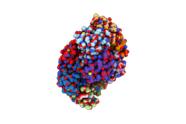
Crystal Structure Of P. Aeruginosa Catalase, Kata Tetramer
Organism: Pseudomonas aeruginosa
Method: X-RAY DIFFRACTION Resolution:2.53 Å Release Date: 2013-03-20 Classification: OXIDOREDUCTASE Ligands: HEM, NDP |
Organism: Pseudomonas aeruginosa
Method: X-RAY DIFFRACTION
Release Date: 2013-03-20
Ligands: HEM, NDP
 |
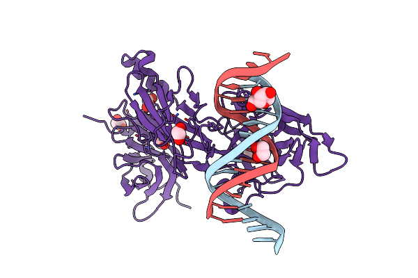
Structure Of Shiga Toxin Type 2 (Stx2) B Pentamer Mutant Q40L
Organism: Escherichia coli
Method: X-RAY DIFFRACTION Resolution:2.49 Å Release Date: 2011-01-12 Classification: TOXIN Ligands: XLS |
Organism: Escherichia coli
Method: X-RAY DIFFRACTION
Release Date: 2011-01-12
Ligands: XLS
 |
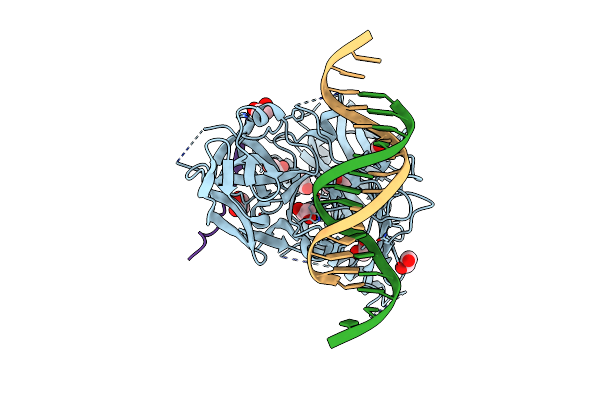
Csl (Rbp-Jk) Bound To Hes-1 Nonconsensus Site
Organism: Mus musculus
Method: X-RAY DIFFRACTION Resolution:2.00 Å Release Date: 2009-11-10 Classification: TRANSCRIPTION/DNA Ligands: EDO, XYL |
Organism: Mus musculus
Method: X-RAY DIFFRACTION
Release Date: 2009-11-10
Ligands: EDO, XYL
 |
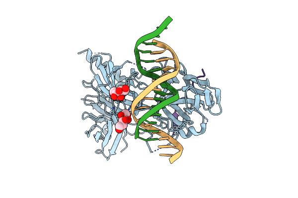
Csl (Lag-1) Bound To Dna With Lin-12 Ram Peptide, P212121
Organism: Caenorhabditis elegans
Method: X-RAY DIFFRACTION Resolution:2.21 Å Release Date: 2008-04-01 Classification: DNA BINDING PROTEIN/DNA Ligands: EDO |
Organism: Caenorhabditis elegans
Method: X-RAY DIFFRACTION
Release Date: 2008-04-01
Ligands: EDO
 |
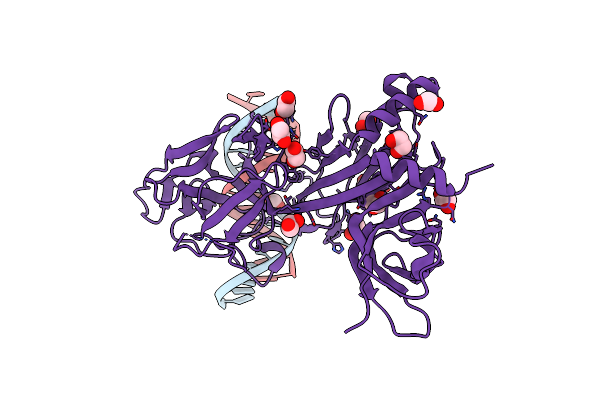
Csl (Lag-1) Bound To Dna With Lin-12 Ram Peptide, C2221
Organism: Caenorhabditis elegans
Method: X-RAY DIFFRACTION Resolution:2.47 Å Release Date: 2008-04-01 Classification: DNA BINDING PROTEIN/DNA Ligands: SOR |
Organism: Caenorhabditis elegans
Method: X-RAY DIFFRACTION
Release Date: 2008-04-01
Ligands: SOR
 |
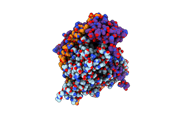
Csl (Rbp-Jk) Bound To Dna
Organism: Mus musculus
Method: X-RAY DIFFRACTION Resolution:2.20 Å Release Date: 2008-04-01 Classification: DNA BINDING PROTEIN/DNA Ligands: EDO |
Organism: Mus musculus
Method: X-RAY DIFFRACTION
Release Date: 2008-04-01
Ligands: EDO
 |
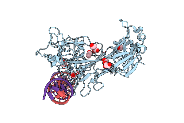
Crystal Structure Of The Csl-Notch-Mastermind Ternary Complex Bound To Dna
Organism: Caenorhabditis elegans
Method: X-RAY DIFFRACTION Resolution:3.12 Å Release Date: 2006-03-21 Classification: GENE REGULATION/SIGNALLING PROTEIN/DNA |
Organism: Caenorhabditis elegans
Method: X-RAY DIFFRACTION
Release Date: 2006-03-21
 |
Crystal Structure Of Csl Bound To Dna
Organism: Caenorhabditis elegans
Method: X-RAY DIFFRACTION Resolution:2.85 Å Release Date: 2004-08-31 Classification: TRANSCRIPTION Ligands: EDO |
Organism: Caenorhabditis elegans
Method: X-RAY DIFFRACTION
Release Date: 2004-08-31
Ligands: EDO
 |
Crystal Structure Of Human Alpha-Tocopherol Transfer Protein Bound To Its Ligand
Organism: Homo sapiens
Method: X-RAY DIFFRACTION Resolution:1.50 Å Release Date: 2003-11-25 Classification: TRANSPORT PROTEIN Ligands: VIV |
Organism: Homo sapiens
Method: X-RAY DIFFRACTION
Release Date: 2003-11-25
Ligands: VIV