

Search Count: 12
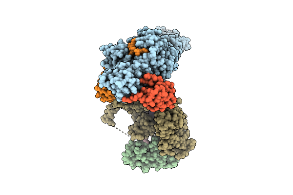 |
Structure Of Rbr Binding E2 Variant Crosslinked With Nedd8-Cul5-Rbx2 Bound Arih2 And Ub
Organism: Homo sapiens, Synthetic construct
Method: ELECTRON MICROSCOPY Resolution:2.97 Å Release Date: 2025-12-24 Classification: LIGASE Ligands: ZN, SY8 |
Organism: Homo sapiens, Synthetic construct
Method: ELECTRON MICROSCOPY
Release Date: 2025-12-24
Ligands: ZN, SY8
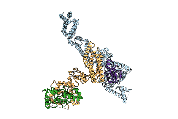 |
Structure Of Rbr E2 Variant Binding To Cul5-Rbx2 Bound Arih2
Organism: Synthetic construct, Homo sapiens
Method: ELECTRON MICROSCOPY Resolution:3.06 Å Release Date: 2025-12-24 Classification: LIGASE Ligands: ZN |
Organism: Synthetic construct, Homo sapiens
Method: ELECTRON MICROSCOPY
Release Date: 2025-12-24
Ligands: ZN
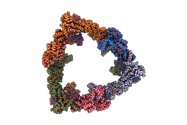 |
Structure Of Cul9-Rbx1 Ubiquitin E3 Ligase Complex - Hexameric Assembly
|
Organism: Homo sapiens
Method: ELECTRON MICROSCOPY
Release Date: 2024-04-17
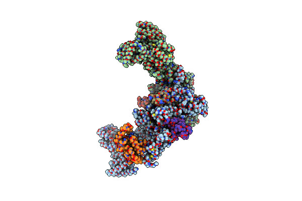 |
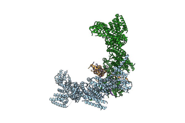
Structure Of Cul9-Rbx1 Ubiquitin E3 Ligase Complex In Unneddylated And Neddylated Conformation - Focused Cullin Dimer
Organism: Homo sapiens
Method: ELECTRON MICROSCOPY Release Date: 2024-04-17 Classification: LIGASE Ligands: ZN |
Organism: Homo sapiens
Method: ELECTRON MICROSCOPY
Release Date: 2024-04-17
Ligands: ZN
 |
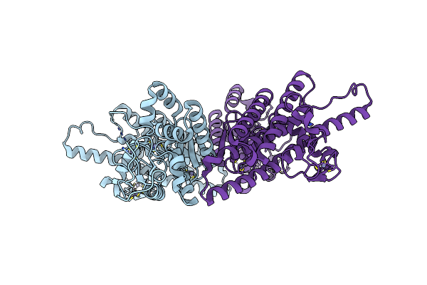
Structure Of Cul9-Rbx1 Ubiquitin E3 Ligase Complex In Unneddylated Conformation - Symmetry Expanded Unneddylated Dimer
Organism: Homo sapiens
Method: ELECTRON MICROSCOPY Release Date: 2024-04-17 Classification: LIGASE Ligands: ZN |
Organism: Homo sapiens
Method: ELECTRON MICROSCOPY
Release Date: 2024-04-17
Ligands: ZN
 |
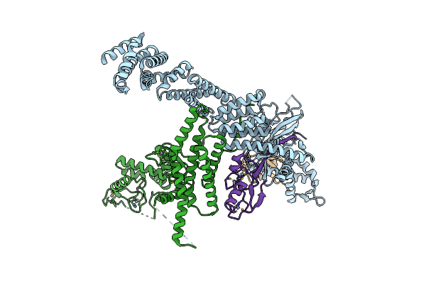
Crystal Structure Of Rbr Ubiquitin Ligase Arih2
Organism: Homo sapiens
Method: X-RAY DIFFRACTION Resolution:2.45 Å Release Date: 2021-09-15 Classification: LIGASE Ligands: ZN |
Organism: Homo sapiens
Method: X-RAY DIFFRACTION
Release Date: 2021-09-15
Ligands: ZN
 |
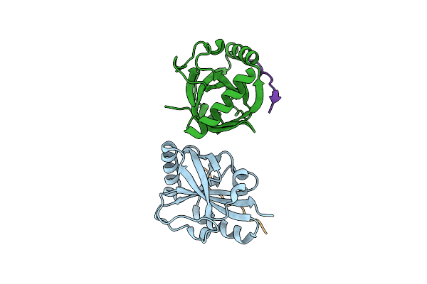
Structure Of Neddylated Cul5 C-Terminal Region-Rbx2-Arih2*
Organism: Homo sapiens
Method: ELECTRON MICROSCOPY Release Date: 2021-09-15 Classification: LIGASE Ligands: ZN |
Organism: Homo sapiens
Method: ELECTRON MICROSCOPY
Release Date: 2021-09-15
Ligands: ZN
 |
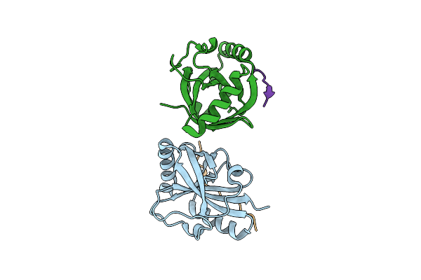
Crystal Structure Of The Spoc Domain Of Human Phf3 In Complex With Rna Polymerase Ii Ctd Diheptapeptide Phosphorylated On Ser2
Organism: Homo sapiens
Method: X-RAY DIFFRACTION Resolution:1.93 Å Release Date: 2020-07-15 Classification: PROTEIN BINDING |
Organism: Homo sapiens
Method: X-RAY DIFFRACTION
Release Date: 2020-07-15
 |
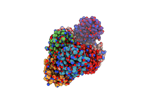
Crystal Structure Of The Spoc Domain Of Human Phf3 In Complex With Rna Polymerase Ii Ctd Diheptapeptide Phosphorylated On Ser2Ser7
Organism: Homo sapiens
Method: X-RAY DIFFRACTION Resolution:1.75 Å Release Date: 2020-07-15 Classification: TRANSCRIPTION |
Organism: Homo sapiens
Method: X-RAY DIFFRACTION
Release Date: 2020-07-15
 |
Crystal Structure Of Spoc Domain Of Human Phd-Finger Protein 3 (Phf3)
Organism: Homo sapiens
Method: X-RAY DIFFRACTION Resolution:2.59 Å Release Date: 2020-07-15 Classification: TRANSCRIPTION Ligands: GOL |
Organism: Homo sapiens
Method: X-RAY DIFFRACTION
Release Date: 2020-07-15
Ligands: GOL
 |
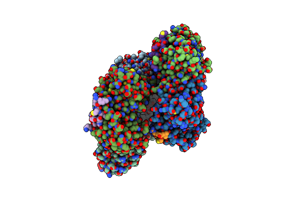
Crystal Structure Of The Spoc Domain Of Human Phf3 In Complex With Rna Polymerase Ii Ctd Diheptapeptide Phosphorylated On Ser2Ser5
Organism: Homo sapiens
Method: X-RAY DIFFRACTION Resolution:2.85 Å Release Date: 2020-07-15 Classification: TRANSCRIPTION |
Organism: Homo sapiens
Method: X-RAY DIFFRACTION
Release Date: 2020-07-15
 |
Crystal Structure Of Human Pcna In Complex With Parg (Poly(Adp-Ribose) Glycohydrolase) Peptide.
Organism: Homo sapiens
Method: X-RAY DIFFRACTION Resolution:2.58 Å Release Date: 2017-08-02 Classification: TRANSCRIPTION |
Organism: Homo sapiens
Method: X-RAY DIFFRACTION
Release Date: 2017-08-02