

Search Count: 8
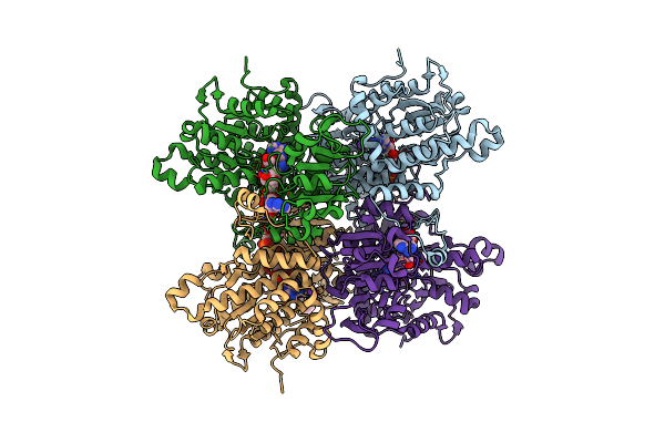 |
Crystal Structure Of The F324A Mutant Of S-Adenosyl-L-Homocysteine Hydrolase From Pseudomonas Aeruginosa Cocrystallized With Adenosine In The Presence Of K+ Cations
Organism: Pseudomonas aeruginosa pao1
Method: X-RAY DIFFRACTION Resolution:1.68 Å Release Date: 2023-08-16 Classification: HYDROLASE Ligands: NAD, ADN, K, CL, PEG |
Organism: Pseudomonas aeruginosa pao1
Method: X-RAY DIFFRACTION
Release Date: 2023-08-16
Ligands: NAD, ADN, K, CL, PEG
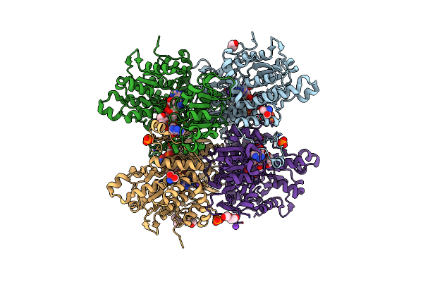 |
Crystal Structure Of The H323A Mutant Of S-Adenosyl-L-Homocysteine Hydrolase From Pseudomonas Aeruginosa Cocrystallized With Adenosine In The Presence Of K+ Cations
Organism: Pseudomonas aeruginosa pao1
Method: X-RAY DIFFRACTION Resolution:1.50 Å Release Date: 2023-08-16 Classification: HYDROLASE Ligands: ADN, NAD, TRS, PEG, K, PO4, CL |
Organism: Pseudomonas aeruginosa pao1
Method: X-RAY DIFFRACTION
Release Date: 2023-08-16
Ligands: ADN, NAD, TRS, PEG, K, PO4, CL
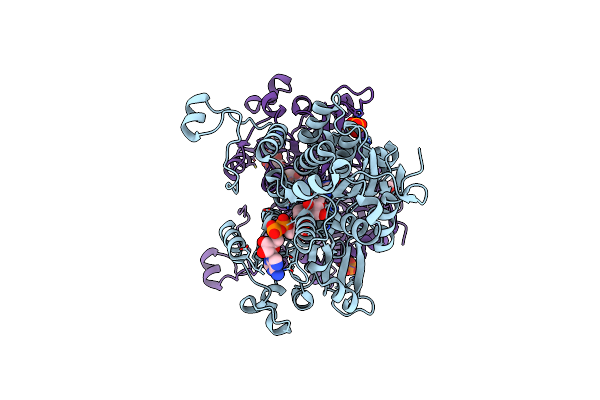 |
Crystal Structure Of The Q65A Mutant Of S-Adenosyl-L-Homocysteine Hydrolase From Pseudomonas Aeruginosa Cocrystallized With Adenosine In The Presence Of K+ Cations
Organism: Pseudomonas aeruginosa pao1
Method: X-RAY DIFFRACTION Resolution:1.65 Å Release Date: 2023-08-16 Classification: HYDROLASE Ligands: PO4, NAD, ADN, GOL |
Organism: Pseudomonas aeruginosa pao1
Method: X-RAY DIFFRACTION
Release Date: 2023-08-16
Ligands: PO4, NAD, ADN, GOL
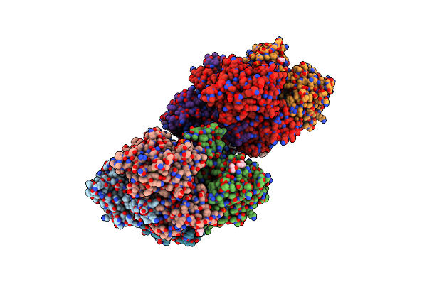 |
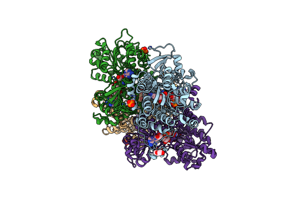
Crystal Structure Of The Q65N Mutant Of S-Adenosyl-L-Homocysteine Hydrolase From Pseudomonas Aeruginosa Crystallized In The Presence Of K+ Cations
Organism: Pseudomonas aeruginosa pao1
Method: X-RAY DIFFRACTION Resolution:1.90 Å Release Date: 2023-08-16 Classification: HYDROLASE Ligands: NAD, GOL, PO4, K, PEG |
Organism: Pseudomonas aeruginosa pao1
Method: X-RAY DIFFRACTION
Release Date: 2023-08-16
Ligands: NAD, GOL, PO4, K, PEG
 |
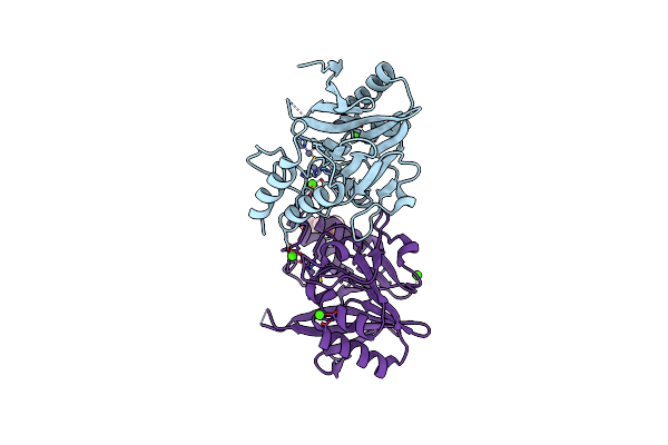
Crystal Structure Of The Q65N Mutant Of S-Adenosyl-L-Homocysteine Hydrolase From Pseudomonas Aeruginosa Cocrystallized With Adenosine In The Presence Of K+ Cations
Organism: Pseudomonas aeruginosa pao1
Method: X-RAY DIFFRACTION Resolution:1.82 Å Release Date: 2023-08-16 Classification: HYDROLASE Ligands: NAD, ADN, PEG, PO4 |
Organism: Pseudomonas aeruginosa pao1
Method: X-RAY DIFFRACTION
Release Date: 2023-08-16
Ligands: NAD, ADN, PEG, PO4
 |
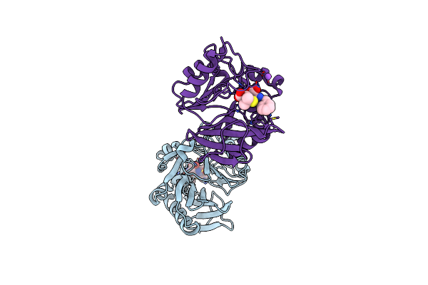
Crystal Structure Of N-Terminally Truncated Ndm-1 Metallo-Beta-Lactamase
Organism: Klebsiella pneumoniae
Method: X-RAY DIFFRACTION Resolution:0.95 Å Release Date: 2020-05-13 Classification: HYDROLASE Ligands: ZN, CA, CL, EPE |
Organism: Klebsiella pneumoniae
Method: X-RAY DIFFRACTION
Release Date: 2020-05-13
Ligands: ZN, CA, CL, EPE
 |
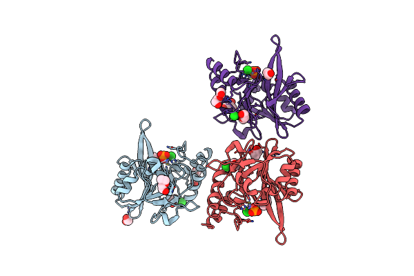
Crystal Structure Of Ndm-12 Metallo-Beta-Lactamase In Complex With Hydrolyzed Ampicillin
Organism: Escherichia coli
Method: X-RAY DIFFRACTION Resolution:2.10 Å Release Date: 2020-04-22 Classification: hydrolase/antibiotic Ligands: ZN, CL, NA, ZZ7 |
Organism: Escherichia coli
Method: X-RAY DIFFRACTION
Release Date: 2020-04-22
Ligands: ZN, CL, NA, ZZ7
 |
Crystal Structure Of Ndm-9 Metallo-Beta-Lactamase
Organism: Escherichia coli
Method: X-RAY DIFFRACTION Resolution:1.43 Å Release Date: 2020-04-15 Classification: HYDROLASE Ligands: ZN, CL, PO4, EDO, PEG, PGE, GOL |
Organism: Escherichia coli
Method: X-RAY DIFFRACTION
Release Date: 2020-04-15
Ligands: ZN, CL, PO4, EDO, PEG, PGE, GOL