

Search Count: 133
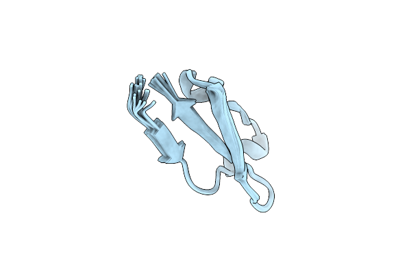 |
Solution Structure Of Toxin, U-Rdtx-Pp19, From Assassin Bug Pristhesancus Plagipennis
Organism: Pristhesancus plagipennis
Method: SOLUTION NMR Release Date: 2024-05-29 Classification: TOXIN |
Organism: Pristhesancus plagipennis
Method: SOLUTION NMR
Release Date: 2024-05-29
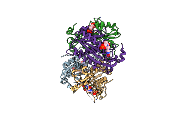 |
Structure Of Human Hypoxanthine Guanine Phosphoribzosyltransferase In Complex With [2S,4R]-4-Guanin-9-Yl-2-(2-Phosphonoethoxymethyl)-1-N-(3-Phosphonopropionyl)Pyrrolidine
Organism: Homo sapiens
Method: X-RAY DIFFRACTION Resolution:2.27 Å Release Date: 2024-05-08 Classification: TRANSFERASE Ligands: JEI, MG |
Organism: Homo sapiens
Method: X-RAY DIFFRACTION
Release Date: 2024-05-08
Ligands: JEI, MG
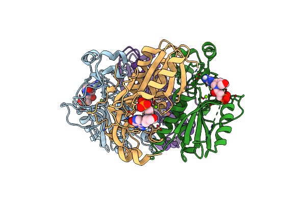 |
Structure Of Human Hypoxanthine Guanine Phosphoribzosyltransferase In Complex With [2S,4R] 4-Guanin-9-Yl-2-Hydroxymethyl-1-N-(3-Phosphonopropionyl)Pyrrolidine
Organism: Homo sapiens
Method: X-RAY DIFFRACTION Resolution:2.50 Å Release Date: 2024-05-08 Classification: TRANSFERASE Ligands: JG6, MG |
Organism: Homo sapiens
Method: X-RAY DIFFRACTION
Release Date: 2024-05-08
Ligands: JG6, MG
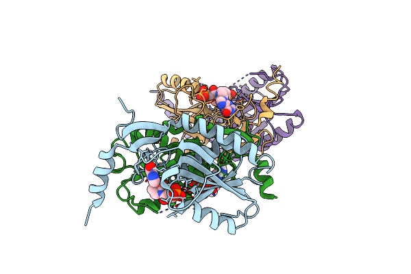 |
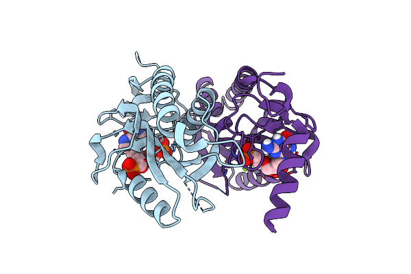
Crystal Structure Of Trypanosome Brucei Hypoxanthine Guanine Phosphopribosyltransferase In Complex With [2S,4R]-4-Guanin-9-Yl-2-(2- Phosphonoethoxymethyl)-1-N-(3-Phosphonopropionyl)Pyrrolidine
Organism: Trypanosoma brucei
Method: X-RAY DIFFRACTION Resolution:2.46 Å Release Date: 2024-05-08 Classification: TRANSFERASE Ligands: JEI |
Organism: Trypanosoma brucei
Method: X-RAY DIFFRACTION
Release Date: 2024-05-08
Ligands: JEI
 |
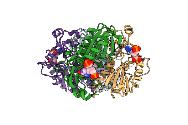
Crystal Structure Of T. Brucei Hypoxanthine Guanine Phosphoribosyltransferase In Complex With [2S,4S]-4-Guanin-9-Yl-2-(2-Phosphonoethoxymethyl)-1-N-(3-Phosphonopropionyl)Pyrrolidine
Organism: Trypanosoma brucei
Method: X-RAY DIFFRACTION Resolution:2.20 Å Release Date: 2024-05-08 Classification: TRANSFERASE Ligands: MG, KFF |
Organism: Trypanosoma brucei
Method: X-RAY DIFFRACTION
Release Date: 2024-05-08
Ligands: MG, KFF
 |
Crystal Structure Of Human Hypoxanthine Guanine Phosphoribzosyltransferase In Complex With (4S,7S)-7-Hydroxy-4-((Guanin-9-Yl)Methyl)-2,5-Dioxaheptan-1,7-Diphosphonate
Organism: Homo sapiens
Method: X-RAY DIFFRACTION Resolution:2.58 Å Release Date: 2022-05-18 Classification: TRANSFERASE/TRANSFERASE INHIBITOR Ligands: 8QI |
Organism: Homo sapiens
Method: X-RAY DIFFRACTION
Release Date: 2022-05-18
Ligands: 8QI
 |
Solution Structure Of Tarantula Toxin Omega-Avsp1A
Organism: Avicularia sp. avic29fpm
Method: SOLUTION NMR Release Date: 2022-03-02 Classification: TOXIN |
Organism: Avicularia sp. avic29fpm
Method: SOLUTION NMR
Release Date: 2022-03-02
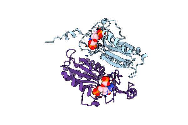 |
Crystal Structure Of T. Brucei Hypoxanthine Guanine Phosphoribosyltransferase In Complex With (4S,7S)-7-Hydroxy-4-((Guanin-9-Yl)Methyl)-2,5-Dioxaheptan-1,7-Diphosphonate
Organism: Trypanosoma brucei brucei
Method: X-RAY DIFFRACTION Resolution:2.65 Å Release Date: 2022-03-02 Classification: TRANSFERASE Ligands: 8QI |
Organism: Trypanosoma brucei brucei
Method: X-RAY DIFFRACTION
Release Date: 2022-03-02
Ligands: 8QI
 |
Crystal Structure Of Trypanosome Brucei Hypoxanthine-Guanine-Xanthine Phosphoribzosyltransferase In Complex With (4S,7S)-7-Hydroxy-4-((Guanin-9-Yl)Methyl)-2,5-Dioxaheptan-1,7-Diphosphonate
Organism: Trypanosoma brucei brucei
Method: X-RAY DIFFRACTION Resolution:2.12 Å Release Date: 2022-03-02 Classification: TRANSFERASE/INHIBITOR Ligands: 8QI |
Organism: Trypanosoma brucei brucei
Method: X-RAY DIFFRACTION
Release Date: 2022-03-02
Ligands: 8QI
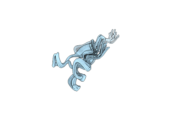 |
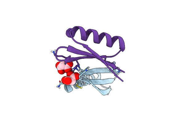
Solution Structure Of Peptide Toxin Miitx2-Mg1A From The Venom Of The Australian Giant Red Bull Ant Myrmecia Gulosa
|
Organism: Myrmecia gulosa
Method: SOLUTION NMR
Release Date: 2022-02-09
 |
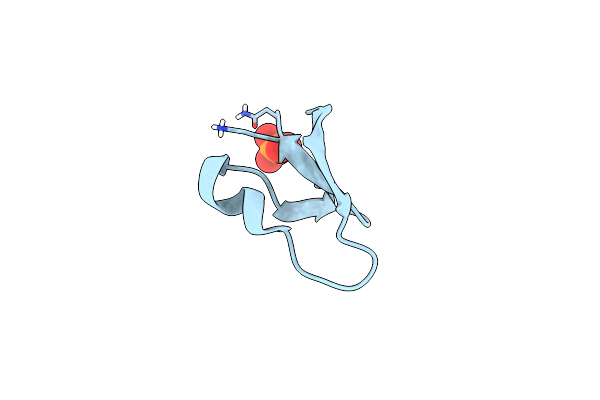
Mopd-1 Mutant-L47W
Organism: Synthetic construct
Method: X-RAY DIFFRACTION Resolution:1.80 Å Release Date: 2021-10-27 Classification: ANTITUMOR PROTEIN Ligands: ZN, MLI |
Organism: Synthetic construct
Method: X-RAY DIFFRACTION
Release Date: 2021-10-27
Ligands: ZN, MLI
 |
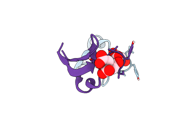
[I11L]Hyen D Crystal Structure
Organism: Hybanthus enneaspermus
Method: X-RAY DIFFRACTION Resolution:1.22 Å Release Date: 2021-09-29 Classification: PLANT PROTEIN Ligands: PO4 |
Organism: Hybanthus enneaspermus
Method: X-RAY DIFFRACTION
Release Date: 2021-09-29
Ligands: PO4
 |
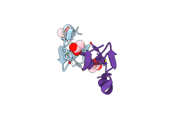
Hyen D
Organism: Hybanthus enneaspermus
Method: X-RAY DIFFRACTION Resolution:1.35 Å Release Date: 2021-09-22 Classification: PLANT PROTEIN Ligands: FLC |
Organism: Hybanthus enneaspermus
Method: X-RAY DIFFRACTION
Release Date: 2021-09-22
Ligands: FLC
 |
[I11G]Hyen D
Organism: Hybanthus enneaspermus
Method: X-RAY DIFFRACTION Resolution:1.30 Å Release Date: 2021-09-22 Classification: PLANT PROTEIN Ligands: ACT |
Organism: Hybanthus enneaspermus
Method: X-RAY DIFFRACTION
Release Date: 2021-09-22
Ligands: ACT
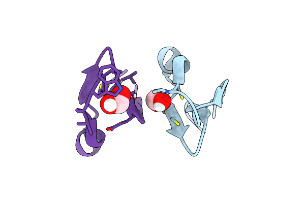 |
Crystal Structure Of Cycloviolacin O2
Organism: Viola odorata
Method: X-RAY DIFFRACTION Resolution:1.17 Å Release Date: 2021-09-22 Classification: PLANT PROTEIN Ligands: FMT |
Organism: Viola odorata
Method: X-RAY DIFFRACTION
Release Date: 2021-09-22
Ligands: FMT
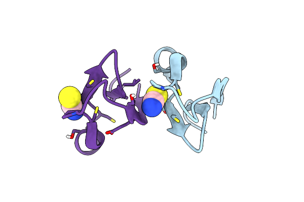 |
Crystal Structure Of [I11L]Cycloviolacin O2
Organism: Viola odorata
Method: X-RAY DIFFRACTION Resolution:1.04 Å Release Date: 2021-09-22 Classification: PLANT PROTEIN Ligands: SCN |
Organism: Viola odorata
Method: X-RAY DIFFRACTION
Release Date: 2021-09-22
Ligands: SCN
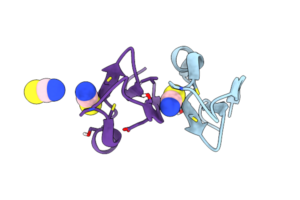 |
Crystal Structure Of [I11G]Cycloviolacin O2
Organism: Viola odorata
Method: X-RAY DIFFRACTION Resolution:1.10 Å Release Date: 2021-09-22 Classification: PLANT PROTEIN Ligands: SCN |
Organism: Viola odorata
Method: X-RAY DIFFRACTION
Release Date: 2021-09-22
Ligands: SCN
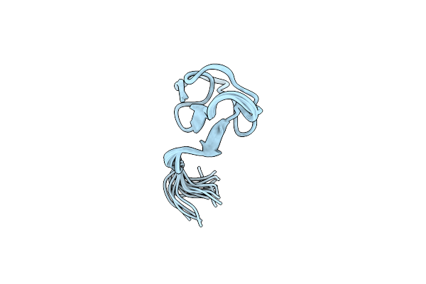 |
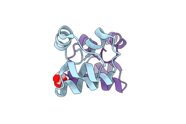
The Solution Structure Of Omega-Theraphotoxin-Pm1B Isolated From King Baboon Spider
|
Organism: Pelinobius muticus
Method: SOLUTION NMR
Release Date: 2021-09-08
 |
Crystal Structure Of Plantacyclin B21Ag
Organism: Lactobacillus plantarum
Method: X-RAY DIFFRACTION Resolution:1.80 Å Release Date: 2021-01-06 Classification: ANTIMICROBIAL PROTEIN Ligands: MLI |
Organism: Lactobacillus plantarum
Method: X-RAY DIFFRACTION
Release Date: 2021-01-06
Ligands: MLI
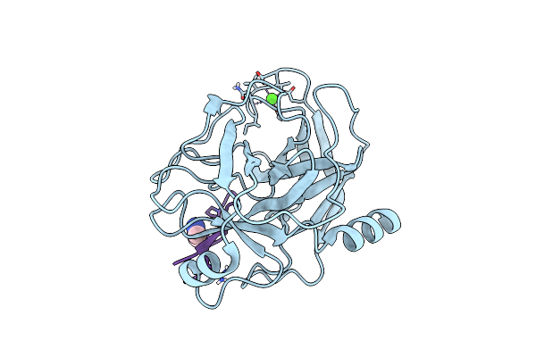 |
Crystal Structure Of Sfti-Triazole Inhibitor In Complex With Beta-Trypsin
Organism: Helianthus annuus, Bos taurus
Method: X-RAY DIFFRACTION Resolution:1.42 Å Release Date: 2020-07-01 Classification: HYDROLASE/INHIBITOR Ligands: CA, WMH |
Organism: Helianthus annuus, Bos taurus
Method: X-RAY DIFFRACTION
Release Date: 2020-07-01
Ligands: CA, WMH