

Search Count: 32
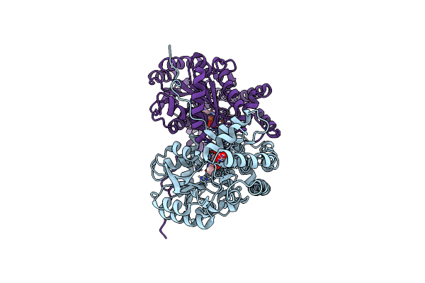 |
Joint X-Ray/Neutron Structure Of Aspastate Aminotransferase (Aat) In Complex With Pyridoxamine 5'-Phosphate (Pmp)
Organism: Sus scrofa
Method: X-RAY DIFFRACTION, NEUTRON DIFFRACTION Resolution:1.70 Å, 2.22 Å Release Date: 2022-09-28 Classification: TRANSFERASE Ligands: PLA, PMP, DOD |
Organism: Sus scrofa
Method: X-RAY DIFFRACTION, NEUTRON DIFFRACTION
Release Date: 2022-09-28
Ligands: PLA, PMP, DOD
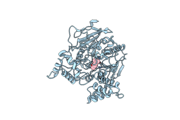 |
Room Temperature Structure Of Hache In Complex With Substrate Analog 4K-Tma
Organism: Homo sapiens
Method: X-RAY DIFFRACTION Resolution:2.80 Å Release Date: 2021-09-22 Classification: HYDROLASE Ligands: NWA |
Organism: Homo sapiens
Method: X-RAY DIFFRACTION
Release Date: 2021-09-22
Ligands: NWA
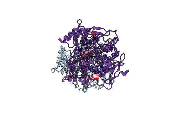 |
Low Temperature Structure Of Hache In Complex With Substrate Analog 4K-Tma
Organism: Homo sapiens
Method: X-RAY DIFFRACTION Resolution:2.40 Å Release Date: 2021-09-22 Classification: HYDROLASE Ligands: NWA, GOL, NO3 |
Organism: Homo sapiens
Method: X-RAY DIFFRACTION
Release Date: 2021-09-22
Ligands: NWA, GOL, NO3
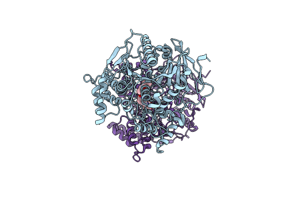 |
Room Temperature Structure Of Hache In Complex With Substrate Analog 4K-Tma And Mmb4 Oxime
Organism: Homo sapiens
Method: X-RAY DIFFRACTION Resolution:2.60 Å Release Date: 2021-09-22 Classification: HYDROLASE Ligands: NWA, 3VI |
Organism: Homo sapiens
Method: X-RAY DIFFRACTION
Release Date: 2021-09-22
Ligands: NWA, 3VI
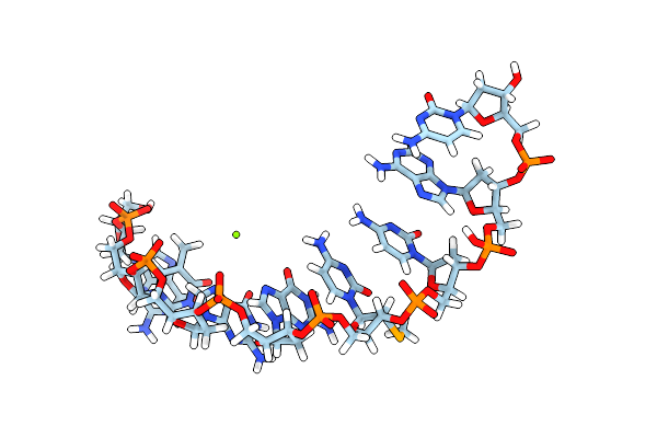 |
Joint X-Ray/Neutron Structure Of Dna Oligonucleotide D(Gtggccac)2 With 2'-Sech3 Modification On Cyt5
Organism: Synthetic construct
Method: X-RAY DIFFRACTION, NEUTRON DIFFRACTION Resolution:1.56 Å, 2.00 Å Release Date: 2018-10-17 Classification: DNA Ligands: MG, DOD |
Organism: Synthetic construct
Method: X-RAY DIFFRACTION, NEUTRON DIFFRACTION
Release Date: 2018-10-17
Ligands: MG, DOD
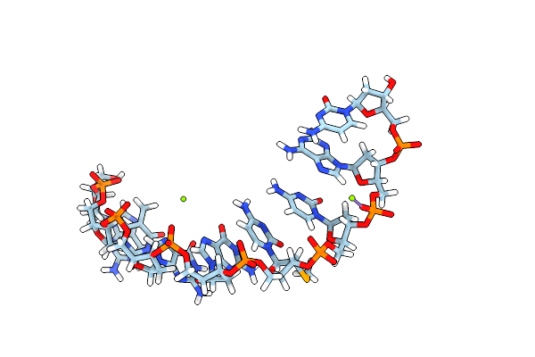 |
Low Temperature Joint X-Ray/Neutron Structure Of Dna Oligonucleotide D(Gtggccac)2 With 2'-Sech3 Modification On Cyt5
Organism: Synthetic construct
Method: X-RAY DIFFRACTION, NEUTRON DIFFRACTION Resolution:1.65 Å, 1.90 Å Release Date: 2018-10-17 Classification: DNA Ligands: MG, DOD |
Organism: Synthetic construct
Method: X-RAY DIFFRACTION, NEUTRON DIFFRACTION
Release Date: 2018-10-17
Ligands: MG, DOD
 |
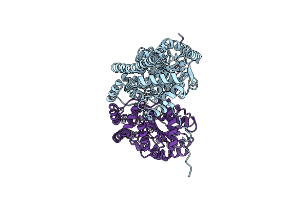
Joint X-Ray/Neutron Structure Of Aspartate Aminotransferase With Alpha-Methyl-Aspartate At Ph 7.5
Organism: Sus scrofa
Method: NEUTRON DIFFRACTION, X-RAY DIFFRACTION Resolution:2.00 Å, 2.21 Å Release Date: 2017-11-01 Classification: TRANSFERASE Ligands: PLA, DOD |
Organism: Sus scrofa
Method: NEUTRON DIFFRACTION, X-RAY DIFFRACTION
Release Date: 2017-11-01
Ligands: PLA, DOD
 |
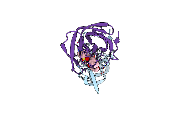
Aspartate Aminotransferase Ph 4.0
Organism: Sus scrofa
Method: X-RAY DIFFRACTION Resolution:1.90 Å Release Date: 2017-11-01 Classification: TRANSFERASE |
Organism: Sus scrofa
Method: X-RAY DIFFRACTION
Release Date: 2017-11-01
 |
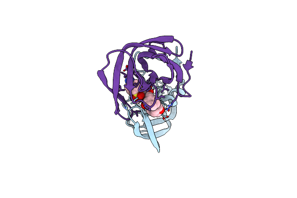
Joint X-Ray/Neutron Structure Of Hiv-1 Protease Triple Mutant (V32I,I47V,V82I) With Amprenavir At Ph 6.0
Organism: Human immunodeficiency virus 1
Method: NEUTRON DIFFRACTION, X-RAY DIFFRACTION Resolution:1.85 Å, 2.20 Å Release Date: 2017-03-01 Classification: HYDROLASE/HYDROLASE INHIBITOR Ligands: 478, DOD |
Organism: Human immunodeficiency virus 1
Method: NEUTRON DIFFRACTION, X-RAY DIFFRACTION
Release Date: 2017-03-01
Ligands: 478, DOD
 |
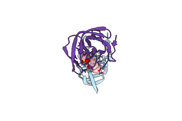
Joint X-Ray/Neutron Structure Of Hiv-1 Protease Triple Mutant (V32I,I47V,V82I) With Darunavir At Ph 6.0
Organism: Human immunodeficiency virus 1
Method: X-RAY DIFFRACTION, NEUTRON DIFFRACTION Resolution:1.85 Å, 2.00 Å Release Date: 2016-05-04 Classification: hydrolase/hydrolase inhibitor Ligands: 017, DOD |
Organism: Human immunodeficiency virus 1
Method: X-RAY DIFFRACTION, NEUTRON DIFFRACTION
Release Date: 2016-05-04
Ligands: 017, DOD
 |
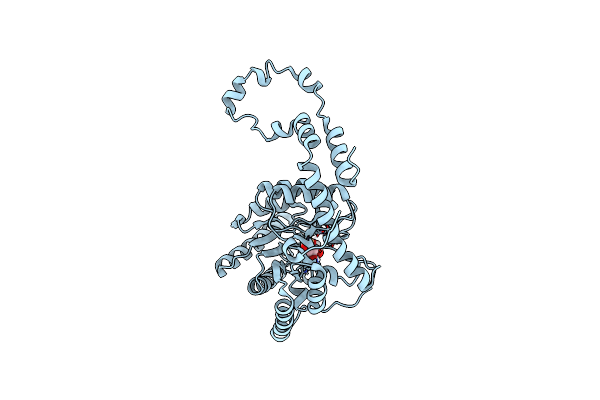
Joint X-Ray/Neutron Structure Of Hiv-1 Protease Triple Mutant (V32I,I47V,V82I) With Darunavir At Ph 4.3
Organism: Human immunodeficiency virus type 1 group m subtype b
Method: X-RAY DIFFRACTION, NEUTRON DIFFRACTION Resolution:1.75 Å, 2.30 Å Release Date: 2016-05-04 Classification: Hydrolase/hydrolase inhibitor Ligands: 017, DOD |
Organism: Human immunodeficiency virus type 1 group m subtype b
Method: X-RAY DIFFRACTION, NEUTRON DIFFRACTION
Release Date: 2016-05-04
Ligands: 017, DOD
 |
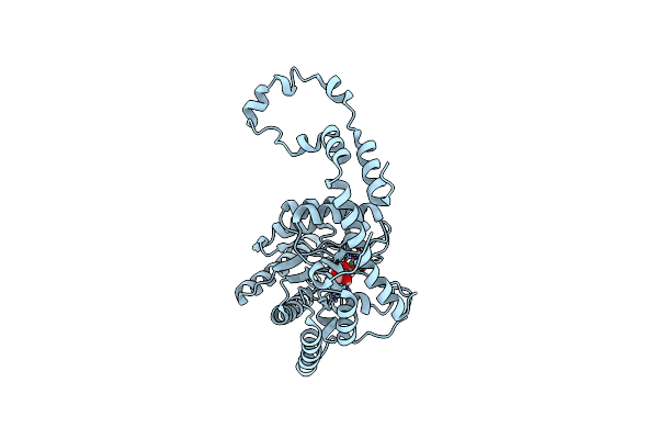
Joint X-Ray And Neutron Structure Of Streptomyces Rubiginosus D-Xylose Isomerase In Complex With Two Cd2+ Ions And Cyclic Beta-L-Arabinose
Organism: Streptomyces rubiginosus
Method: NEUTRON DIFFRACTION, X-RAY DIFFRACTION Resolution:2.00 Å Release Date: 2014-09-03 Classification: ISOMERASE Ligands: CD, ARB, DOD |
Organism: Streptomyces rubiginosus
Method: NEUTRON DIFFRACTION, X-RAY DIFFRACTION
Release Date: 2014-09-03
Ligands: CD, ARB, DOD
 |
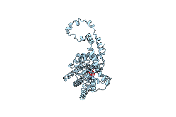
Joint X-Ray And Neutron Structure Of Streptomyces Rubiginosus D-Xylose Isomerase In Complex With Two Ni2+ Ions And Linear L-Arabinose
Organism: Streptomyces rubiginosus
Method: X-RAY DIFFRACTION, NEUTRON DIFFRACTION Resolution:1.80 Å Release Date: 2014-09-03 Classification: ISOMERASE Ligands: NI, LAI, DOD |
Organism: Streptomyces rubiginosus
Method: X-RAY DIFFRACTION, NEUTRON DIFFRACTION
Release Date: 2014-09-03
Ligands: NI, LAI, DOD
 |
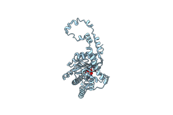
Room Temperature X-Ray Structure Of D-Xylose Isomerase In Complex With Two Cd2+ Ions And L-Ribulose
Organism: Streptomyces rubiginosus
Method: X-RAY DIFFRACTION Resolution:1.55 Å Release Date: 2014-09-03 Classification: ISOMERASE Ligands: CD, RUU |
Organism: Streptomyces rubiginosus
Method: X-RAY DIFFRACTION
Release Date: 2014-09-03
Ligands: CD, RUU
 |
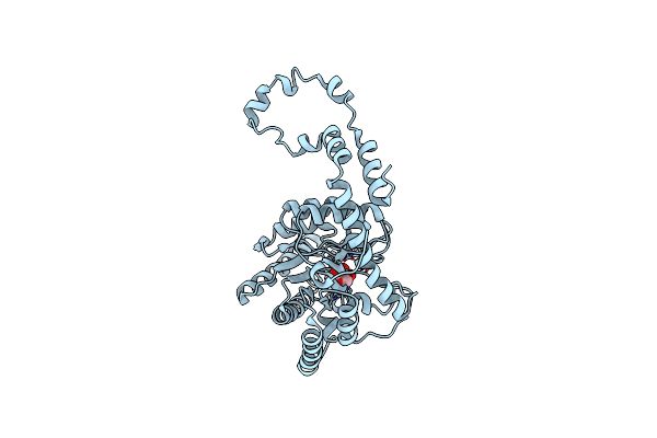
Room Temperature X-Ray Structure Of D-Xylose Isomerase In Complex With Two Ni2+ Ions And L-Ribulose
Organism: Streptomyces rubiginosus
Method: X-RAY DIFFRACTION Resolution:1.70 Å Release Date: 2014-09-03 Classification: ISOMERASE Ligands: NI, 34V |
Organism: Streptomyces rubiginosus
Method: X-RAY DIFFRACTION
Release Date: 2014-09-03
Ligands: NI, 34V
 |
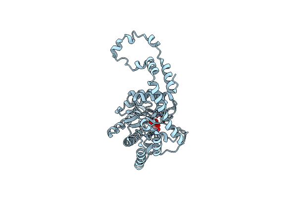
Room Temperature X-Ray Structure Of D-Xylose Isomerase In Complex With Two Mg2+ Ions And L-Ribulose
Organism: Streptomyces rubiginosus
Method: X-RAY DIFFRACTION Resolution:1.56 Å Release Date: 2014-09-03 Classification: ISOMERASE Ligands: MG, RUU |
Organism: Streptomyces rubiginosus
Method: X-RAY DIFFRACTION
Release Date: 2014-09-03
Ligands: MG, RUU
 |
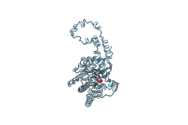
Room Temperature X-Ray Structure Of D-Xylose Isomerase In Complex With Two Ni2+ Ions And L-Ribose
Organism: Streptomyces rubiginosus
Method: X-RAY DIFFRACTION Resolution:1.60 Å Release Date: 2014-09-03 Classification: ISOMERASE Ligands: NI, Z6J |
Organism: Streptomyces rubiginosus
Method: X-RAY DIFFRACTION
Release Date: 2014-09-03
Ligands: NI, Z6J
 |
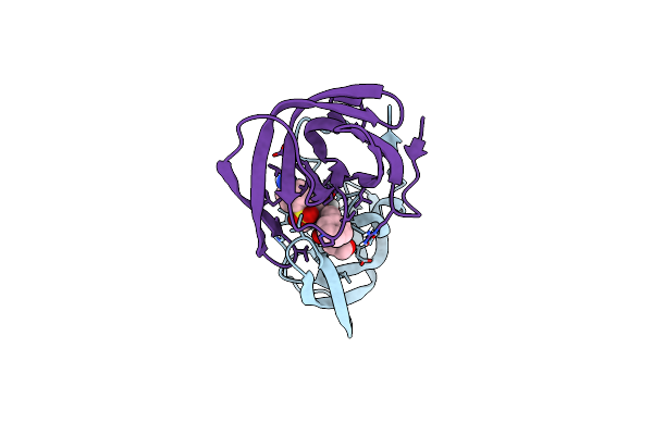
Room Temperature X-Ray Structure Of D-Xylose Isomerase In Complex With Two Mg2+ Ions And L-Ribose
Organism: Streptomyces rubiginosus
Method: X-RAY DIFFRACTION Resolution:1.55 Å Release Date: 2014-09-03 Classification: ISOMERASE Ligands: MG, 32O |
Organism: Streptomyces rubiginosus
Method: X-RAY DIFFRACTION
Release Date: 2014-09-03
Ligands: MG, 32O
 |
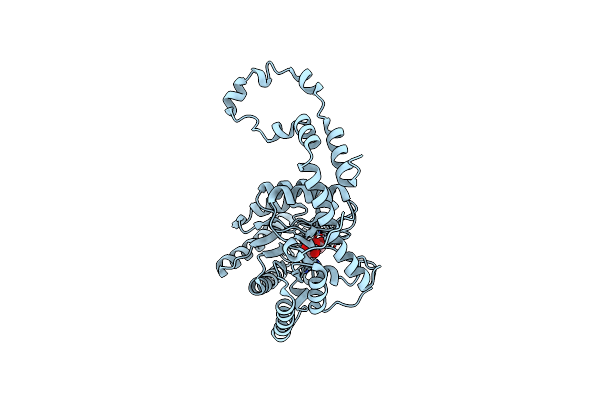
Joint Neutron And X-Ray Structure Of Per-Deuterated Hiv-1 Protease In Complex With Clinical Inhibitor Amprenavir
Organism: Human immunodeficiency virus type 1
Method: NEUTRON DIFFRACTION, X-RAY DIFFRACTION Resolution:2.01 Å, 2.00 Å Release Date: 2013-07-24 Classification: HYDROLASE/HYDROLASE INHIBITOR Ligands: CL, 478, DOD |
Organism: Human immunodeficiency virus type 1
Method: NEUTRON DIFFRACTION, X-RAY DIFFRACTION
Release Date: 2013-07-24
Ligands: CL, 478, DOD
 |
Room-Temperature X-Ray Structure Of D-Xylose Isomerase In Complex With 2Mg2+ Ions And Xylitol At Ph 7.7
Organism: Streptomyces rubiginosus
Method: X-RAY DIFFRACTION Resolution:2.00 Å Release Date: 2012-08-29 Classification: isomerase/isomerase inhibitor Ligands: MG, XYL |
Organism: Streptomyces rubiginosus
Method: X-RAY DIFFRACTION
Release Date: 2012-08-29
Ligands: MG, XYL