

Search Count: 111
 |
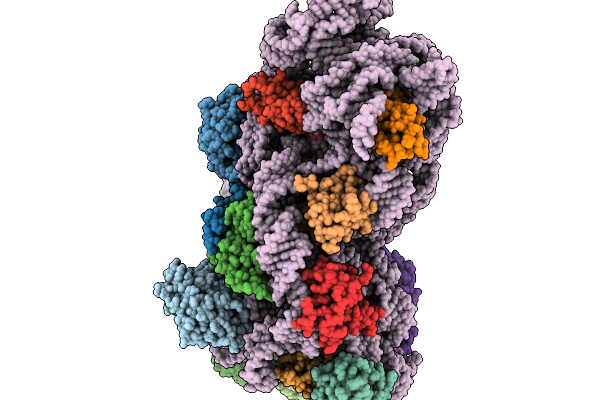
Hodarchaeales Hc1 Actin Filament
Organism: Candidatus hodarchaeales
Method: ELECTRON MICROSCOPY Release Date: 2026-06-24 Classification: PROTEIN FIBRIL |
Organism: Candidatus hodarchaeales
Method: ELECTRON MICROSCOPY
Release Date: 2026-06-24
 |
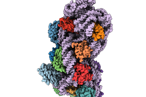
30S Ribosomal Subunit Rimm-Ko With If1 And If3 (State I)
Organism: Escherichia coli
Method: ELECTRON MICROSCOPY Release Date: 2026-02-04 Classification: RIBOSOME |
Organism: Escherichia coli
Method: ELECTRON MICROSCOPY
Release Date: 2026-02-04
 |
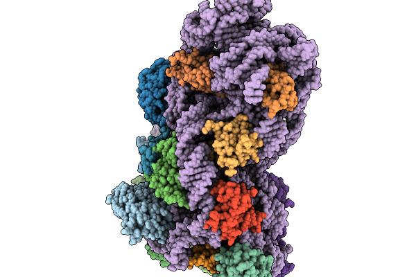
30S Ribosomal Subunit Rimm-Ko With If1 And If3 (State Ii)
Organism: Escherichia coli
Method: ELECTRON MICROSCOPY Release Date: 2026-02-04 Classification: RIBOSOME |
Organism: Escherichia coli
Method: ELECTRON MICROSCOPY
Release Date: 2026-02-04
 |
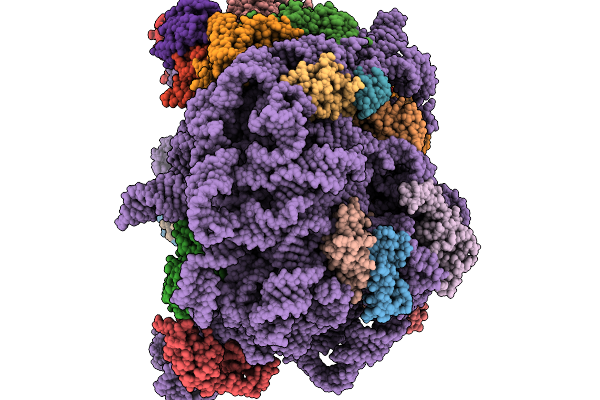
30S Ribosomal Subunit Rimm-Ko With If1 And If3 (State Iii)
Organism: Escherichia coli
Method: ELECTRON MICROSCOPY Release Date: 2026-02-04 Classification: RIBOSOME |
Organism: Escherichia coli
Method: ELECTRON MICROSCOPY
Release Date: 2026-02-04
 |
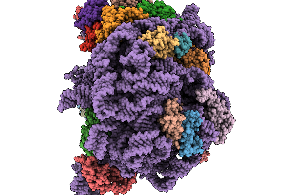
Pre-50S Ribosomal Subunit In Rimm-Ko With Rsfs (-H68)
Organism: Escherichia coli
Method: ELECTRON MICROSCOPY Release Date: 2026-02-04 Classification: RIBOSOME |
Organism: Escherichia coli
Method: ELECTRON MICROSCOPY
Release Date: 2026-02-04
 |
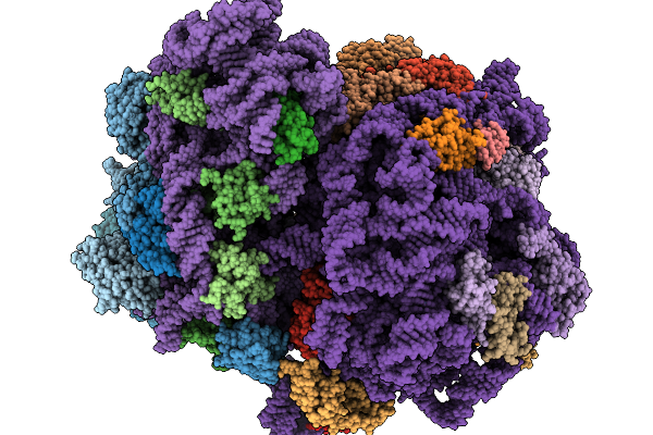
50S Ribosomal Subunit In Rimm-Ko With Rsfs (+H68)
Organism: Escherichia coli
Method: ELECTRON MICROSCOPY Release Date: 2026-02-04 Classification: RIBOSOME |
Organism: Escherichia coli
Method: ELECTRON MICROSCOPY
Release Date: 2026-02-04
 |
70S Ribosome From Rimm-Ko
Organism: Escherichia coli
Method: ELECTRON MICROSCOPY Release Date: 2026-02-04 Classification: RIBOSOME |
Organism: Escherichia coli
Method: ELECTRON MICROSCOPY
Release Date: 2026-02-04
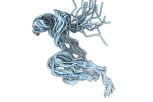 |
N-Terminal Half Domain Of Epc3
Organism: Colletotrichum orbiculare
Method: SOLUTION NMR Release Date: 2025-11-19 Classification: VIRAL PROTEIN |
Organism: Colletotrichum orbiculare
Method: SOLUTION NMR
Release Date: 2025-11-19
 |
Round 18 Arylesterase Variant Of Apo-Phosphotriesterase Measured At 13 Kev
Organism: Brevundimonas diminuta
Method: X-RAY DIFFRACTION Resolution:1.50 Å Release Date: 2024-04-03 Classification: HYDROLASE Ligands: MPD, CL |
Organism: Brevundimonas diminuta
Method: X-RAY DIFFRACTION
Release Date: 2024-04-03
Ligands: MPD, CL
 |
Round 18 Arylesterase Variant Of Apo-Phosphotriesterase Measured At 9.5 Kev
Organism: Brevundimonas diminuta
Method: X-RAY DIFFRACTION Resolution:1.52 Å Release Date: 2024-04-03 Classification: HYDROLASE Ligands: MPD, CL |
Organism: Brevundimonas diminuta
Method: X-RAY DIFFRACTION
Release Date: 2024-04-03
Ligands: MPD, CL
 |
Round 18 Arylesterase Variant Of Phosphotriesterase Bound To Europium(Iii) Measured At 9.5 Kev
Organism: Brevundimonas diminuta
Method: X-RAY DIFFRACTION Resolution:1.78 Å Release Date: 2024-04-03 Classification: HYDROLASE Ligands: MPD, EU3, CL |
Organism: Brevundimonas diminuta
Method: X-RAY DIFFRACTION
Release Date: 2024-04-03
Ligands: MPD, EU3, CL
 |
Round 18 Arylesterase Variant Of Phosphotriesterase Bound To Gadolinium(Iii) Measured At 9.5 Kev
Organism: Brevundimonas diminuta
Method: X-RAY DIFFRACTION Resolution:1.61 Å Release Date: 2024-04-03 Classification: HYDROLASE Ligands: MPD, GD3, CL |
Organism: Brevundimonas diminuta
Method: X-RAY DIFFRACTION
Release Date: 2024-04-03
Ligands: MPD, GD3, CL
 |
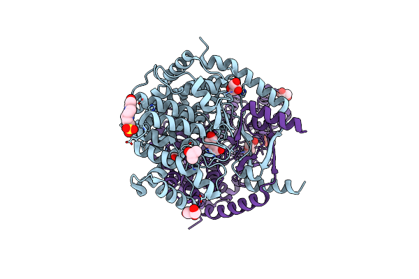
Crystal Structure Of Mitochondrial Citrate Synthase (Cit1) From Saccharomyces Cerevisiae
Organism: Saccharomyces cerevisiae
Method: X-RAY DIFFRACTION Resolution:1.42 Å Release Date: 2023-04-26 Classification: TRANSFERASE |
Organism: Saccharomyces cerevisiae
Method: X-RAY DIFFRACTION
Release Date: 2023-04-26
 |
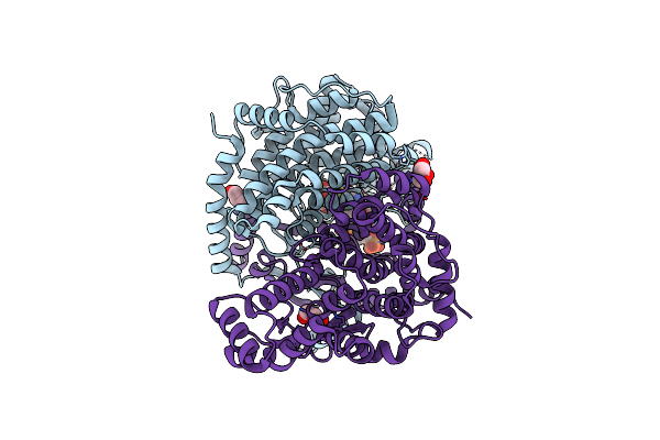
Crystal Structure Of Peroxisomal Citrate Synthase (Cit2) From Saccharomycescerevisiae
Organism: Saccharomyces cerevisiae
Method: X-RAY DIFFRACTION Resolution:2.39 Å Release Date: 2023-04-26 Classification: TRANSFERASE Ligands: CL, GOL, EPE, PEG |
Organism: Saccharomyces cerevisiae
Method: X-RAY DIFFRACTION
Release Date: 2023-04-26
Ligands: CL, GOL, EPE, PEG
 |
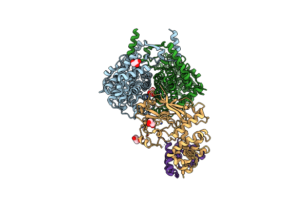
Crystal Structure Of Peroxisomal Citrate Synthase (Cit2) From Saccharomyces Cerevisiae In Complex With Oxaloacetate And Coenzyme-A
Organism: Saccharomyces cerevisiae
Method: X-RAY DIFFRACTION Resolution:1.48 Å Release Date: 2023-04-26 Classification: TRANSFERASE Ligands: COA, GOL, K, CL, OAA |
Organism: Saccharomyces cerevisiae
Method: X-RAY DIFFRACTION
Release Date: 2023-04-26
Ligands: COA, GOL, K, CL, OAA
 |
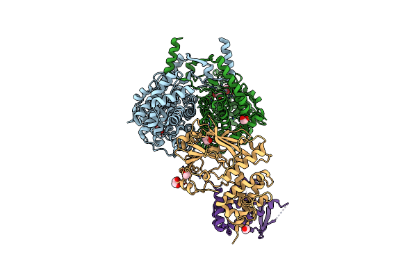
F-Box Protein In Complex With Skp1(Fl) And Substrate
Organism: Saccharomyces cerevisiae
Method: X-RAY DIFFRACTION Resolution:2.30 Å Release Date: 2023-04-26 Classification: TRANSFERASE/LIGASE Ligands: GOL |
Organism: Saccharomyces cerevisiae
Method: X-RAY DIFFRACTION
Release Date: 2023-04-26
Ligands: GOL
 |
Crystal Structure Of F-Box Protein In The Ternary Complex With Adaptor Protein Skp1(Dl) And Its Substrate
Organism: Saccharomyces cerevisiae
Method: X-RAY DIFFRACTION Resolution:2.53 Å Release Date: 2023-04-26 Classification: TRANSFERASE/LIGASE Ligands: EDO |
Organism: Saccharomyces cerevisiae
Method: X-RAY DIFFRACTION
Release Date: 2023-04-26
Ligands: EDO
 |
Crystal Structure Of Antibody Fab Fragment In Complex With Cd147(Emmpirin)
Organism: Homo sapiens, Synthetic construct
Method: X-RAY DIFFRACTION Resolution:2.30 Å Release Date: 2022-11-23 Classification: PROTEIN BINDING |
Organism: Homo sapiens, Synthetic construct
Method: X-RAY DIFFRACTION
Release Date: 2022-11-23
 |
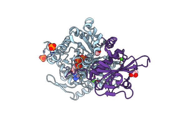
Crystal Structure Of Fragmin Domain-1 In Complex With Actin (Amppnp-Form)
Organism: Physarum polycephalum, Gallus gallus
Method: X-RAY DIFFRACTION Resolution:1.15 Å Release Date: 2022-10-26 Classification: CONTRACTILE PROTEIN Ligands: ANP, MG, PO4, EDO, CA |
Organism: Physarum polycephalum, Gallus gallus
Method: X-RAY DIFFRACTION
Release Date: 2022-10-26
Ligands: ANP, MG, PO4, EDO, CA
 |
Crystal Structure Of Fragmin Domain-1 In Complex With Actin (Adp-Pi-Form)
Organism: Physarum polycephalum, Gallus gallus
Method: X-RAY DIFFRACTION Resolution:1.15 Å Release Date: 2022-10-26 Classification: CONTRACTILE PROTEIN Ligands: ADP, PO4, MG, EDO, CA |
Organism: Physarum polycephalum, Gallus gallus
Method: X-RAY DIFFRACTION
Release Date: 2022-10-26
Ligands: ADP, PO4, MG, EDO, CA