
Search Count: 14
 |
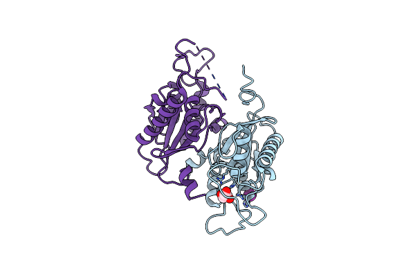
Rosc(R33A) - Riboflavin Complex
Organism: Streptomyces davaonensis jcm 4913
Method: X-RAY DIFFRACTION Resolution:1.50 Å Release Date: 2025-03-19 Classification: HYDROLASE Ligands: RBF, GOL |
 |
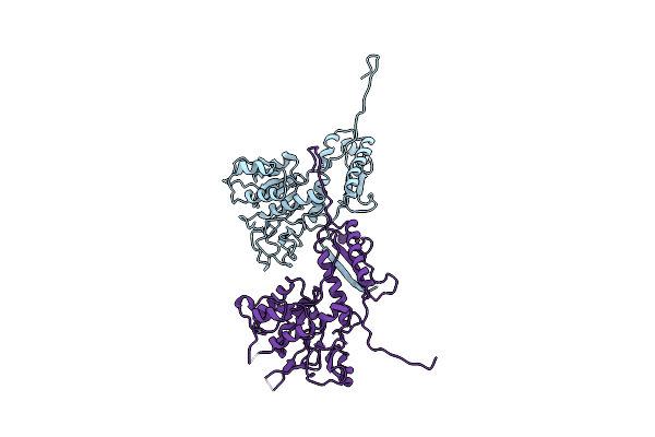
Rosc
Organism: Streptomyces davaonensis
Method: X-RAY DIFFRACTION Resolution:1.70 Å Release Date: 2025-03-12 Classification: HYDROLASE Ligands: IOD, NA, GOL |
 |
Rosc-8.Demethyl-8-Amino-Riboflavin Complex
Organism: Streptomyces davaonensis
Method: X-RAY DIFFRACTION Resolution:1.25 Å Release Date: 2025-03-12 Classification: HYDROLASE Ligands: RS3, GOL |
 |
Rosc-8-Demethyl-8-Amino-Fmn - Phosphate Complex
Organism: Streptomyces davaonensis
Method: X-RAY DIFFRACTION Resolution:1.40 Å Release Date: 2024-09-04 Classification: HYDROLASE Ligands: RS3, PO4, GOL |
 |
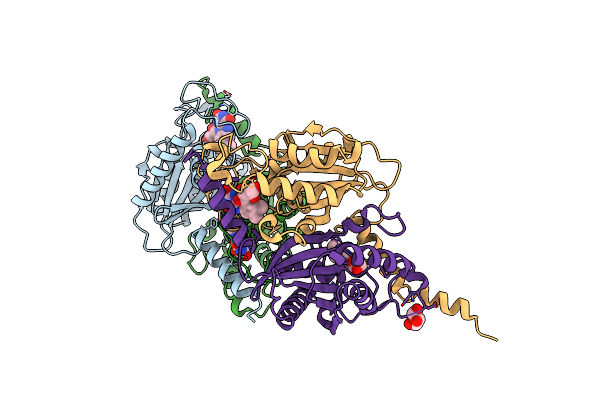
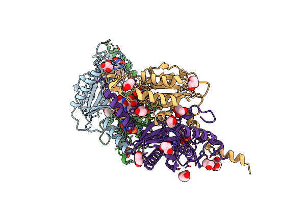
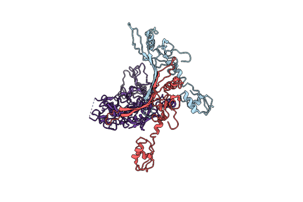
26S Proteasome Wt-Ubp6-Ubvs Complex In The Si State (Atpases, Rpn1, Ubp6, And Ubvs)
Organism: Saccharomyces cerevisiae, Homo sapiens
Method: ELECTRON MICROSCOPY Release Date: 2022-06-01 Classification: MOTOR PROTEIN Ligands: ATP, MG, ADP |
 |
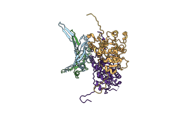
Structure Of The 26S Proteasome-Ubp6 Complex In The Si State (Core Particle And Lid)
Organism: Saccharomyces cerevisiae
Method: ELECTRON MICROSCOPY Release Date: 2022-04-13 Classification: MOTOR PROTEIN |
 |
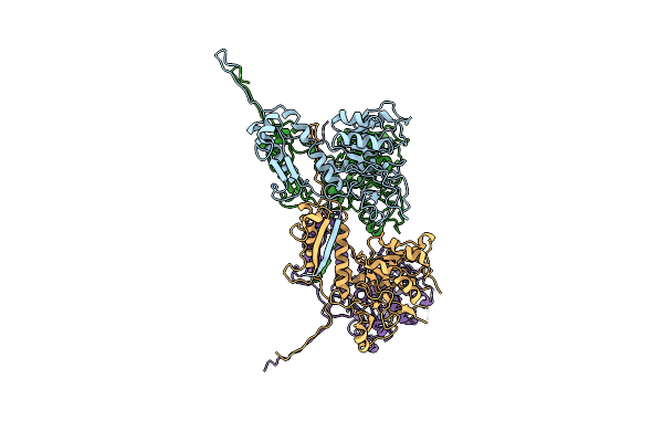
26S Proteasome Rpt1-Rk -Ubp6-Ubvs Complex In The Si State
Organism: Saccharomyces cerevisiae, Homo sapiens
Method: ELECTRON MICROSCOPY Release Date: 2022-03-16 Classification: MOTOR PROTEIN Ligands: ATP, MG, ADP |
 |
26S Proteasome Rpt1-Rk -Ubp6-Ubvs Complex In The S2 State
Organism: Saccharomyces cerevisiae, Homo sapiens
Method: ELECTRON MICROSCOPY Release Date: 2022-03-16 Classification: MOTOR PROTEIN Ligands: ATP, MG, ADP |
 |
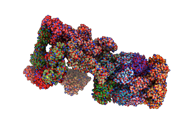
Cryo-Em Structure Of The Anti-Feeding Prophage (Afp) Baseplate In Extended State, 3-Fold Symmetrised
Organism: Serratia entomophila
Method: ELECTRON MICROSCOPY Release Date: 2019-04-24 Classification: VIRUS LIKE PARTICLE |
 |
Cryo-Em Structure Of The Anti-Feeding Prophage (Afp) Helical Sheath-Tube Complex In Extended State
Organism: Serratia entomophila
Method: ELECTRON MICROSCOPY Release Date: 2019-04-24 Classification: VIRUS LIKE PARTICLE |
 |
Cryo-Em Structure Of The Anti-Feeding Prophage (Afp) Baseplate In Contracted State
Organism: Serratia entomophila
Method: ELECTRON MICROSCOPY Release Date: 2019-04-24 Classification: VIRUS LIKE PARTICLE |
 |
Cryo-Em Structure Of The Anti-Feeding Prophage (Afp) Baseplate, 6-Fold Symmetrised
Organism: Serratia entomophila
Method: ELECTRON MICROSCOPY Release Date: 2019-04-17 Classification: VIRUS LIKE PARTICLE |
 |
Cryo-Em Structure Of The Anti-Feeding Prophage Cap (Afp Tube Terminating Cap)
Organism: Serratia entomophila
Method: ELECTRON MICROSCOPY Release Date: 2019-04-17 Classification: VIRUS LIKE PARTICLE |
 |
Cryo-Em Structure Of The Anti-Feeding Prophage (Afp) Helical Sheath In Contracted State
Organism: Serratia entomophila
Method: ELECTRON MICROSCOPY Release Date: 2019-04-17 Classification: VIRUS LIKE PARTICLE |