

Search Count: 10
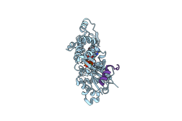 |
Structures Of Actin-Bound Wh2 Domains Of Spire And The Implication For Filament Nucleation
Organism: Drosophila melanogaster
Method: X-RAY DIFFRACTION Resolution:2.80 Å Release Date: 2010-06-02 Classification: Contractile Protein/Protein binding Ligands: CA, ATP |
Organism: Drosophila melanogaster
Method: X-RAY DIFFRACTION
Release Date: 2010-06-02
Ligands: CA, ATP
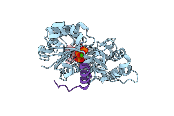 |
Structures Of Actin-Bound Wh2 Domains Of Spire And The Implication For Filament Nucleation
Organism: Drosophila melanogaster, Oryctolagus cuniculus
Method: X-RAY DIFFRACTION Resolution:1.50 Å Release Date: 2010-06-02 Classification: Contractile Protein/Protein binding Ligands: CA, ATP, LAB |
Organism: Drosophila melanogaster, Oryctolagus cuniculus
Method: X-RAY DIFFRACTION
Release Date: 2010-06-02
Ligands: CA, ATP, LAB
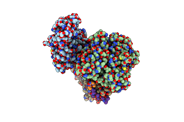 |
Structures Of Actin-Bound Wh2 Domains Of Spire And The Implication For Filament Nucleation
Organism: Drosophila melanogaster
Method: X-RAY DIFFRACTION Resolution:2.00 Å Release Date: 2010-06-02 Classification: Contractile Protein/Protein binding Ligands: CA, ATP |
Organism: Drosophila melanogaster
Method: X-RAY DIFFRACTION
Release Date: 2010-06-02
Ligands: CA, ATP
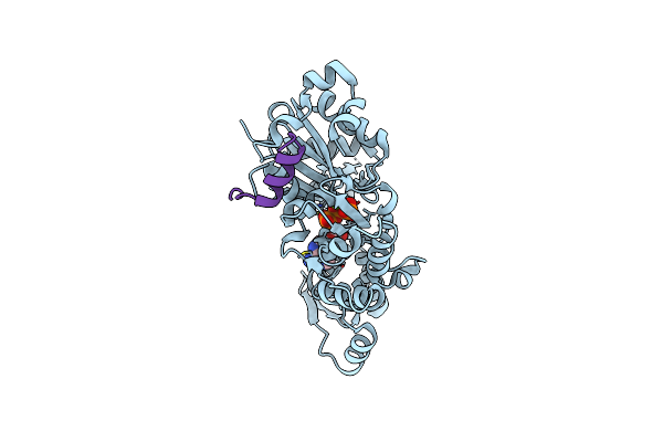 |
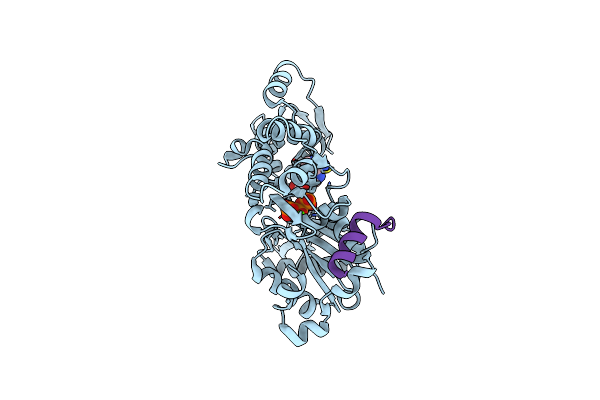
Structures Of Actin-Bound Wh2 Domains Of Spire And The Implication For Filament Nucleation
Organism: Drosophila melanogaster
Method: X-RAY DIFFRACTION Resolution:2.00 Å Release Date: 2010-05-26 Classification: Contractile Protein/Protein binding Ligands: ATP, CA |
Organism: Drosophila melanogaster
Method: X-RAY DIFFRACTION
Release Date: 2010-05-26
Ligands: ATP, CA
 |
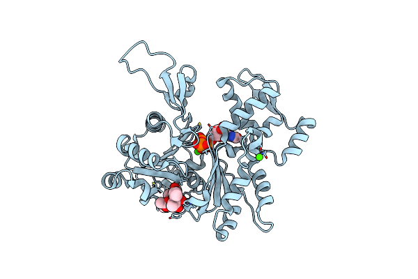
Structures Of Actin-Bound Wh2 Domains Of Spire And The Implication For Filament Nucleation
Organism: Drosophila melanogaster
Method: X-RAY DIFFRACTION Resolution:2.00 Å Release Date: 2010-05-26 Classification: Contractile Protein/Protein binding Ligands: ATP, CA |
Organism: Drosophila melanogaster
Method: X-RAY DIFFRACTION
Release Date: 2010-05-26
Ligands: ATP, CA
 |
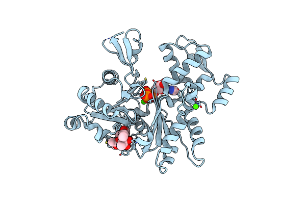
Crystal Structure Of Monomeric Actin Bound To Cytochalasin D
Organism: Drosophila melanogaster
Method: X-RAY DIFFRACTION Resolution:1.80 Å Release Date: 2008-10-07 Classification: CONTRACTILE PROTEIN Ligands: ATP, CA, CY9 |
Organism: Drosophila melanogaster
Method: X-RAY DIFFRACTION
Release Date: 2008-10-07
Ligands: ATP, CA, CY9
 |
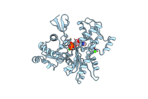
Crystal Structure Of Monomeric Actin Bound To Cytochalasin D
Organism: Drosophila melanogaster
Method: X-RAY DIFFRACTION Resolution:2.50 Å Release Date: 2008-10-07 Classification: CONTRACTILE PROTEIN Ligands: ATP, CA, CY9 |
Organism: Drosophila melanogaster
Method: X-RAY DIFFRACTION
Release Date: 2008-10-07
Ligands: ATP, CA, CY9
 |
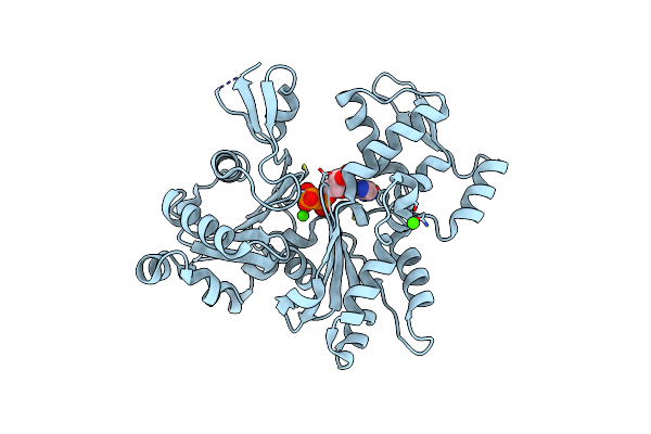
Crystal Structure Of Monomeric Actin Bound To Ca-Atp
Organism: Drosophila melanogaster
Method: X-RAY DIFFRACTION Resolution:2.50 Å Release Date: 2008-10-07 Classification: CONTRACTILE PROTEIN Ligands: ATP, CA |
Organism: Drosophila melanogaster
Method: X-RAY DIFFRACTION
Release Date: 2008-10-07
Ligands: ATP, CA
 |
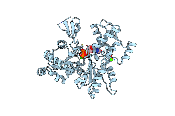
Crystal Structure Of Monomeric Actin In The Adp Bound State
Organism: Drosophila melanogaster
Method: X-RAY DIFFRACTION Resolution:1.80 Å Release Date: 2006-08-29 Classification: CONTRACTILE PROTEIN Ligands: CA, ADP |
Organism: Drosophila melanogaster
Method: X-RAY DIFFRACTION
Release Date: 2006-08-29
Ligands: CA, ADP
 |
Crystal Structure Of Monomeric Actin In Its Atp-Bound State
Organism: Drosophila melanogaster
Method: X-RAY DIFFRACTION Resolution:1.80 Å Release Date: 2006-08-29 Classification: CONTRACTILE PROTEIN Ligands: CA, ATP |
Organism: Drosophila melanogaster
Method: X-RAY DIFFRACTION
Release Date: 2006-08-29
Ligands: CA, ATP