
Search Count: 202
 |
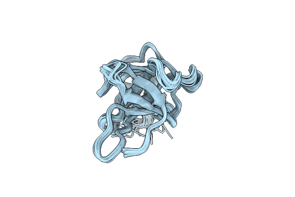
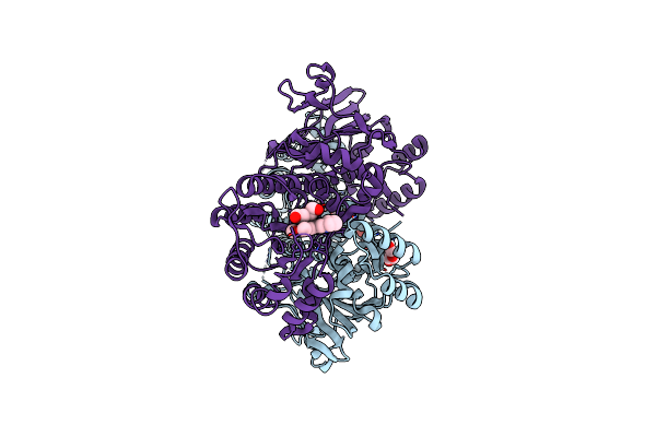
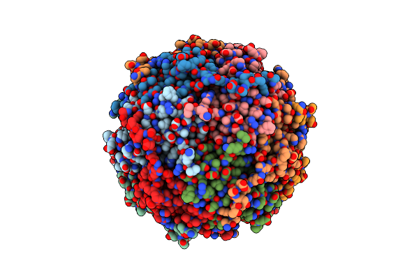
Mfa1 Type V Pilus From P.Gingivalis
Organism: Porphyromonas gingivalis atcc 33277
Method: ELECTRON MICROSCOPY Release Date: 2026-07-08 Classification: CELL ADHESION Ligands: CA |
Organism: Porphyromonas gingivalis atcc 33277
Method: ELECTRON MICROSCOPY
Release Date: 2026-07-08
Ligands: CA
 |
Solution Structure Of Tx33.1 From Conus Textile
|
Organism: Conus textile
Method: SOLUTION NMR
Release Date: 2025-07-02
 |
Alpha-Hemolysin Heptameric Late Pre-Pore State Derived From 10:0 Pc/Sphingomyelin Liposomes
Organism: Staphylococcus aureus
Method: ELECTRON MICROSCOPY Release Date: 2025-06-11 Classification: LIPID BINDING PROTEIN |
Organism: Staphylococcus aureus
Method: ELECTRON MICROSCOPY
Release Date: 2025-06-11
 |
Alpha-Hemolysin Heptameric Pre-Pore State Derived From 10:0 Pc Liposomes.
Organism: Staphylococcus aureus
Method: ELECTRON MICROSCOPY Release Date: 2025-06-11 Classification: LIPID BINDING PROTEIN |
Organism: Staphylococcus aureus
Method: ELECTRON MICROSCOPY
Release Date: 2025-06-11
 |
Alpha-Hemolysin Heptameric Pore State Derived From Egg Pc/Cholesterol (3:1 Molar Ratio) Liposomes
Organism: Staphylococcus aureus
Method: ELECTRON MICROSCOPY Release Date: 2025-06-11 Classification: LIPID BINDING PROTEIN |
Organism: Staphylococcus aureus
Method: ELECTRON MICROSCOPY
Release Date: 2025-06-11
 |
Alpha-Hemolysin Heptameric Pore State Derived From 10:0 Pc Liposomes
Organism: Staphylococcus aureus
Method: ELECTRON MICROSCOPY Release Date: 2025-06-11 Classification: LIPID BINDING PROTEIN |
Organism: Staphylococcus aureus
Method: ELECTRON MICROSCOPY
Release Date: 2025-06-11
 |
Alpha-Hemolysin Heptameric Popc Bound Pore State Derived From Egg Pc/Cholesterol (3:1 Molar Ratio) Liposomes
Organism: Staphylococcus aureus
Method: ELECTRON MICROSCOPY Resolution:3.00 Å Release Date: 2025-06-11 Classification: LIPID BINDING PROTEIN Ligands: POV |
Organism: Staphylococcus aureus
Method: ELECTRON MICROSCOPY
Release Date: 2025-06-11
Ligands: POV
 |
Alpha-Hemolysin Heptameric Pore State Bound To 10:0 Pc Lipid Chains Derived From 10:0 Pc Liposomes
Organism: Staphylococcus aureus
Method: ELECTRON MICROSCOPY Resolution:2.80 Å Release Date: 2025-06-11 Classification: LIPID BINDING PROTEIN Ligands: P1O |
Organism: Staphylococcus aureus
Method: ELECTRON MICROSCOPY
Release Date: 2025-06-11
Ligands: P1O
 |
Alpha-Hemolysin Heptameric Pre-Pore State Bound To 10:Pc Lipid Chains Derived From 10:0 Pc Liposomes.
Organism: Staphylococcus aureus
Method: ELECTRON MICROSCOPY Resolution:2.90 Å Release Date: 2025-06-11 Classification: LIPID BINDING PROTEIN Ligands: P1O |
Organism: Staphylococcus aureus
Method: ELECTRON MICROSCOPY
Release Date: 2025-06-11
Ligands: P1O
 |
Alpha-Hemolysin Heptameric Late Pre-Pore State With Bound Lipids Derived From 10:0 Pc/Sphingomyelin Liposomes
Organism: Staphylococcus aureus
Method: ELECTRON MICROSCOPY Resolution:2.60 Å Release Date: 2025-06-11 Classification: LIPID BINDING PROTEIN Ligands: P1O |
Organism: Staphylococcus aureus
Method: ELECTRON MICROSCOPY
Release Date: 2025-06-11
Ligands: P1O
 |
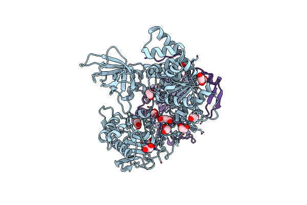
Crystal Structure Of Nampt-Pf403
Organism: Homo sapiens
Method: X-RAY DIFFRACTION Resolution:1.86 Å Release Date: 2025-02-19 Classification: BIOSYNTHETIC PROTEIN Ligands: A1D52, PEG |
Organism: Homo sapiens
Method: X-RAY DIFFRACTION
Release Date: 2025-02-19
Ligands: A1D52, PEG
 |
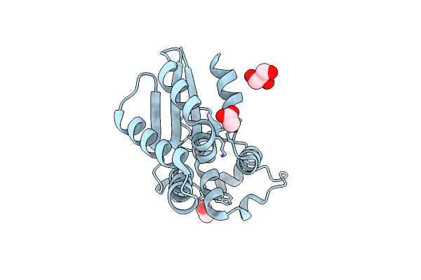
Sfx Structure Of An Mn-Carbonyl Complex Immobilized In Hen Egg White Lysozyme Microcrystals, 100 Ns After Photoexcitation At Rt.
Organism: Gallus gallus
Method: X-RAY DIFFRACTION Resolution:1.60 Å Release Date: 2024-09-04 Classification: HYDROLASE Ligands: CL, NA, XTX |
Organism: Gallus gallus
Method: X-RAY DIFFRACTION
Release Date: 2024-09-04
Ligands: CL, NA, XTX
 |
Crystal Structure Of Saccharomyces Cerevisiae Nmd4 Protein Bound To Upf1 Helicase Domain
Organism: Saccharomyces cerevisiae s288c
Method: X-RAY DIFFRACTION Resolution:2.40 Å Release Date: 2024-08-14 Classification: RNA BINDING PROTEIN Ligands: EDO, SIN, FMT, MLA |
Organism: Saccharomyces cerevisiae s288c
Method: X-RAY DIFFRACTION
Release Date: 2024-08-14
Ligands: EDO, SIN, FMT, MLA
 |
Crystal Structure Of Saccharomyces Cerevisiae Nmd4 Protein Involved In Nonsense Mediated Mrna Decay
Organism: Saccharomyces cerevisiae s288c
Method: X-RAY DIFFRACTION Resolution:1.80 Å Release Date: 2024-08-14 Classification: RNA BINDING PROTEIN Ligands: GOL, ACY |
Organism: Saccharomyces cerevisiae s288c
Method: X-RAY DIFFRACTION
Release Date: 2024-08-14
Ligands: GOL, ACY
 |
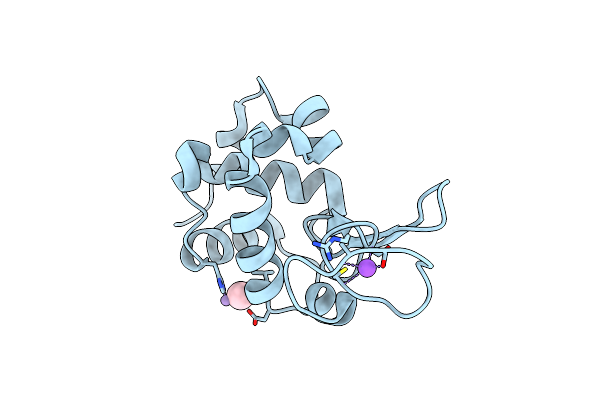
Dark State Sfx Structure Of Hen Egg White Lysozyme Microcrystals Immobilized With A Mn Carbonyl Complex At Rt.
Organism: Gallus gallus
Method: X-RAY DIFFRACTION Resolution:1.60 Å Release Date: 2024-07-31 Classification: HYDROLASE Ligands: CL, NA, XTX |
Organism: Gallus gallus
Method: X-RAY DIFFRACTION
Release Date: 2024-07-31
Ligands: CL, NA, XTX
 |
Sfx Structure Of An Mn-Carbonyl Complex Immobilized In Hen Egg White Lysozyme Microcrystals, 10 Ns After Photoexcitation At Rt.
Organism: Gallus gallus
Method: X-RAY DIFFRACTION Resolution:1.60 Å Release Date: 2024-07-31 Classification: HYDROLASE Ligands: CL, NA, XTX |
Organism: Gallus gallus
Method: X-RAY DIFFRACTION
Release Date: 2024-07-31
Ligands: CL, NA, XTX
 |
Sfx Structure Of An Mn-Carbonyl Complex Immobilized In Hen Egg White Lysozyme Microcrystals, 1 Microsecond After Photoexcitation At Rt.
Organism: Gallus gallus
Method: X-RAY DIFFRACTION Resolution:1.60 Å Release Date: 2024-07-31 Classification: HYDROLASE Ligands: CL, NA, XTX |
Organism: Gallus gallus
Method: X-RAY DIFFRACTION
Release Date: 2024-07-31
Ligands: CL, NA, XTX
 |
Sfx Structure Of An Mn-Carbonyl Complex Immobilized In Hen Egg White Lysozyme Microcrystals, 1 Microsecond After Photoexcitation With 40 Microjoules Laser Intensity At Rt.
Organism: Gallus gallus
Method: X-RAY DIFFRACTION Resolution:1.60 Å Release Date: 2024-07-31 Classification: HYDROLASE Ligands: CL, NA, XTX |
Organism: Gallus gallus
Method: X-RAY DIFFRACTION
Release Date: 2024-07-31
Ligands: CL, NA, XTX
 |
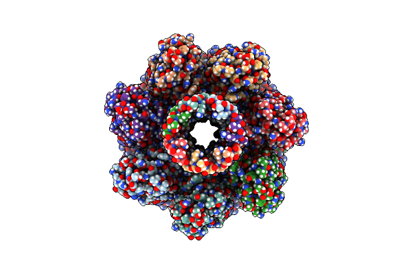
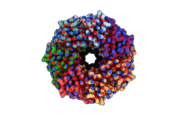
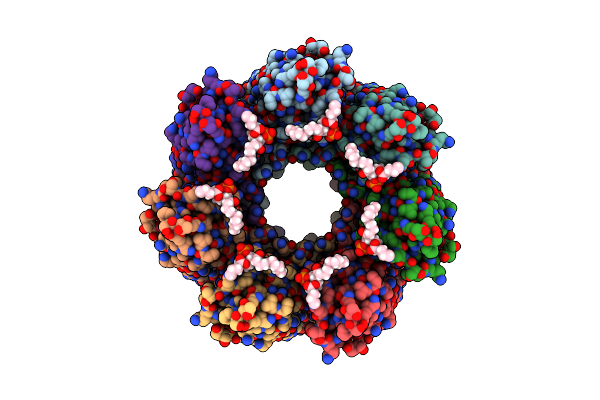
Cryo-Em Structure Of Delta N15 Msdps2 Of Mycobacterium Smegmatis
Organism: Mycolicibacterium smegmatis mc2 155
Method: ELECTRON MICROSCOPY Release Date: 2024-07-10 Classification: DNA BINDING PROTEIN |
Organism: Mycolicibacterium smegmatis mc2 155
Method: ELECTRON MICROSCOPY
Release Date: 2024-07-10
 |
Cryo-Em Structure Of Msdps2 From Mycobacterium Smegmatis
Organism: Mycolicibacterium smegmatis mc2 155
Method: ELECTRON MICROSCOPY Release Date: 2024-07-10 Classification: DNA BINDING PROTEIN |
Organism: Mycolicibacterium smegmatis mc2 155
Method: ELECTRON MICROSCOPY
Release Date: 2024-07-10